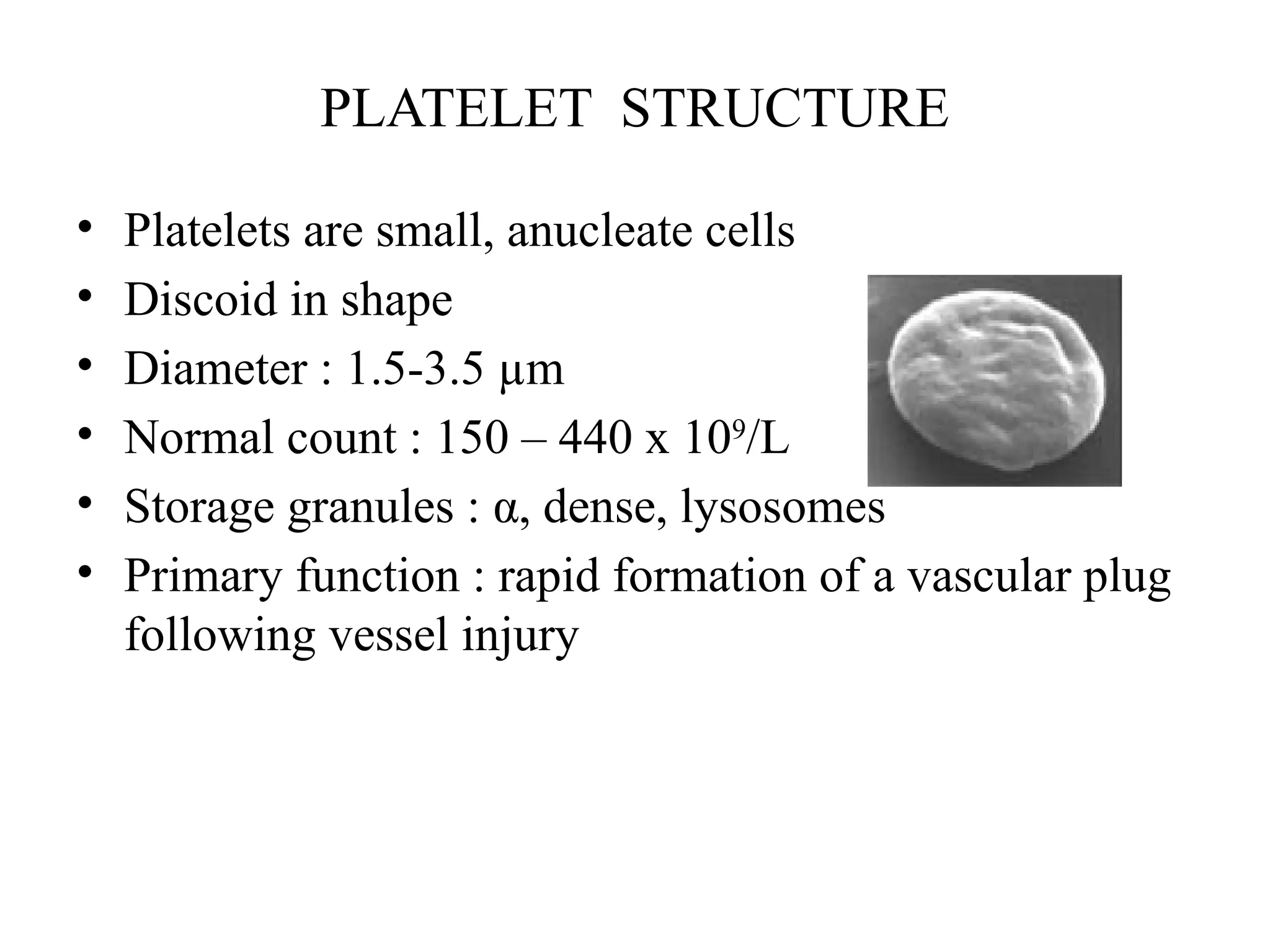
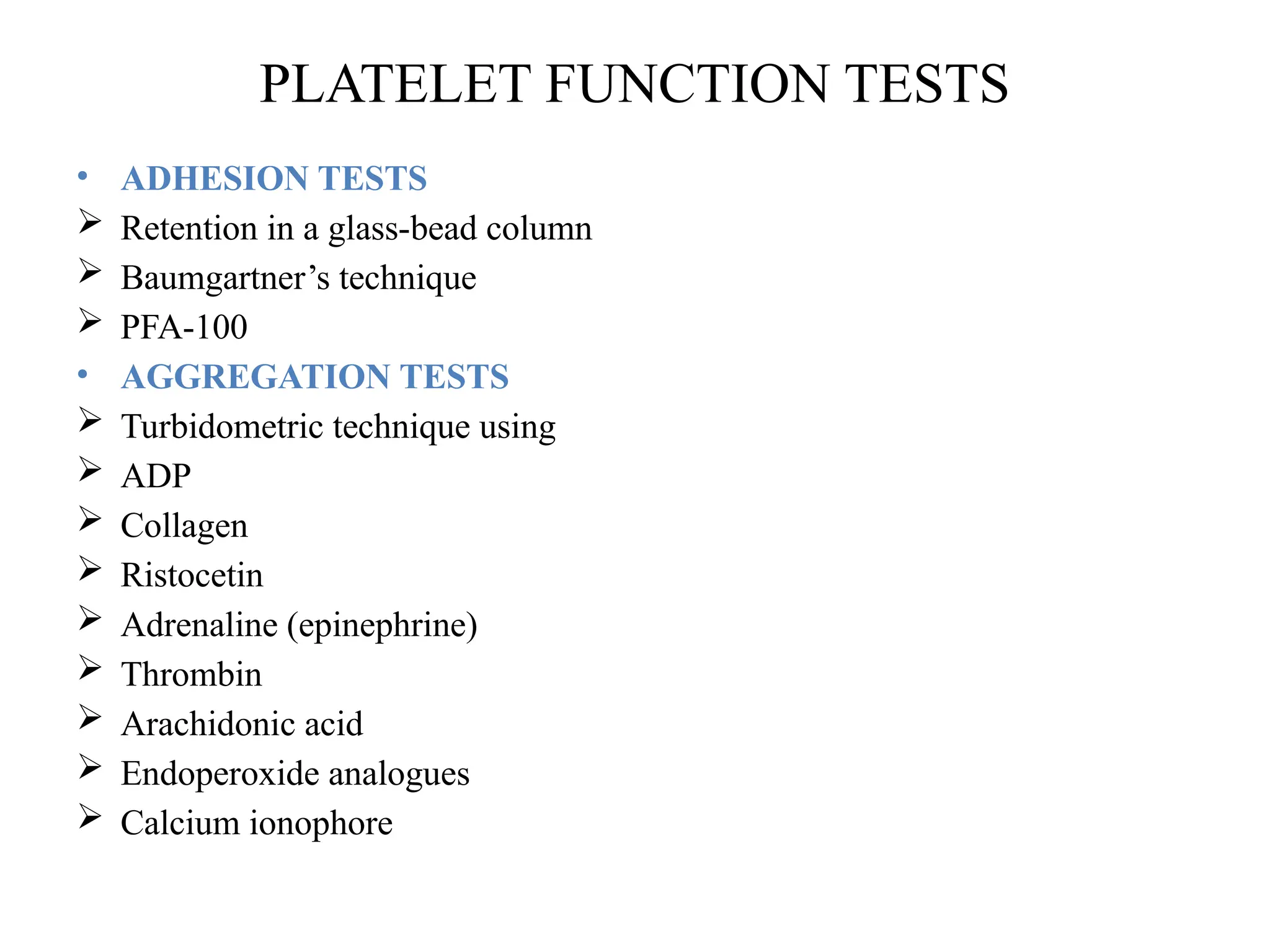
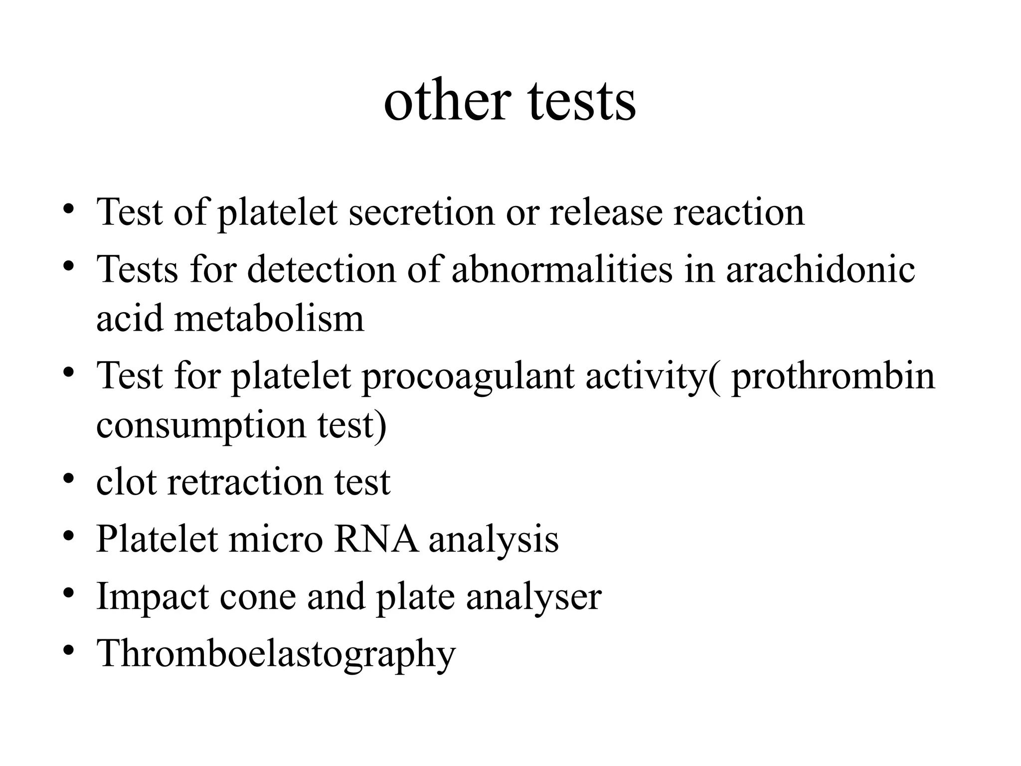
PLATELET FUNCTION TESTS
INTRODUCTION
• Definition: Small, colourless,anucleated cellular
fragment formed from megakaryocyte
• Platelets are 2nd
major component of the hemostatic
system
• There role is first established by Bizzozero in 1892
PLATELET FORMATION
• From megakaryocytes in the bone marrow
• To generate 2000 – 3000 platelets per megakaryocyte
PLATELET STRUCTURE
• Platelets are small, anucleate cells
• Discoid in shape
• Diameter : 1.5-3.5 µm
• Normal count : 150 – 440 x 109
/L
• Storage granules : α, dense, lysosomes
• Primary function : rapid formation of a vascular plug
following vessel injury
Peripheral zone
Structural zone
Organelle zone
Membrane systems
α Granules
Group 1- hemostatic proteins
• Fibrinogen
• Factor v
• Von willebrand factor
• Plasminogen
• Plasminogen activator
inhibitor(PAI-1)
• α2antiplasmin
Group II- non hemostatic proteins
• Platelet specific
 Β thromboglobulin
 Platelet factor 4
 Platelet derived growth factor
• Non platelet specific
 Albumin
 Thrombospondin
 Fibronectin
PLATELET FUNCTIONS
• Formation of primary haemostatic plug
• Surface for coagulation factor to make secondary
haemostatic plug
• Aid in healing of injured tissue
1. Adhesion
2. Shape change
3. Aggregation
4. Secretion
PLATELET FUNCTION TEST
• Group of test that measure the ability of
platelet to aggregate and promote clotting in
sample.
• OR it measures any dysfunction in platelet.
PLATELET FUNCTION TESTS
• ADHESION TESTS
 Retention in a glass-bead column
 Baumgartner’s technique
 PFA-100
• AGGREGATION TESTS
 Turbidometric technique using
 ADP
 Collagen
 Ristocetin
 Adrenaline (epinephrine)
 Thrombin
 Arachidonic acid
 Endoperoxide analogues
 Calcium ionophore
• INVESTIGATION OF GRANULE CONTENT AND
RELEASE
 Dense bodies
 Electron microscopy
 ADP and ATP content (bioluminescence)
 Serotonin release
• GRANULES
 b-thromboglobulin
 Platelet factor 4
 VWF
 Fluorescence by flow cytometry
• PROSTAGLANDIN PATHWAYS
 TXA2 radioimmunoassay
• PLATELET COAGULANT ACTIVITY
 Prothrombin consumption index
• FLOW CYTOMETRY
 Glycoprotein surface expression
 Activation
 P selectin (CD62) surface expression
• FIBRINOGEN BINDING
 Annexin binding (to phosphatidyl serine)
 Conformational changes in Gp IIbIIIa
PLATELET FUNCTION ANALYSER
• New generation of rapid whole-blood analyzers
• Screen for platelet function defect
• Whole blood is passed through a hole in a membrane
coated with collagen and either ADP or epinephrine
• Combined influence of high shear rates and VWF,
platelets encountering this membrane spontaneously
adhere to collagen
PFA-100 [Platelet Function Analyser]
• And aggregate because of stimulation by ADP or
epinephrine
• Measures closure time (CT), the amount of time
required for a platelet plug to occlude the aperture
and stop flow
CT is prolonged in
• Severe congenital and acquired platelet dysfunction
• Defects in platelet glycoprotein receptors GPIIb/IIIa
(Glanzmann thrombasthenia) and GPIb (Bernard-
Soulier syndrome)
• Monitoring of patients taking aspirin – aspirin
resistance
ADVANTAGES
• Whole blood test
• Small blood volumes
• Simple
• Rapid
DISADVANTAGES
• Inflexible
• vWf dependent
OTHER ADHESION TESTS :
• Retention in a glass bead column
• Baumgartners technique
Retention in a glass-bead column
AGGREGATION TESTS
• Measures the ability of various agonists of platelets to
induce in vitro activation and platelet-to-platelet
aggregation
• Instrument: Aggregometer
• PRINCIPLE:
• PRP has high optical density and transmits light
poorly because platelets prevent passage of light.
• PPP has been depleted of platelets and is able to
transmit light well (low optical density)
• When substance that promotes platelet activation and
aggregation (agonist) is added to PRP, normal
platelets are activated and aggregate.
• Platelet aggregometer measures change in
optical density for light transmittance) over
time of stirred PRP in cuvettes at 37°C after
addition of agonists.
• Aggregation of platelets in PRP results in
decrease in optical density (increased light
transmittance) as aggregated platelets fall out
of path of light.
SEMINAR
STRONG AGONISTS
• Directly induce platelet
aggregation, TxA2
synthesis and platelet
granule secretion
• Eg : Collagen
Thrombin
TxA2
WEAK AGONISTS
• Induce platelet
aggregation without
inducing secretion
• Eg : ADP
Epinephrine
20/08/2018
SECRETION ASSAYS
• Measured by 2 different ways
 Measuring ATP secretion from platelets
 14
C-labelled serotonin
 ATP secretion from platelets
• PRP or whole blood is incubated with firefly
luciferase and its substrate luciferin
• Addition of platelet agonists causes release of ATP, a
cofactor in a light-producing luciferin-luciferase
assay
• Resulting light can be measured with a photodetector
system
• Often performed simultaneously with a platelet
aggregation assay
FLOW CYTOMETRY
• Measure multiple properties of single cells in
suspension
• Specific monoclonal antibodies are used to detect
antigens or proteins in these cells
• Antibodies are bind with fluorescent dyes that are
activated as the cells pass in a single-file stream
through the flow cytometer
• Light emitted from each fluorochrome can be
individually detected and its intensity measured for each
cell
Diagnosis of:
• inherited defects in platelet surface glycoproteins
• Decreased expression or absence of GPIb - Bernard-
Soulier syndrome
• GPIIb/IIIa - Glanzmann thrombasthenia
• Multiple antibodies against different GPIIb/IIIa epitopes
- acquired Glanzmann thrombasthenia
other tests
• Test of platelet secretion or release reaction
• Tests for detection of abnormalities in arachidonic
acid metabolism
• Test for platelet procoagulant activity( prothrombin
consumption test)
• clot retraction test
• Platelet micro RNA analysis
• Impact cone and plate analyser
• Thromboelastography
REFERENCES
• 1.Hoffbrand A V, Catovsky D, Tuddenham E G D, editors.
Postgraduate Hematology.5th
ed. Massachusetts:Blackwell
Publishing Ltd; 2005.P.808-24.
• 2. Seegmiller A, Sarode R. Laboratory Evaluation of Platelet
Function. Hematol Oncol Clin N Am 2007;21:731–42.
• 3. Quinn M, Fitzgerald D, Platelet function, assesment,
diagnosis and treatment. New Jersy:Humana press;2005:P.202-
328.
• 4. Platelet Function Testing: Light Transmission Aggregometry
[LTA], 29-4-2012, Available from http://www.platelet-
research.org/3/aggregometry.htm.
• 5.Laffan M A, Manning R , Investigation of hemostasis, Bain
B.J, Bates I, Laffan M.A, Lewis S.M, Dacie and Lewis Practical
Haematology. 11th
ed. London: 2012. P.424-39.
Thank you!!!

pft.platelet function test haematology..

  • 1.
  • 2.
    INTRODUCTION • Definition: Small,colourless,anucleated cellular fragment formed from megakaryocyte • Platelets are 2nd major component of the hemostatic system • There role is first established by Bizzozero in 1892
  • 3.
    PLATELET FORMATION • Frommegakaryocytes in the bone marrow • To generate 2000 – 3000 platelets per megakaryocyte
  • 4.
    PLATELET STRUCTURE • Plateletsare small, anucleate cells • Discoid in shape • Diameter : 1.5-3.5 µm • Normal count : 150 – 440 x 109 /L • Storage granules : α, dense, lysosomes • Primary function : rapid formation of a vascular plug following vessel injury
  • 5.
  • 6.
    α Granules Group 1-hemostatic proteins • Fibrinogen • Factor v • Von willebrand factor • Plasminogen • Plasminogen activator inhibitor(PAI-1) • α2antiplasmin Group II- non hemostatic proteins • Platelet specific  Β thromboglobulin  Platelet factor 4  Platelet derived growth factor • Non platelet specific  Albumin  Thrombospondin  Fibronectin
  • 7.
    PLATELET FUNCTIONS • Formationof primary haemostatic plug • Surface for coagulation factor to make secondary haemostatic plug • Aid in healing of injured tissue 1. Adhesion 2. Shape change 3. Aggregation 4. Secretion
  • 8.
    PLATELET FUNCTION TEST •Group of test that measure the ability of platelet to aggregate and promote clotting in sample. • OR it measures any dysfunction in platelet.
  • 9.
    PLATELET FUNCTION TESTS •ADHESION TESTS  Retention in a glass-bead column  Baumgartner’s technique  PFA-100 • AGGREGATION TESTS  Turbidometric technique using  ADP  Collagen  Ristocetin  Adrenaline (epinephrine)  Thrombin  Arachidonic acid  Endoperoxide analogues  Calcium ionophore
  • 10.
    • INVESTIGATION OFGRANULE CONTENT AND RELEASE  Dense bodies  Electron microscopy  ADP and ATP content (bioluminescence)  Serotonin release • GRANULES  b-thromboglobulin  Platelet factor 4  VWF  Fluorescence by flow cytometry
  • 11.
    • PROSTAGLANDIN PATHWAYS TXA2 radioimmunoassay • PLATELET COAGULANT ACTIVITY  Prothrombin consumption index • FLOW CYTOMETRY  Glycoprotein surface expression  Activation  P selectin (CD62) surface expression • FIBRINOGEN BINDING  Annexin binding (to phosphatidyl serine)  Conformational changes in Gp IIbIIIa
  • 12.
    PLATELET FUNCTION ANALYSER •New generation of rapid whole-blood analyzers • Screen for platelet function defect • Whole blood is passed through a hole in a membrane coated with collagen and either ADP or epinephrine • Combined influence of high shear rates and VWF, platelets encountering this membrane spontaneously adhere to collagen
  • 13.
  • 14.
    • And aggregatebecause of stimulation by ADP or epinephrine • Measures closure time (CT), the amount of time required for a platelet plug to occlude the aperture and stop flow
  • 15.
    CT is prolongedin • Severe congenital and acquired platelet dysfunction • Defects in platelet glycoprotein receptors GPIIb/IIIa (Glanzmann thrombasthenia) and GPIb (Bernard- Soulier syndrome) • Monitoring of patients taking aspirin – aspirin resistance
  • 16.
    ADVANTAGES • Whole bloodtest • Small blood volumes • Simple • Rapid DISADVANTAGES • Inflexible • vWf dependent
  • 17.
    OTHER ADHESION TESTS: • Retention in a glass bead column • Baumgartners technique
  • 18.
    Retention in aglass-bead column
  • 19.
    AGGREGATION TESTS • Measuresthe ability of various agonists of platelets to induce in vitro activation and platelet-to-platelet aggregation • Instrument: Aggregometer
  • 20.
    • PRINCIPLE: • PRPhas high optical density and transmits light poorly because platelets prevent passage of light. • PPP has been depleted of platelets and is able to transmit light well (low optical density) • When substance that promotes platelet activation and aggregation (agonist) is added to PRP, normal platelets are activated and aggregate.
  • 22.
    • Platelet aggregometermeasures change in optical density for light transmittance) over time of stirred PRP in cuvettes at 37°C after addition of agonists. • Aggregation of platelets in PRP results in decrease in optical density (increased light transmittance) as aggregated platelets fall out of path of light.
  • 23.
    SEMINAR STRONG AGONISTS • Directlyinduce platelet aggregation, TxA2 synthesis and platelet granule secretion • Eg : Collagen Thrombin TxA2 WEAK AGONISTS • Induce platelet aggregation without inducing secretion • Eg : ADP Epinephrine 20/08/2018
  • 24.
    SECRETION ASSAYS • Measuredby 2 different ways  Measuring ATP secretion from platelets  14 C-labelled serotonin
  • 25.
     ATP secretionfrom platelets • PRP or whole blood is incubated with firefly luciferase and its substrate luciferin • Addition of platelet agonists causes release of ATP, a cofactor in a light-producing luciferin-luciferase assay • Resulting light can be measured with a photodetector system • Often performed simultaneously with a platelet aggregation assay
  • 26.
    FLOW CYTOMETRY • Measuremultiple properties of single cells in suspension • Specific monoclonal antibodies are used to detect antigens or proteins in these cells • Antibodies are bind with fluorescent dyes that are activated as the cells pass in a single-file stream through the flow cytometer
  • 27.
    • Light emittedfrom each fluorochrome can be individually detected and its intensity measured for each cell Diagnosis of: • inherited defects in platelet surface glycoproteins • Decreased expression or absence of GPIb - Bernard- Soulier syndrome • GPIIb/IIIa - Glanzmann thrombasthenia • Multiple antibodies against different GPIIb/IIIa epitopes - acquired Glanzmann thrombasthenia
  • 29.
    other tests • Testof platelet secretion or release reaction • Tests for detection of abnormalities in arachidonic acid metabolism • Test for platelet procoagulant activity( prothrombin consumption test) • clot retraction test • Platelet micro RNA analysis • Impact cone and plate analyser • Thromboelastography
  • 30.
    REFERENCES • 1.Hoffbrand AV, Catovsky D, Tuddenham E G D, editors. Postgraduate Hematology.5th ed. Massachusetts:Blackwell Publishing Ltd; 2005.P.808-24. • 2. Seegmiller A, Sarode R. Laboratory Evaluation of Platelet Function. Hematol Oncol Clin N Am 2007;21:731–42. • 3. Quinn M, Fitzgerald D, Platelet function, assesment, diagnosis and treatment. New Jersy:Humana press;2005:P.202- 328. • 4. Platelet Function Testing: Light Transmission Aggregometry [LTA], 29-4-2012, Available from http://www.platelet- research.org/3/aggregometry.htm. • 5.Laffan M A, Manning R , Investigation of hemostasis, Bain B.J, Bates I, Laffan M.A, Lewis S.M, Dacie and Lewis Practical Haematology. 11th ed. London: 2012. P.424-39.
  • 31.