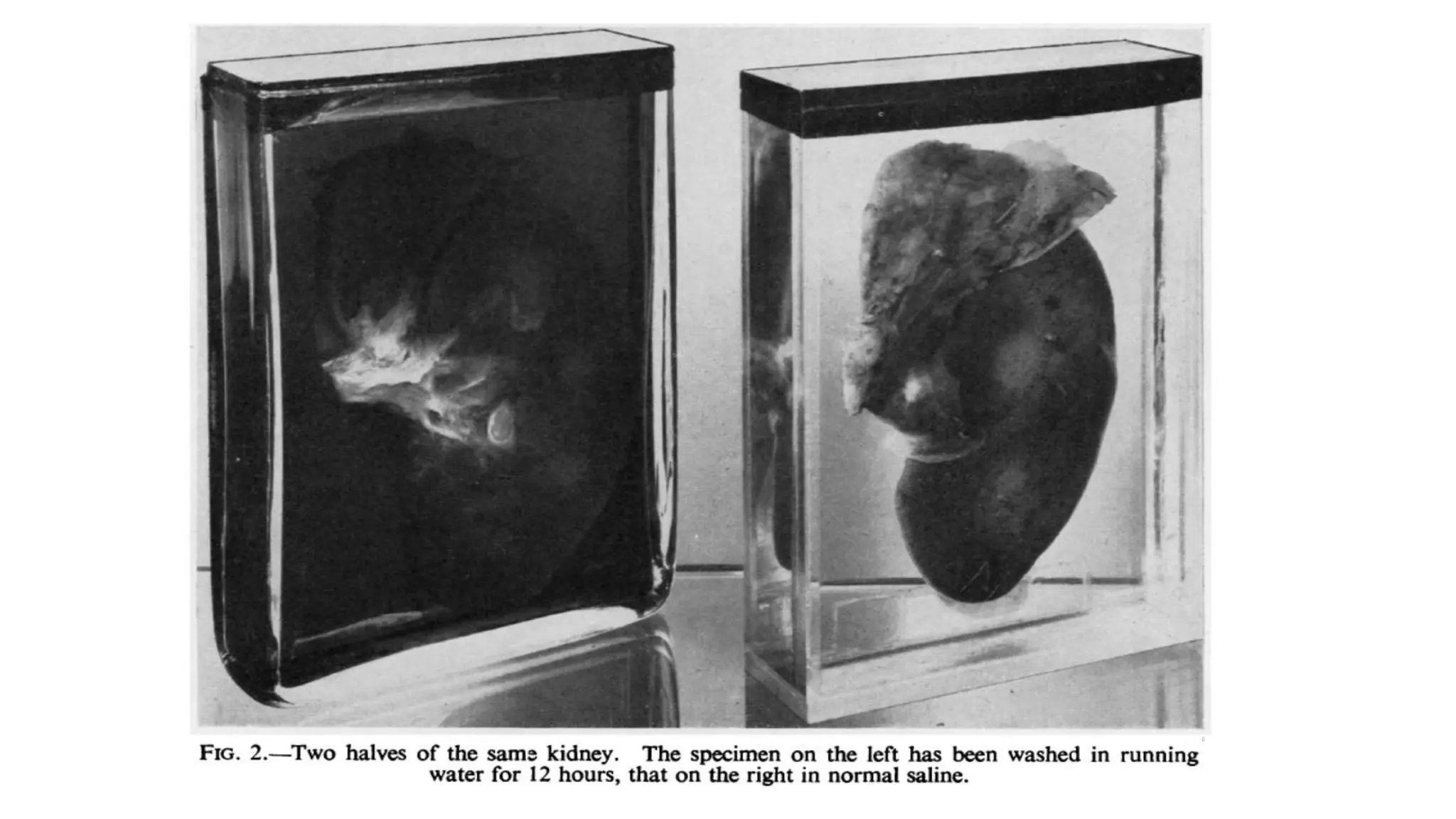
Dr. Anju Shrestha
PGY III, Pathology
CMCTH
MUSEUM TECHNIQUES
Introduction
• Pathological museums are in part historical, representing the pioneer
work of diagnosticians and therapists.
• Presents records of past states, not now encountered, or conditions of
great rarity.
• Provide the student with the basic material of his current teaching.
Basic museum techniques
• Reception
• Preparation
• Fixation
• Color restoration
• Preservation
• Mounting
• Special methods
• Presentation
Reception
• Museum specimens are collected from teaching hospitals.
• Necropsy specimens from postmortem room
• Research laboratories
Specimen should be received with full details of the patient/lesion
Preparation of the specimen
• One of the commonest causes of inferior specimens is contact with tap
water. The resultant haemolysis greatly reduces their value.
• Specimens should be washed only with saline, and should be kept in
saline before demonstration. Drying ruins surface appearance.
• Should not be kept in saline for more than 2 hours as autolysis sets in.
Fixation of the specimen
• Objective: To preserve cells and tissue constituents in as close to life
like state as possible
• Fixation stops autolysis and bacterial decomposition, and stabilizing
the cellular and tissue constituents
• Fixatives are based on formalin fixative technique.
• They are derived from Kaiserling technique and its modification.
• Kaiserling recommended that the initial fixation should be in neutral
formalin (KI) solution and then preserving glycerine solution (KIII)
for long term display.
• These solutions also preserve color.
Principle of fixation
• Specimens containing bile or stained by bile must be fixed and stored apart from
others, as they will stain them.
• Specimens undergoing fixation must not touch other specimens, or the sides of jars;
they must either lie on washed lint or be suspended by linen thread.
• Flat flaps of tissues like stomach, intestine etc. should be fixed to cork bread and left
in formalin so that they are not crumpled and irregularly fixed.
• Cystic cavities- if unopened, should be injected with fixatives. Opened ones should
be packed with cotton wool.
• Solid viscera should be fixed by vascular injection. For eg, brain is
fixed by injecting fixative through basilar artery.
• Lungs and limbs should be fixed by vascular injection.
Fixation technique
• Most widely used techniques are modification of methods described
by Kaiserling (1897).
• Originally 3 solutions were used
- First for fixing
- Second for restoring color
- Third as a mounting fluid
Kaiserling No. I- fixing fluid
• Formalin (40%) 400 ml.
• Pot. nitrate 30 g.
• Pot. acetate 60 g.
• Tap water 2,000 ml.
Tissue is fixed in Kaiserling No.I solution for 24 hours to few weeks
depending on the size of the specimen.
Kaiserling No. II solution
• The specimen is placed in 80% ethyl alcohol solution for an optimal
period for 1 hour ( upto 4 hours depending on the size of specimen), if
the specimen is discolored.
• If the specimen is left in alcohol for too long the color will fade, and
the effect is irreversible.
• This step is not required if mounting fluid used is sodium hydrogen
sulfite.
Color restoration
• The fixed specimen is transferred to a jar containg industrial
methylated spirit until the color is fully restored.
• The alcohol penetrates the tissue rapidly.
• Floating specimen cover with surgical gauze. The vessel should be
closed to prevent evaporation.
• Color restoration is complete in 2-8 hours, depending on the size and
character of the specimen.
• Restoration can be achieved by adding reducing agent- sodium
hydrogen sulfite to the mounting fluid (Pulvertaft, 1936).
• Specimen mounted show remarkably little fading even after 25 yrs.
Original Kaiserling No. III solution
• Glycerine 500 ml.
• Arsenious acid 1% 200 ml.
• Pot. acetate 250 g.
• Thymol 2.5 g.
Pulvertaft – Kaiserling mounting fluid III
• Glycerine 300 ml
• 10% sodium acetate 100 g
• 10% formalin 5 ml
• Tap water 1000 ml
• Camphor/ thymol- prevents the growth of moulds.
• Immediately before sealing 0.4% sodium hydrosulfite is added.
• Solution should be filtered through paper pulp under negative pressure
to remove impurities.
• Carbon monoxide has also been employed as a colour-retaining agent.
Schultz (1931) introduced the technique, which gives brilliant colour
contrast, but entails the risks of poisoning and explosion.
• Pure liquid paraffin can be used as final mountant after color
restoration with alcohol. It reduces discoloration of mounting fluid by
pigments in the specimen.
Hollow viscera
• Cut hollow viscera should be padded with cotton wool.
• Uncut viscera can be pressure inflated. Eg
- Through urethra into the bladder
- Through urethra into pelvicalyceal system
- Through trachea into the lungs
- Direct injection in case of cysts
The fixatives can be injected into such organs by Higginson syringe or
with conventional hypodermic syringe.
Higginson syringe
Preservation
• The specimen together with a duplicate label, is wrapped in gauze or
muslin and the label is attached with a piece of linen thread.
• Specimens are preserved in a rectangular earthenware tanks.
• The fluid used can be Kaiserling fixing fluid I for a period of six
months.
• After 6 months, the specimen should be treated with 80% alcohol to
restore color.
Mounting
• Specimens are trimmed to desired size and shape so that they fit in the
jar. All unwanted and non representative tissues are removed after
careful dissection.
• If the specimen do not remain in natural position after removal of
cotton wool packing, fill the cavities with arsenious acid- gelatin.
• Regular cuts given keeping in anatomical position.
• Friable specimens can be covered by thin layer of arsenious acid
gelatin.
• Bile stained specimen are soaked in solution of calcium chloride for
24 hours to avoid discoloration of mounting fluid.
Mounting procedure
• Museum jars and boxes
• Center plates
• Stitching specimens to center plate
• Fixing the center plate
• Filling and sealing
Factors affecting fixation
• Buffering
• Penetration
• Volume
• Temperature
• Concentration
• Time interval
• Position of the tissue
Special methods
• Maceration
- Used to demonstrate bony lesions eg; osteogenic sarcomas, osteomas and
tuberculosis.
• Calculi
- Calculi are cut in half with fine fretsaw and cut surface is polished with
sand paper.
- Dry mounting in closed jars.
• Amyloid
- Iodine technique
- Congo red technique
Presentation
• Museum specimen should be clearly labelled and a system of
cataloging should be employed which allows easy and rapid access.
Labelling
• Rectangle of Perspex sheet 1/16th of an inch in thickness which is
cemented in the center at the bottom of the outside of the box or the
bottom of the center plate.
References
• MUSEUM TECHNIQUES: A REVIEW BY R. J. V. PULVERTAFT From the
Westminster Hospital School of Medicine, London
Thank you….

museum techniques in pathology.pptx

  • 1.
    Dr. Anju Shrestha PGYIII, Pathology CMCTH MUSEUM TECHNIQUES
  • 2.
    Introduction • Pathological museumsare in part historical, representing the pioneer work of diagnosticians and therapists. • Presents records of past states, not now encountered, or conditions of great rarity. • Provide the student with the basic material of his current teaching.
  • 3.
    Basic museum techniques •Reception • Preparation • Fixation • Color restoration • Preservation • Mounting • Special methods • Presentation
  • 4.
    Reception • Museum specimensare collected from teaching hospitals. • Necropsy specimens from postmortem room • Research laboratories Specimen should be received with full details of the patient/lesion
  • 5.
    Preparation of thespecimen • One of the commonest causes of inferior specimens is contact with tap water. The resultant haemolysis greatly reduces their value. • Specimens should be washed only with saline, and should be kept in saline before demonstration. Drying ruins surface appearance. • Should not be kept in saline for more than 2 hours as autolysis sets in.
  • 7.
    Fixation of thespecimen • Objective: To preserve cells and tissue constituents in as close to life like state as possible • Fixation stops autolysis and bacterial decomposition, and stabilizing the cellular and tissue constituents • Fixatives are based on formalin fixative technique. • They are derived from Kaiserling technique and its modification.
  • 8.
    • Kaiserling recommendedthat the initial fixation should be in neutral formalin (KI) solution and then preserving glycerine solution (KIII) for long term display. • These solutions also preserve color.
  • 9.
    Principle of fixation •Specimens containing bile or stained by bile must be fixed and stored apart from others, as they will stain them. • Specimens undergoing fixation must not touch other specimens, or the sides of jars; they must either lie on washed lint or be suspended by linen thread. • Flat flaps of tissues like stomach, intestine etc. should be fixed to cork bread and left in formalin so that they are not crumpled and irregularly fixed. • Cystic cavities- if unopened, should be injected with fixatives. Opened ones should be packed with cotton wool.
  • 10.
    • Solid viscerashould be fixed by vascular injection. For eg, brain is fixed by injecting fixative through basilar artery. • Lungs and limbs should be fixed by vascular injection.
  • 11.
    Fixation technique • Mostwidely used techniques are modification of methods described by Kaiserling (1897). • Originally 3 solutions were used - First for fixing - Second for restoring color - Third as a mounting fluid
  • 12.
    Kaiserling No. I-fixing fluid • Formalin (40%) 400 ml. • Pot. nitrate 30 g. • Pot. acetate 60 g. • Tap water 2,000 ml. Tissue is fixed in Kaiserling No.I solution for 24 hours to few weeks depending on the size of the specimen.
  • 13.
    Kaiserling No. IIsolution • The specimen is placed in 80% ethyl alcohol solution for an optimal period for 1 hour ( upto 4 hours depending on the size of specimen), if the specimen is discolored. • If the specimen is left in alcohol for too long the color will fade, and the effect is irreversible. • This step is not required if mounting fluid used is sodium hydrogen sulfite.
  • 14.
    Color restoration • Thefixed specimen is transferred to a jar containg industrial methylated spirit until the color is fully restored. • The alcohol penetrates the tissue rapidly. • Floating specimen cover with surgical gauze. The vessel should be closed to prevent evaporation. • Color restoration is complete in 2-8 hours, depending on the size and character of the specimen.
  • 15.
    • Restoration canbe achieved by adding reducing agent- sodium hydrogen sulfite to the mounting fluid (Pulvertaft, 1936). • Specimen mounted show remarkably little fading even after 25 yrs.
  • 16.
    Original Kaiserling No.III solution • Glycerine 500 ml. • Arsenious acid 1% 200 ml. • Pot. acetate 250 g. • Thymol 2.5 g.
  • 17.
    Pulvertaft – Kaiserlingmounting fluid III • Glycerine 300 ml • 10% sodium acetate 100 g • 10% formalin 5 ml • Tap water 1000 ml
  • 18.
    • Camphor/ thymol-prevents the growth of moulds. • Immediately before sealing 0.4% sodium hydrosulfite is added. • Solution should be filtered through paper pulp under negative pressure to remove impurities.
  • 19.
    • Carbon monoxidehas also been employed as a colour-retaining agent. Schultz (1931) introduced the technique, which gives brilliant colour contrast, but entails the risks of poisoning and explosion. • Pure liquid paraffin can be used as final mountant after color restoration with alcohol. It reduces discoloration of mounting fluid by pigments in the specimen.
  • 20.
    Hollow viscera • Cuthollow viscera should be padded with cotton wool. • Uncut viscera can be pressure inflated. Eg - Through urethra into the bladder - Through urethra into pelvicalyceal system - Through trachea into the lungs - Direct injection in case of cysts The fixatives can be injected into such organs by Higginson syringe or with conventional hypodermic syringe.
  • 21.
  • 22.
    Preservation • The specimentogether with a duplicate label, is wrapped in gauze or muslin and the label is attached with a piece of linen thread. • Specimens are preserved in a rectangular earthenware tanks. • The fluid used can be Kaiserling fixing fluid I for a period of six months. • After 6 months, the specimen should be treated with 80% alcohol to restore color.
  • 24.
    Mounting • Specimens aretrimmed to desired size and shape so that they fit in the jar. All unwanted and non representative tissues are removed after careful dissection. • If the specimen do not remain in natural position after removal of cotton wool packing, fill the cavities with arsenious acid- gelatin. • Regular cuts given keeping in anatomical position.
  • 25.
    • Friable specimenscan be covered by thin layer of arsenious acid gelatin. • Bile stained specimen are soaked in solution of calcium chloride for 24 hours to avoid discoloration of mounting fluid.
  • 26.
    Mounting procedure • Museumjars and boxes • Center plates • Stitching specimens to center plate • Fixing the center plate • Filling and sealing
  • 29.
    Factors affecting fixation •Buffering • Penetration • Volume • Temperature • Concentration • Time interval • Position of the tissue
  • 30.
    Special methods • Maceration -Used to demonstrate bony lesions eg; osteogenic sarcomas, osteomas and tuberculosis. • Calculi - Calculi are cut in half with fine fretsaw and cut surface is polished with sand paper. - Dry mounting in closed jars. • Amyloid - Iodine technique - Congo red technique
  • 32.
    Presentation • Museum specimenshould be clearly labelled and a system of cataloging should be employed which allows easy and rapid access.
  • 33.
    Labelling • Rectangle ofPerspex sheet 1/16th of an inch in thickness which is cemented in the center at the bottom of the outside of the box or the bottom of the center plate.
  • 34.
    References • MUSEUM TECHNIQUES:A REVIEW BY R. J. V. PULVERTAFT From the Westminster Hospital School of Medicine, London
  • 35.