Downloaded 67 times


![• JOHN BRAXTON HICKS(LONDON,1868): First to
experiment with chemical methods to prevent
coagulation of blood ; phosphate of sodas(blood kept
fluid but patient died of shock)
• Swiss Physiologists ARTHUR & PAGES(1890):
Connect calcium with blood clotting on addition of
any small amount of organic salts
• LANDOIS(1892): Suggested Hirudin from leeches
• A.E.WRIGHT(1894): Suggested non-toxic citrates;
that bind enough ca without causing convulsions [21
years back before it was used]
• SATTERLEE & HOOKER(1914): Used Hirudin but
found to have a narrow therapeutic window&
difficulty in obtaining](https://image.slidesharecdn.com/anticoagulants-170608144752/75/Anticoagulants-6-2048.jpg)


![HEPARIN
• Prevents coagulation by inactivating the
prophylactic activity of thrombin after combining
with AT 111 and thrombin.
• 1000 IU of heparin is equal to 10 mg (commercially
available as IU)
• Dose of heparin for anticoagulation is 0.5-2.0 IU/ml
of blood (approx.500 IU of heparin for 500 ml of
blood)
• Heparinized blood should be used within 24 hours.
• Earlier heparinized blood was used in pen heart
surgery but now usually it is not used as
extracorporeal pumps are now usually primed with
crystalloids and not with blood.
•The effect of heparin can be neutralized with
Protamine sulphate
•1 mg of protamine sulphate neutralizes 1 mg of
heparin (to neutralize 5000 units of heparin [50 mg], 5
ml of 1 % solution of protamine sulphate will be
needed)
•Heparin is used in cord blood collection apart from
CPD](https://image.slidesharecdn.com/anticoagulants-170608144752/75/Anticoagulants-22-2048.jpg)


![RED CELL FREEZING
• Smith in 1950 reported that glycerol could prevent
freezing injury in human red cells and that red cells,
mixed with glycerol could be frozen without damage
• Glycerol, Dimethyl sulfoxide (DMSO) & Hydroxyethyl
Starch(HES) (cryoprotective agent) is added to red
cells they can be frozen and thawed without damage
by intracellular ice formation and hypertonicity
• Glycerol limits ice formation and provides liquid
phase in which salts are distributed as cooling
proceeds thereby avoiding excessive hypertonicity
• Frozen red cells are primarily used for autologous
transfusion and the storage of rare group blood
•Cryoprotective agent is added to red cells that are less
than 6 days old
• Glycerol (used commonly) is added to the red cells
slowly with vigorous shaking so that glycerol
permeates into the red cells
•The cells are rapidly frozen and stored in a freezer
•The freezing and storage temperature depends on the
concentration of glycerol.
•High concentration glycerol [40% weight in volume]
and a low concentration glycerol [20% weight in
volume] in the final concentration of cryopreservative
• Most blood banks use the high glycerol technique.](https://image.slidesharecdn.com/anticoagulants-170608144752/75/Anticoagulants-58-2048.jpg)


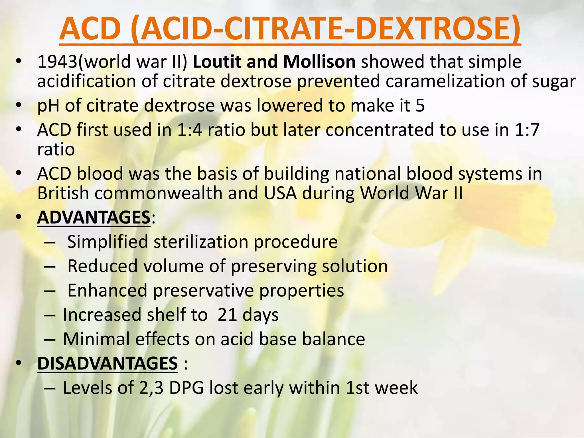
This document discusses the history of anticoagulants used for blood transfusion and storage. It describes some of the key developments including: - Early attempts in the 1800s using defibrinated blood or direct transfusion before anticoagulants were discovered. - The first chemical anticoagulant experimented with was sodium phosphate by John Braxton Hicks in 1868. - Important early anticoagulants developed were sodium citrate in 1914 and acid-citrate-dextrose solution in 1943 which allowed blood to be stored for longer periods. - Common anticoagulants now used include sodium citrate, heparin, EDTA, and oxalates


