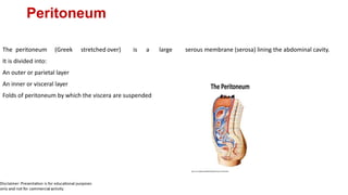
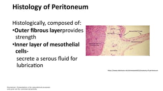
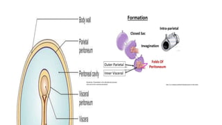
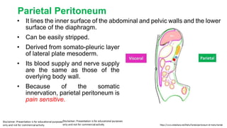
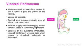
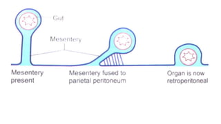
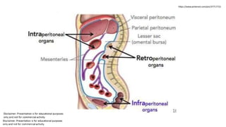
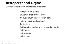
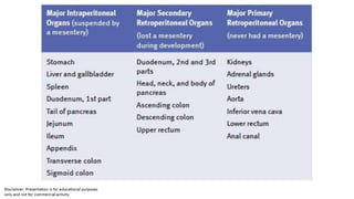
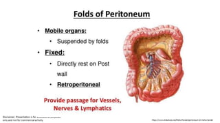
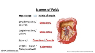
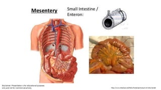
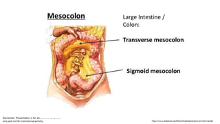
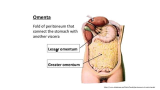
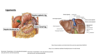
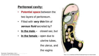
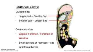
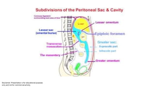
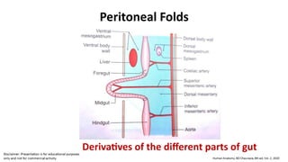
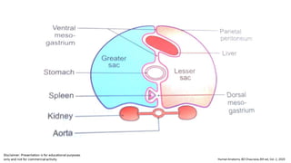
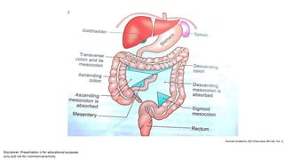
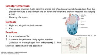
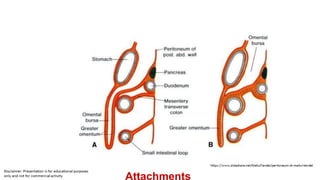
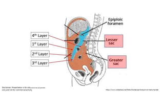
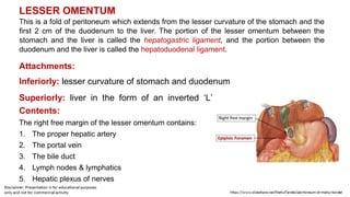
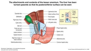
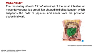
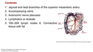
.04.2020
Peritoneum
The peritoneum (Greek stretched over) is a large serous membrane (serosa) lining the abdominal cavity.
It is divided into:
An outer or parietal layer
An inner or visceral layer
Folds of peritoneum by which the viscera are suspended (lect
1
Histology of Peritoneum
Histologically, composed of:
•Outer fibrous layerprovides
strength
•Inner layer of mesothelial
cells-
secrete a serous fluid for
lubrication
https://www.slideshare.net/ahmedsalah925/anatomy-of-peritoneum
Diagrammatic transverse section of the abdomen showing the
arrangement of the peritoneum
Parietal Peritoneum
• It lines the inner surface of the abdominal and pelvic walls and the lower
surface of the diaphragm.
• Can be easily stripped.
• Derived from somato-pleuric layer
of lateral plate mesoderm.
• Its blood supply and nerve supply
are the same as those of the
overlying body wall.
• Because of the somatic
innervation, parietal peritoneum is
pain sensitive.
Visceral Peritoneum
• It lines the outer surface of the viscera. In
fact it forms a part and parcel of the
viscera.
• Cannot be stripped.
• Derived from splanchno-pleuric layer of
lateral plate mesoderm.
• Its blood supply and nerve supply are the
same as those of the underlying viscera.
• Because of the autonomic innervation,
visceral peritoneum evokes pain when
viscera is stretched, ischaemic or
distended.
Folds of Peritoneum
• Many organs within the abdomen are suspended by folds of
peritoneum. Such organs are mobile.
• Other organs are fixed and immobile. Such organs are said to be
retroperitoneal.
• Some are partially covered.
• Few are sub-peritoneal (infra-peritoneal).
• Provide pathways forpassageof vessels, nerves and
lymphatics.
Process of zygosis
https://www.pinterest.com/pin/37717715
609967916/
Intraperitoneal Organs
(completely surrounded by peritoneum)
S= Stomach
A= Appendix
L= Liver
T= Transverse colon
D= Duodenum (1st
part only)
S= Small intestines
P= Pancreas (tail only)
R= Rectum (upper 3rd
only)
S= Sigmoid colon
S= Spleen
Retroperitoneal Organs
(covered by peritoneum on anterior surfaces only)
S= Suprarenal glands
A= Aorta/Inferior Vena Cava
D= Duodenum (except for 1st
part)
P= Pancreas (head and neck)
U= Ureters
C= Colon (ascending and descending parts)
K= Kidneys
E= Esophagus
R= Rectum
/
e
https://www.studyblue.com/notes/note/n/liver-pancreas-spleen/deck/21887610
https://www.proprofs.com/quiz-
school/story.php?title=fsadfds
• Provide passage for nerves, vessels &
lymphatics
CLINICAL ANATOMY
• Ascites: Collection of free fluid in the peritoneal cavity is known as ascites.
Common causes of ascites are cirrhosis of the liver, tubercular peritonitis,
congestive heart failure, and malignant infiltration of the peritoneum. Veins
also get prominent in cirrhosis of liver.
• Peritonitis
• Pneumoperitoneum
• Laparoscopy
• Laparotomy
• Greater omentum limits the spread of infection by sealing off the site of
ruptured vermiform appendix or gastric ulcer and tries to delay the onset of
peritonitis. It is called ‘‘abdominal policeman’’.
• Peritoneal dialysis
Paracentesis
X sites for paracentesis
Peritoneal Folds
Derivatives of the different parts of gut
https://quizlet.com/7308810/stomach-small-intestine-flash-cards/
Greater Omentum
• The greater omentum (Latin apron) is a large fold of peritoneum which hangs down from the
greater curvature of the stomach like an apron and covers the loops of intestines to a varying
extent.
• Made up of 4 layers.
Contents
• Right and left gastroepiploic vessels
• Fat
Functions
1. It is a storehouseof fat
2. It protects the peritoneal cavity against infection
(collection of macrophages form milkyspots) 3. Also
known as “policeman of the abdomen”
https://m.youtube.com/watch?v=_bGjvdc5Sqk
Attachments
LESSER OMENTUM
This is a fold of peritoneum which extends from the lesser curvature of the stomach and the
first 2 cm of the duodenum to the liver. The portion of the lesser omentum between the
stomach and the liver is called the hepatogastric ligament, and the portion between the
duodenum and the liver is called the hepatoduodenal ligament.
Attachments:
Inferiorly: lesser curvature of stomach and duodenum
Superiorly: liver in the form of an inverted ‘L’
Contents:
The right free margin of the lesser omentum contains:
1. The proper hepatic artery
2. The portal vein
3. The bile duct
4. Lymph nodes & lymphatics
5. Hepatic plexus of nerves
The attachments and contents of the lesser omentum. The liver has been
turned upwards so that its posteroinferior surface can be seen
MESENTERY
The mesentery (Greek fold of intestine) of the small intestine or
mesentery proper is a broad, fan-shaped fold of peritoneum which
suspends the coils of jejunum and ileum from the posterior
abdominal wall.
https://www.kenhub.com/en/library/anatomy/mesentery
Borders
• The attached border, or root of the
mesentery, is 15 cm long, it crosses the
following structures:
1.The third part of the duodenum where the
superior mesenteric vessels enter into it
2.The abdominal aorta
3.The inferior vena cava
4.The right ureter
5.The right psoas major
• Free or intestinal border is 6 metres long.
• The breadth of the mesentery is
maximum and is about 20 cm in the
central part, but gradually diminishes
towards both the ends.
https://www.kenhub.com/en/library/anatomy/mesentery
Contents
1. Jejunal and ileal branches of the superior mesenteric artery
2. Accompanying veins
3. Autonomic nerve plexuses
4. Lymphatics or lacteals
5. 100–200 lymph nodes 6. Connective
tissue with fat
https://www.slideshare.net/DrSherifFahmy/jejunum-ileum-anatomy-of-the-abdomen
Mesoappendix
It is a small, triangular fold of peritoneum which
suspends the vermiform appendix from the posterior
surface of the lower end of the mesentery close to the
ileocaecal junction.
Transverse Mesocolon
Attachments
• The root of the transverse mesocolon
is attached to the anterior surface of
the head, and the anterior border of the
body of the pancreas.
Contents
• It contains the middle colic vessels; the
nerves, lymph nodes and lymphatics of
the transverse colon.
https://www.memorangapp.com/flashcards/71585/Inferior+Mesenteric+Artery+and+Large+intestine/
Sigmoid Mesocolon
Features
• This is a triangular (Greek ‘S’ shape) fold of
peritoneum which suspends the sigmoid colon from
the pelvic wall.
Attachments
• The root is shaped like an inverted ‘V’.
Contents
• The sigmoid vessels in the left limb; superior rectal
vessels, nerves, lymph nodes and lymphatics in the
right limb of the sigmoid colon.
THANKYOU

peritoneum.docx

  • 1.
  • 2.
    Peritoneum The peritoneum (Greekstretched over) is a large serous membrane (serosa) lining the abdominal cavity. It is divided into: An outer or parietal layer An inner or visceral layer Folds of peritoneum by which the viscera are suspended (lect 1
  • 3.
    Histology of Peritoneum Histologically,composed of: •Outer fibrous layerprovides strength •Inner layer of mesothelial cells- secrete a serous fluid for lubrication https://www.slideshare.net/ahmedsalah925/anatomy-of-peritoneum
  • 6.
    Diagrammatic transverse sectionof the abdomen showing the arrangement of the peritoneum
  • 7.
    Parietal Peritoneum • Itlines the inner surface of the abdominal and pelvic walls and the lower surface of the diaphragm. • Can be easily stripped. • Derived from somato-pleuric layer of lateral plate mesoderm. • Its blood supply and nerve supply are the same as those of the overlying body wall. • Because of the somatic innervation, parietal peritoneum is pain sensitive.
  • 8.
    Visceral Peritoneum • Itlines the outer surface of the viscera. In fact it forms a part and parcel of the viscera. • Cannot be stripped. • Derived from splanchno-pleuric layer of lateral plate mesoderm. • Its blood supply and nerve supply are the same as those of the underlying viscera. • Because of the autonomic innervation, visceral peritoneum evokes pain when viscera is stretched, ischaemic or distended.
  • 9.
    Folds of Peritoneum •Many organs within the abdomen are suspended by folds of peritoneum. Such organs are mobile. • Other organs are fixed and immobile. Such organs are said to be retroperitoneal. • Some are partially covered. • Few are sub-peritoneal (infra-peritoneal). • Provide pathways forpassageof vessels, nerves and lymphatics. Process of zygosis
  • 11.
  • 12.
  • 15.
    Intraperitoneal Organs (completely surroundedby peritoneum) S= Stomach A= Appendix L= Liver T= Transverse colon D= Duodenum (1st part only) S= Small intestines P= Pancreas (tail only) R= Rectum (upper 3rd only) S= Sigmoid colon S= Spleen
  • 16.
    Retroperitoneal Organs (covered byperitoneum on anterior surfaces only) S= Suprarenal glands A= Aorta/Inferior Vena Cava D= Duodenum (except for 1st part) P= Pancreas (head and neck) U= Ureters C= Colon (ascending and descending parts) K= Kidneys E= Esophagus R= Rectum
  • 18.
  • 24.
  • 25.
  • 29.
  • 31.
    • Provide passagefor nerves, vessels &
  • 32.
    lymphatics CLINICAL ANATOMY • Ascites:Collection of free fluid in the peritoneal cavity is known as ascites. Common causes of ascites are cirrhosis of the liver, tubercular peritonitis, congestive heart failure, and malignant infiltration of the peritoneum. Veins also get prominent in cirrhosis of liver. • Peritonitis • Pneumoperitoneum • Laparoscopy • Laparotomy • Greater omentum limits the spread of infection by sealing off the site of ruptured vermiform appendix or gastric ulcer and tries to delay the onset of peritonitis. It is called ‘‘abdominal policeman’’. • Peritoneal dialysis
  • 33.
  • 34.
    Peritoneal Folds Derivatives ofthe different parts of gut
  • 35.
  • 39.
    Greater Omentum • Thegreater omentum (Latin apron) is a large fold of peritoneum which hangs down from the greater curvature of the stomach like an apron and covers the loops of intestines to a varying extent. • Made up of 4 layers. Contents • Right and left gastroepiploic vessels • Fat Functions 1. It is a storehouseof fat 2. It protects the peritoneal cavity against infection (collection of macrophages form milkyspots) 3. Also known as “policeman of the abdomen”
  • 40.
  • 41.
  • 43.
    LESSER OMENTUM This isa fold of peritoneum which extends from the lesser curvature of the stomach and the first 2 cm of the duodenum to the liver. The portion of the lesser omentum between the stomach and the liver is called the hepatogastric ligament, and the portion between the duodenum and the liver is called the hepatoduodenal ligament. Attachments: Inferiorly: lesser curvature of stomach and duodenum Superiorly: liver in the form of an inverted ‘L’ Contents: The right free margin of the lesser omentum contains: 1. The proper hepatic artery 2. The portal vein 3. The bile duct 4. Lymph nodes & lymphatics 5. Hepatic plexus of nerves
  • 44.
    The attachments andcontents of the lesser omentum. The liver has been turned upwards so that its posteroinferior surface can be seen
  • 45.
    MESENTERY The mesentery (Greekfold of intestine) of the small intestine or mesentery proper is a broad, fan-shaped fold of peritoneum which suspends the coils of jejunum and ileum from the posterior abdominal wall. https://www.kenhub.com/en/library/anatomy/mesentery
  • 46.
    Borders • The attachedborder, or root of the mesentery, is 15 cm long, it crosses the following structures: 1.The third part of the duodenum where the superior mesenteric vessels enter into it 2.The abdominal aorta 3.The inferior vena cava 4.The right ureter 5.The right psoas major • Free or intestinal border is 6 metres long. • The breadth of the mesentery is maximum and is about 20 cm in the central part, but gradually diminishes towards both the ends. https://www.kenhub.com/en/library/anatomy/mesentery
  • 47.
    Contents 1. Jejunal andileal branches of the superior mesenteric artery 2. Accompanying veins 3. Autonomic nerve plexuses 4. Lymphatics or lacteals 5. 100–200 lymph nodes 6. Connective tissue with fat https://www.slideshare.net/DrSherifFahmy/jejunum-ileum-anatomy-of-the-abdomen
  • 48.
    Mesoappendix It is asmall, triangular fold of peritoneum which suspends the vermiform appendix from the posterior surface of the lower end of the mesentery close to the ileocaecal junction.
  • 49.
    Transverse Mesocolon Attachments • Theroot of the transverse mesocolon is attached to the anterior surface of the head, and the anterior border of the body of the pancreas. Contents • It contains the middle colic vessels; the nerves, lymph nodes and lymphatics of the transverse colon. https://www.memorangapp.com/flashcards/71585/Inferior+Mesenteric+Artery+and+Large+intestine/
  • 50.
    Sigmoid Mesocolon Features • Thisis a triangular (Greek ‘S’ shape) fold of peritoneum which suspends the sigmoid colon from the pelvic wall. Attachments • The root is shaped like an inverted ‘V’. Contents • The sigmoid vessels in the left limb; superior rectal vessels, nerves, lymph nodes and lymphatics in the right limb of the sigmoid colon.
  • 51.