Download to read offline
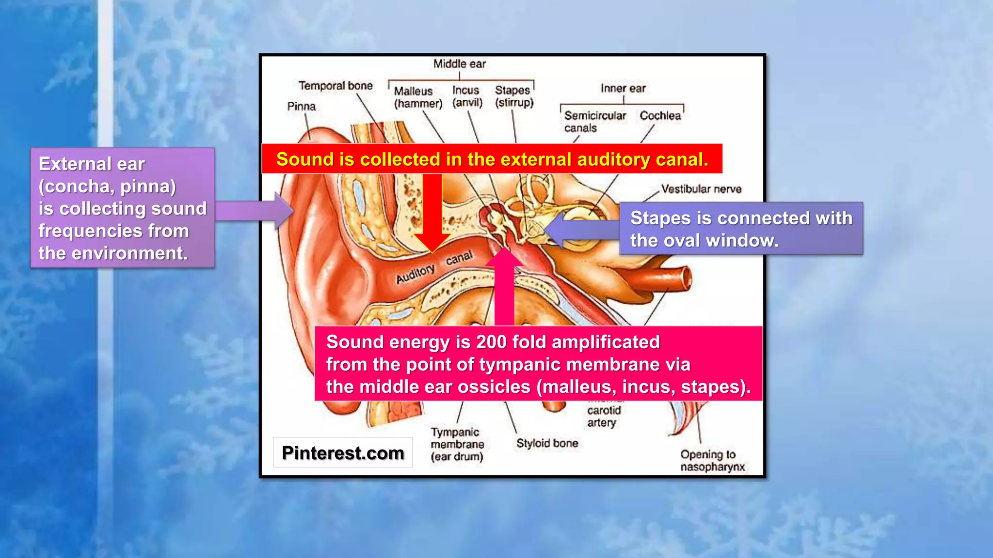
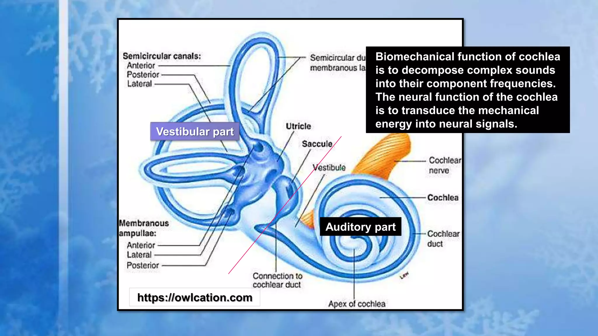
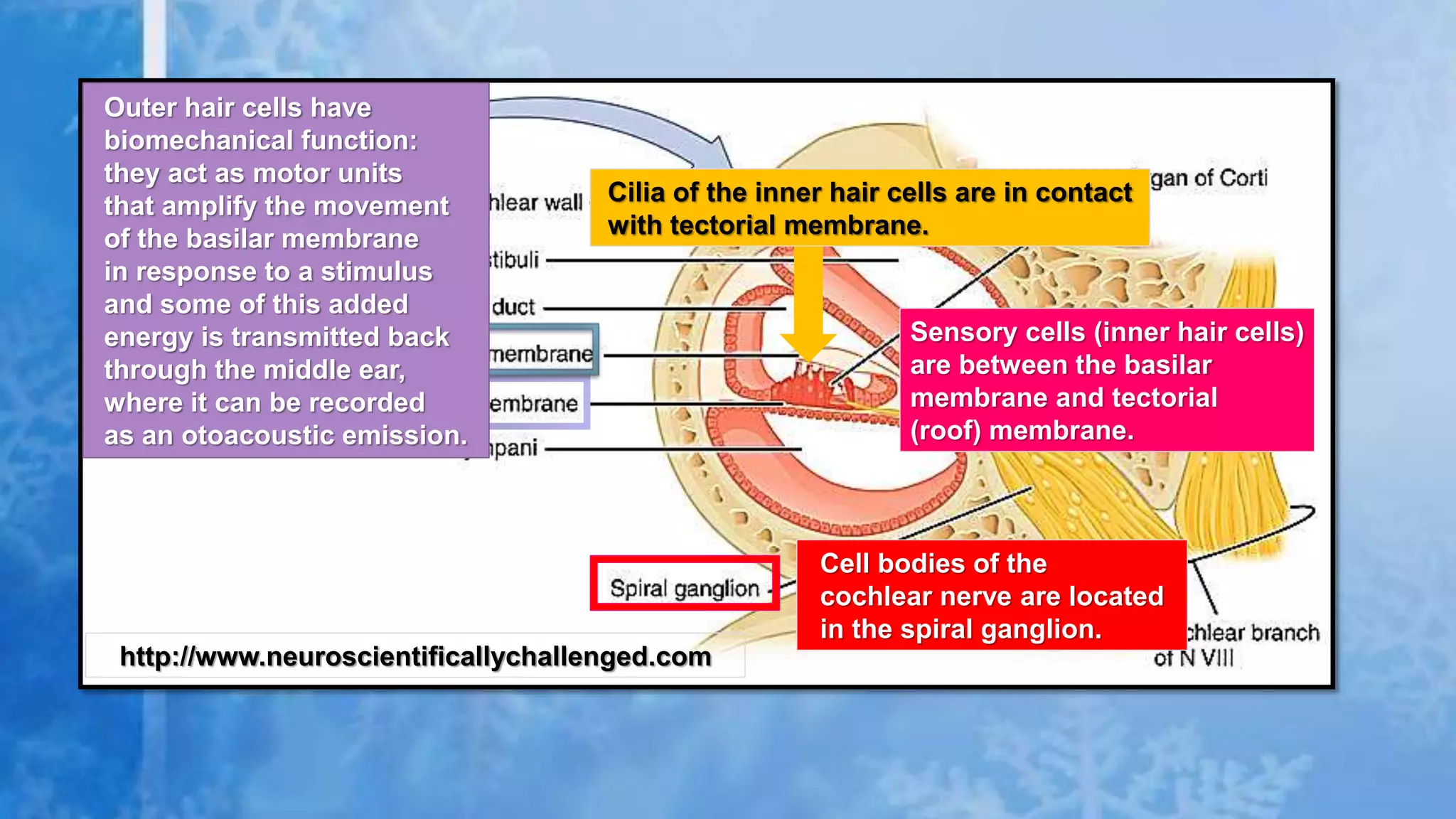
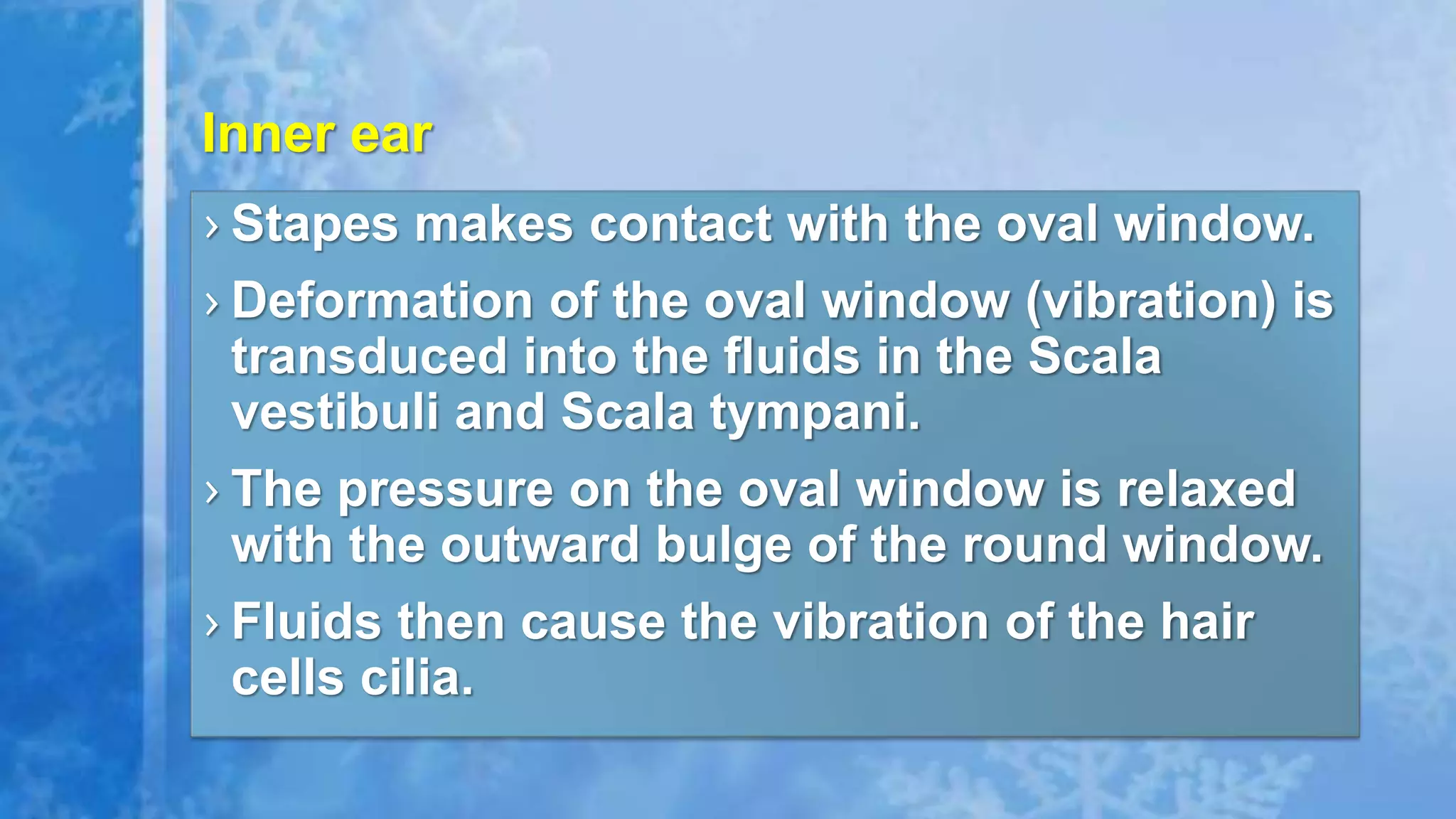
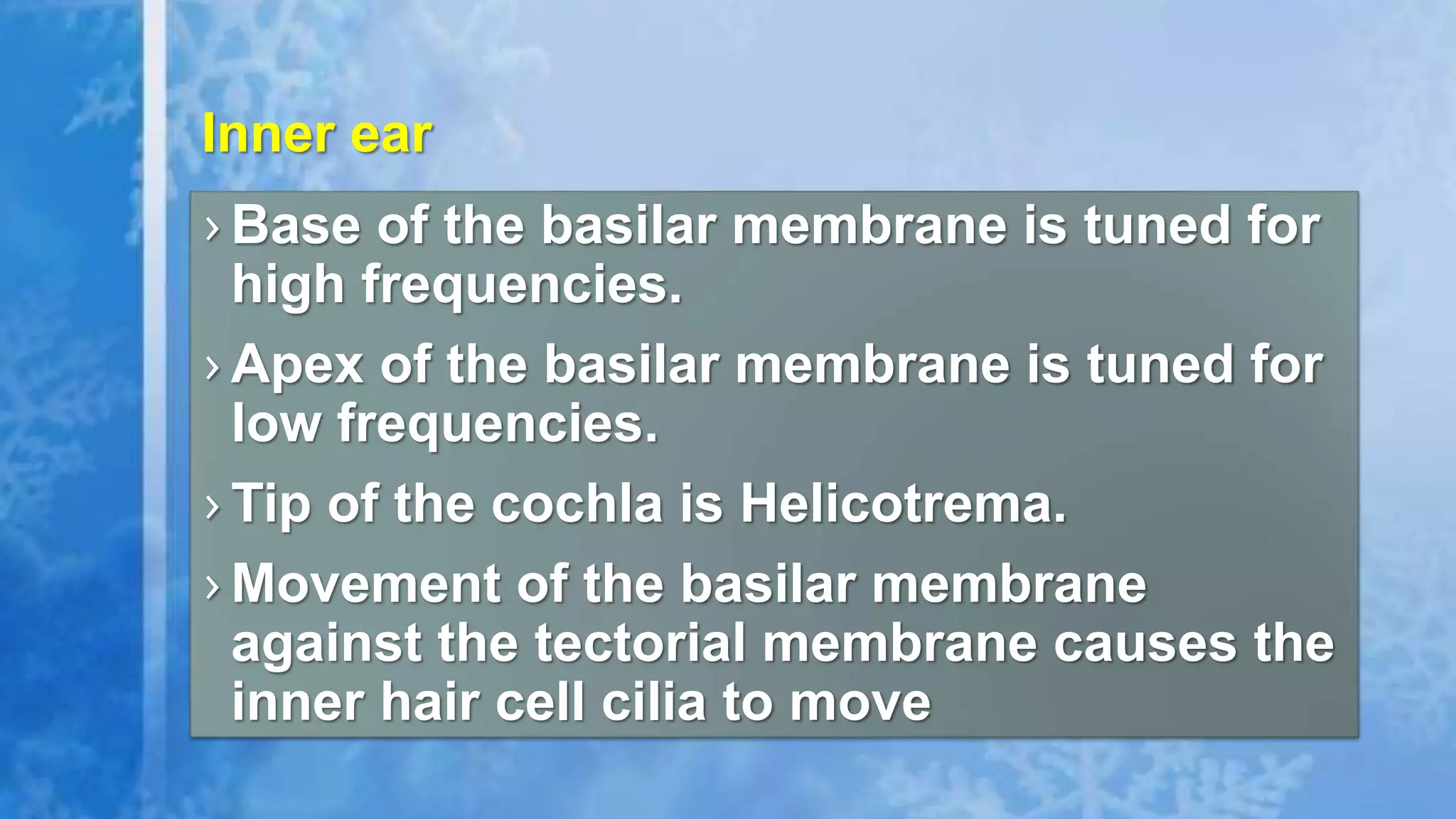
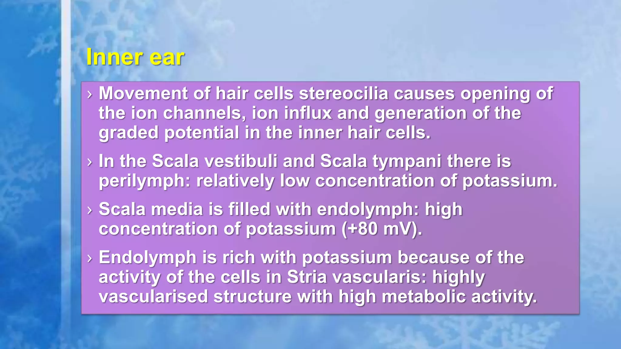
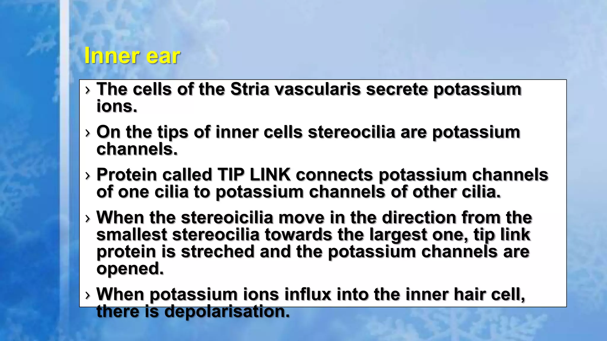
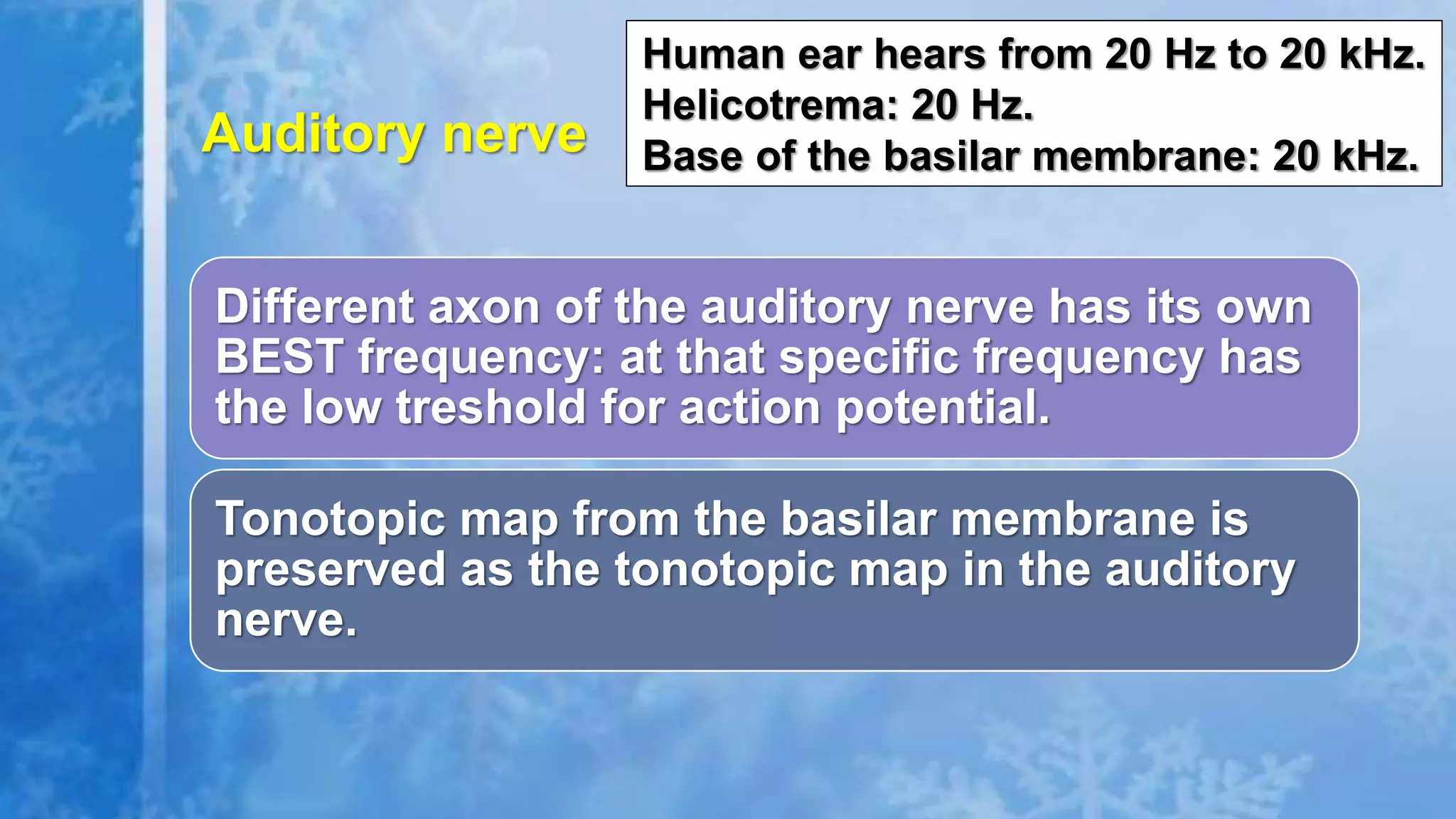


The document summarizes the peripheral auditory system and mechanisms of hearing. Sound waves are collected by the outer and middle ear and transferred to the inner ear. In the inner ear, the cochlea decomposes sound waves into frequencies which are encoded by hair cells. This information is transmitted through the auditory nerve and processed in the brainstem, midbrain, and auditory cortex. The cochlea uses tonotopic organization to transmit frequencies to the brain in an organized way.


![Apporach to lung biopsy [Auto-saved].pptx latest](https://cdn.slidesharecdn.com/ss_thumbnails/apporachtolungbiopsyauto-saved-251211225655-93258539-thumbnail.jpg?width=640&height=640&fit=bounds)

