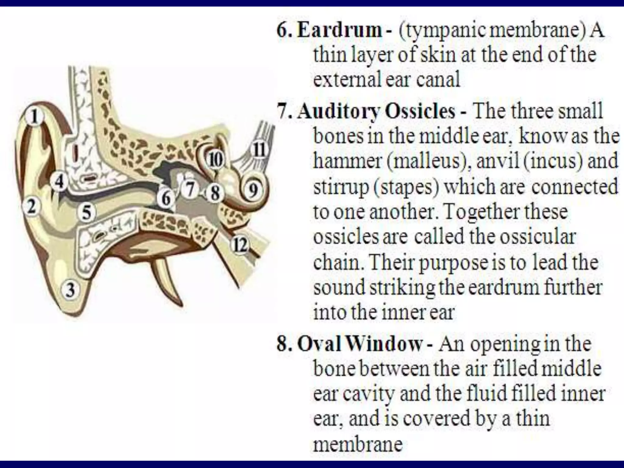
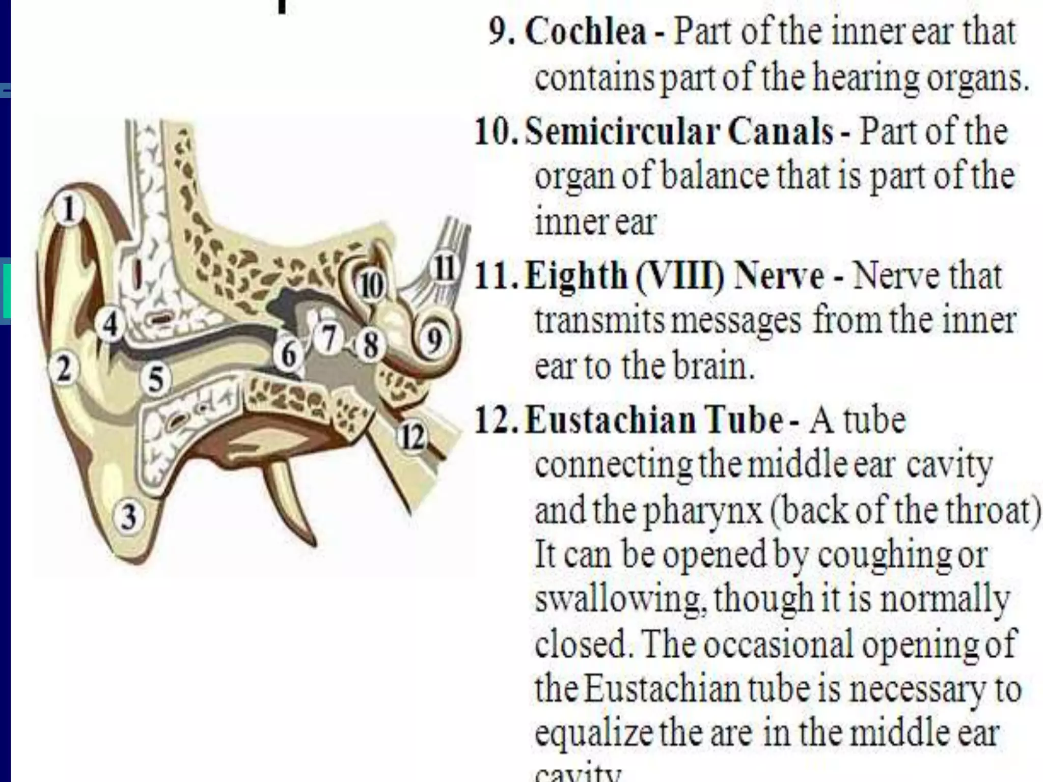
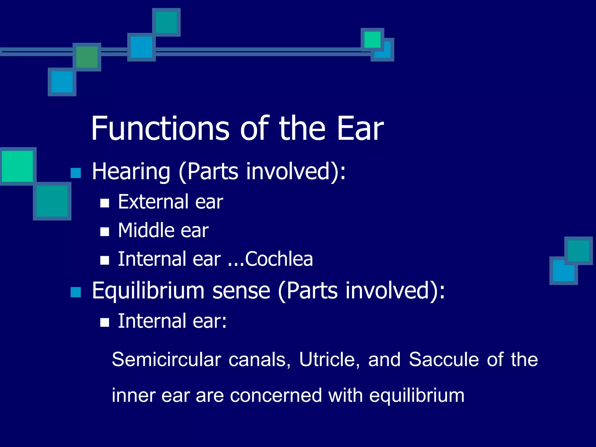
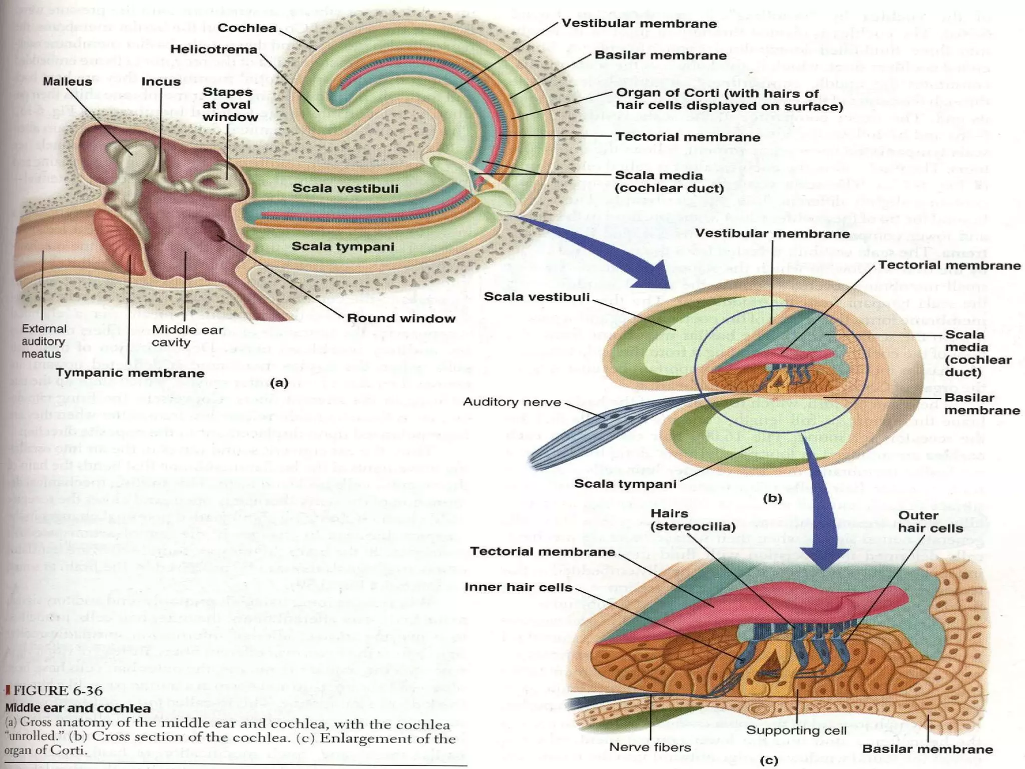
PHYSIOLOGY OF EAR AND MECHANISM
OF HEARING
Group 04
Members:
 Husnain Saddique
 Maimoona Arshad
 Hina Gul
 Naseemullah Wazir
Anatomical consideration
 External Ear:
 Pinna
 External canal
 Tympanic Membrane (funnel shaped,
pointing inward)
Anatomical consideration
 Middle Ear:
 Air filled cavity
 Three bones:
 Mallaus
 Incus
 Stapes (with its foot sitting on the oval window of
the inner ear)
 Inner Ear:
 Bony and membranous labyrinth
Functions of the Ear
 Hearing (Parts involved):
 External ear
 Middle ear
 Internal ear ...Cochlea
 Equilibrium sense (Parts involved):
 Internal ear:
Semicircular canals, Utricle, and Saccule of the
inner ear are concerned with equilibrium
Functions of the ear
 External ear:
 Act as funnel to collect sound
 Sound localisation (front, back, high, low)
 Alter amplitude (Pinna)
 Protection
 Wax
Functions of the Ear
 Middle ear: it is a space between tympanic
membrane and the inner ear (opens via
Eustachian tube into nasopharynx)
 Content:
 1- Air
 2- Ossicles
 Malleus
 Incus
 Stapes
 3- Muscles
 1- Tensor tympani
 2- Stepedius
Tensor tympani and stapedius muscles contract reflexly in
response to constant loud sound
Functions of the Middle Ear
 1- Ossicles:
 Manbrium of the malleus attached to the
back of the tympanic membrane and its short
process attached to the incus.
 The incus then articulates with the head of
the stapes, and its foot plate attached to the
oval window
Functions of the middle ear
 2- Muscles:
 Muscles contract reflexly in response to loud
sound (over 70dB)
 Contraction of the tensor tympani pulls the
manubruim & makes the tympanic m. tens.
Thus decreasing the vibration.
 Contraction of the stapeduis pull the foot
plate outward so that vibration reduced
 (protection from constant loud noise, but not
sudden noise, latency of 40-80 msec.
Inner ear
 Anatomy:
 Cochlea (snail like, coiled tubular system
laying deep in the temporal bone)
 Bony labyrinth
 Membranous labyrinth
Transmission of sound
through the middle ear
 sound waves vibrate the tympanic m.
 Tympanic m moves the handle of malleus
 Incus moves
 Stapes move in & out of the oval
window. The pressure transmitted
through cochlea cause stimulation of hair
cells in the organ of corti, which will
stimulate the auditory nerve
Middle ear magnifying effect
 1- The force from a large surface area
(Tympanic m.) are concentrated to a
small (oval window) the ratio is 17=1
 2- Lever action of ossicles = the lever
action of ossicles increase the force
of movement 1.3 times
 ▲ the total increase 17 X 1.3 = 22 times
Cochlea
 It is a system of three coiled tubes
through its length
 The basilar m. & the reissners m divide it
into three canals:
 Scala Vestibuli
 Scala Media
 Scala Tympani
Composition
 Scala Vestibuli: Na high K low
 Scala Tympani: Na high K low
 Scala Media : Na low K high
Organ of Corti
 Located (resting) on the basilar m.
 Contain inner & outer hair cells
 Extend from base to apex
Hair cells
 Steroclia extend from the top
 Arrangement:
 Three rows of outer hair cells (attached to
the reticular lamina or tectorial m.)
 One row of inner hair cells (not attached to
tectorial m.)
Function of inner hair cells
 Striocellia not embedded in tectorial m.
but bent by fluid movement under the
tectorial m.
 They are primary receptors for sound,
transducing fluid movement in cochlea
into action potential in the auditory nerve
Function of the outer hair cells
 Large number, but stimulate only small
fraction of nerve fibres in the cochlear
nerve
 If damaged, significant loss of hearing
(they control the sensitivity of inner hair
cells to particular sound frequency)
Receptors & Endocochlear
potentials
 Sound transmission into the inner ear
cause upper & lower movements of the
reticular m. (tectorial m.)
 »»»»» produce bending of steriocillia of
the hair cells alternatively open & close
cation channels at the tip of the
steriocillia
 Inward current: Depolarization
 Outward current: Hyperpolarisation
 The net results is depolarization
 Production of cells receptors potentials
 Release of neurotransmitter
 Production of action potentials
Nature of Sound
 Sound is produced from alternate
compression and rarefaction of air
molecules by vibrating body
Characteristics of sound
 1- Pitch (Tone) depend on No. Of cycle / sec.
Human ear can detect sound waves with freq..
20-20000 cycle /sec
 2- Intensity (Loudness) depend on amplitude
 3- Quality depend on the over tone or
interference
NOISE POLLUTION
 Noise pollution is
an environmental
hazard
 Exposure to sound
intensity above
80dB may damage
outer hair cells
Conduction of sound wave
 Air conduction:
 Normal situation of hearing, sound travel in
air causes vibration of Tympanic m.,
transmitted by ossicles to the oval window
Conduction of sound wave
 Bone conduction:
 Sound cause vibration of skull bones directly
transmitting the sound vibration to the
cochlea (eg when placing tuning fork on the
head or mastoid process)
Test of hearing
 Audiometer
 Weber test
 Rinnes test
THANK YOU

Physiology of Hearing(Presentation).pptx

  • 1.
    PHYSIOLOGY OF EARAND MECHANISM OF HEARING Group 04
  • 2.
    Members:  Husnain Saddique Maimoona Arshad  Hina Gul  Naseemullah Wazir
  • 7.
    Anatomical consideration  ExternalEar:  Pinna  External canal  Tympanic Membrane (funnel shaped, pointing inward)
  • 8.
    Anatomical consideration  MiddleEar:  Air filled cavity  Three bones:  Mallaus  Incus  Stapes (with its foot sitting on the oval window of the inner ear)  Inner Ear:  Bony and membranous labyrinth
  • 10.
    Functions of theEar  Hearing (Parts involved):  External ear  Middle ear  Internal ear ...Cochlea  Equilibrium sense (Parts involved):  Internal ear: Semicircular canals, Utricle, and Saccule of the inner ear are concerned with equilibrium
  • 11.
    Functions of theear  External ear:  Act as funnel to collect sound  Sound localisation (front, back, high, low)  Alter amplitude (Pinna)  Protection  Wax
  • 12.
    Functions of theEar  Middle ear: it is a space between tympanic membrane and the inner ear (opens via Eustachian tube into nasopharynx)  Content:  1- Air  2- Ossicles  Malleus  Incus  Stapes  3- Muscles  1- Tensor tympani  2- Stepedius Tensor tympani and stapedius muscles contract reflexly in response to constant loud sound
  • 13.
    Functions of theMiddle Ear  1- Ossicles:  Manbrium of the malleus attached to the back of the tympanic membrane and its short process attached to the incus.  The incus then articulates with the head of the stapes, and its foot plate attached to the oval window
  • 14.
    Functions of themiddle ear  2- Muscles:  Muscles contract reflexly in response to loud sound (over 70dB)  Contraction of the tensor tympani pulls the manubruim & makes the tympanic m. tens. Thus decreasing the vibration.  Contraction of the stapeduis pull the foot plate outward so that vibration reduced  (protection from constant loud noise, but not sudden noise, latency of 40-80 msec.
  • 15.
    Inner ear  Anatomy: Cochlea (snail like, coiled tubular system laying deep in the temporal bone)  Bony labyrinth  Membranous labyrinth
  • 17.
    Transmission of sound throughthe middle ear  sound waves vibrate the tympanic m.  Tympanic m moves the handle of malleus  Incus moves  Stapes move in & out of the oval window. The pressure transmitted through cochlea cause stimulation of hair cells in the organ of corti, which will stimulate the auditory nerve
  • 18.
    Middle ear magnifyingeffect  1- The force from a large surface area (Tympanic m.) are concentrated to a small (oval window) the ratio is 17=1  2- Lever action of ossicles = the lever action of ossicles increase the force of movement 1.3 times  ▲ the total increase 17 X 1.3 = 22 times
  • 19.
    Cochlea  It isa system of three coiled tubes through its length  The basilar m. & the reissners m divide it into three canals:  Scala Vestibuli  Scala Media  Scala Tympani
  • 20.
    Composition  Scala Vestibuli:Na high K low  Scala Tympani: Na high K low  Scala Media : Na low K high
  • 22.
    Organ of Corti Located (resting) on the basilar m.  Contain inner & outer hair cells  Extend from base to apex
  • 25.
    Hair cells  Sterocliaextend from the top  Arrangement:  Three rows of outer hair cells (attached to the reticular lamina or tectorial m.)  One row of inner hair cells (not attached to tectorial m.)
  • 26.
    Function of innerhair cells  Striocellia not embedded in tectorial m. but bent by fluid movement under the tectorial m.  They are primary receptors for sound, transducing fluid movement in cochlea into action potential in the auditory nerve
  • 27.
    Function of theouter hair cells  Large number, but stimulate only small fraction of nerve fibres in the cochlear nerve  If damaged, significant loss of hearing (they control the sensitivity of inner hair cells to particular sound frequency)
  • 28.
    Receptors & Endocochlear potentials Sound transmission into the inner ear cause upper & lower movements of the reticular m. (tectorial m.)  »»»»» produce bending of steriocillia of the hair cells alternatively open & close cation channels at the tip of the steriocillia
  • 29.
     Inward current:Depolarization  Outward current: Hyperpolarisation  The net results is depolarization  Production of cells receptors potentials  Release of neurotransmitter  Production of action potentials
  • 30.
    Nature of Sound Sound is produced from alternate compression and rarefaction of air molecules by vibrating body
  • 31.
    Characteristics of sound 1- Pitch (Tone) depend on No. Of cycle / sec. Human ear can detect sound waves with freq.. 20-20000 cycle /sec  2- Intensity (Loudness) depend on amplitude  3- Quality depend on the over tone or interference
  • 34.
    NOISE POLLUTION  Noisepollution is an environmental hazard  Exposure to sound intensity above 80dB may damage outer hair cells
  • 35.
    Conduction of soundwave  Air conduction:  Normal situation of hearing, sound travel in air causes vibration of Tympanic m., transmitted by ossicles to the oval window
  • 36.
    Conduction of soundwave  Bone conduction:  Sound cause vibration of skull bones directly transmitting the sound vibration to the cochlea (eg when placing tuning fork on the head or mastoid process)
  • 37.
    Test of hearing Audiometer  Weber test  Rinnes test
  • 38.