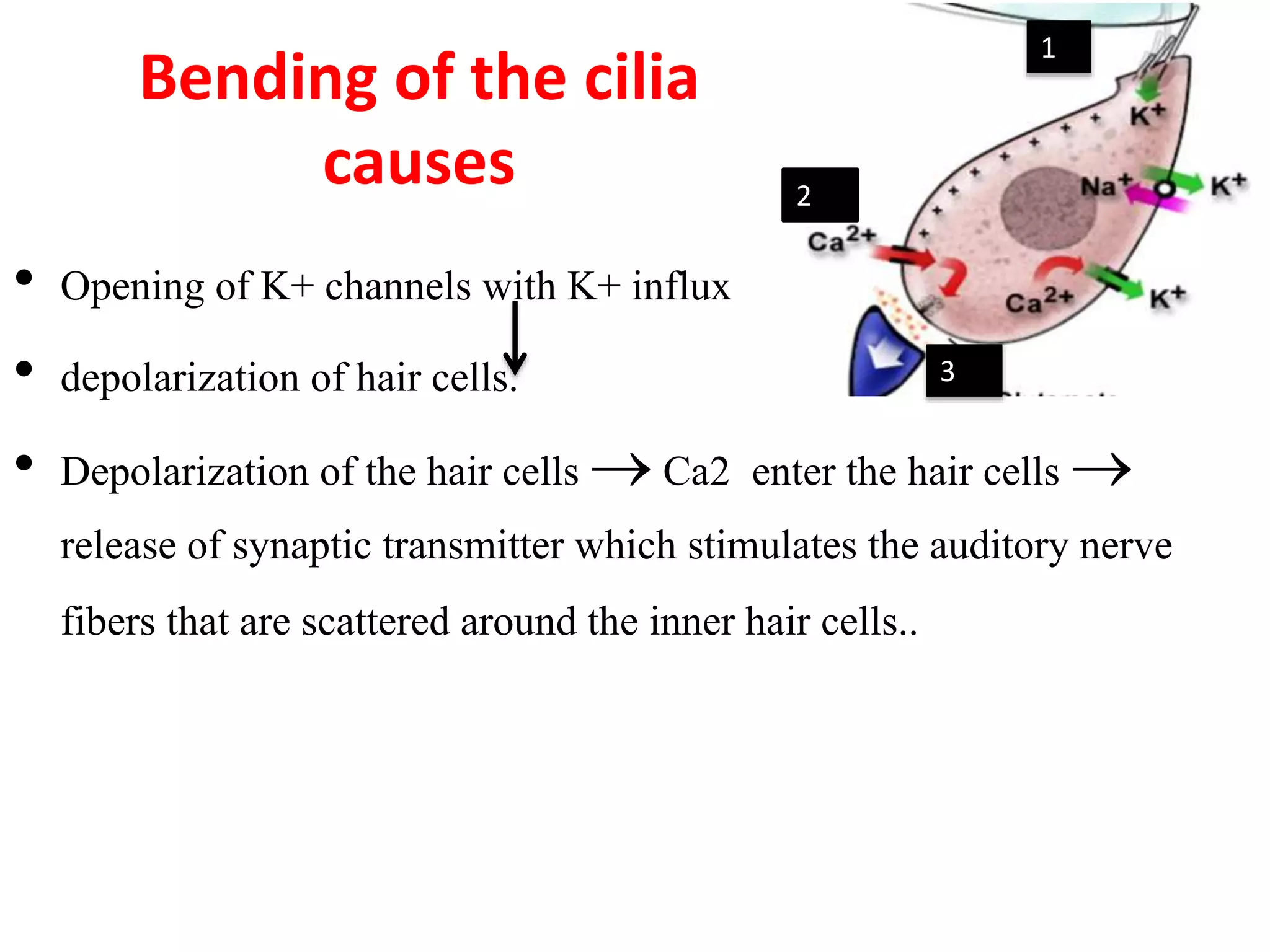
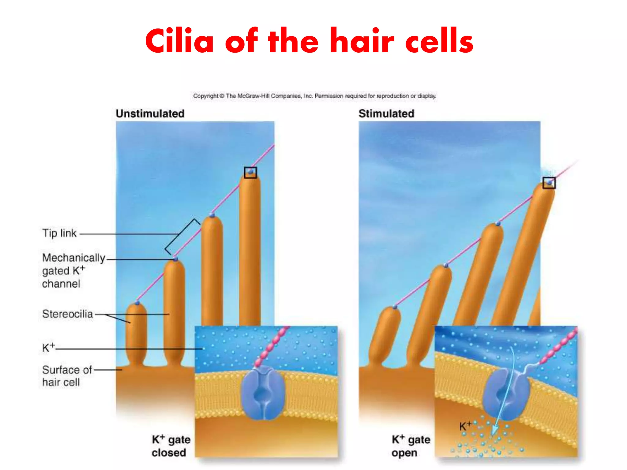
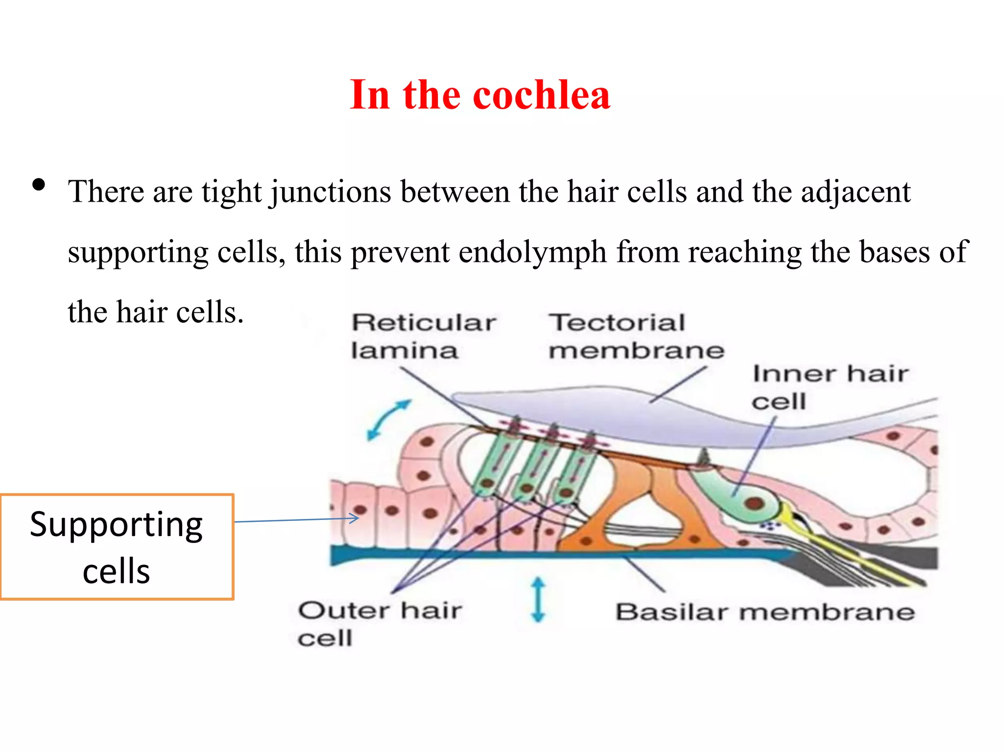
Hearing
Inner ear
part 1
By
Dr. Nanees El-Malkey
Lecturer of medical physiology
Ilos
1. Identify general Structure of inner ear
2. Understand Cochlear structure
3. describe organ of corti
4. Understand mechanism of hearing
Oval window cochlea
Semicircular canals
Round window
•
The labyrinth:
Is a group of canals tunneled in the bone called the bony labyrinth
Bony labyrinth is lined by membranes called the membranous labyrinth
Both are separated by a fluid called the perilymph
Inner ear
(Labyrinth)
perilymph
• the membranous labyrinth is filled by another
fluid called the endolymph.
Thus the inner ear is a fluid filled cavity unlike,
the middle and the external ears which are air
filled cavities.
The membranous labyrinth
• It is subdivided into two parts:
A- Non auditory labyrinth (semicircular canals) for equilibrium.
B- Auditory labyrinth for hearing and is called the "Cochlea".
• The cochlea is a coiled tube which is 35 mm long.
• Throughout its length, the basilar membrane and vestibular membrane divide it
into 3 chambers (scalae).
•
The upper:
scala vestibuli
Contains perilymph
ends at the oval
window
the lower:
scala tympani
Contains perilymph
ends at the round window
The middle:
scala media
Contains endolymph
Scala vestibuli and tympani contain perilymph and
communicate with each other at the apex of the
cochlea (helicotrema).
• The scala media does not communicate with the other 2
scalae.
Organ of Corti
• 4 raws of hair cells that sit on the Basilar membrane
• Tectorial membrane above the hair cells
 It contains the hair cells
which are the auditory
receptors
 There are 20.000 outer hair
cells and 3500 inner hair
cells in each cochlea.
 tips of the hair cells are
embedded in elastic
membrane tectorial
membrane
Tunnel
of corti
The hair cells
• The cell bodies of the
afferent neurons are located
in the spiral ganglion
within the modiolus, the
bony core in the center of
cochlea.
• 90-95% of these afferent
neurons innervate the inner
hair cells
• forming the auditory
(cochlear) division of the
vestibulocochlear
(acoustic) nerve.
Mechanism Of Hearing
The sound waves are collected by the ear pinna which also
determines the direction of the sound.
These waves travel the external auditory tube to be concentrated
on the tympanic membrane which responds to these sound
vibrations
• With the help of the three bonny ossicles, these vibrations are
magnified and produces movement in the oval window.
The movements of the oval window lead to movements in the
perilymph of the scala vestibuli with upward and downward
movements of the vestibular membrane.
The vibrations of this membrane are transmitted to the endolymph
of the scala media vibration of the
basilar membrane
.
The cilia of the hair cells are attached to the tectorial membrane and
both the tectorial and basilar membranes are attached to the limbus;
thus both membranes slide on each other causing what is called
"shearing movement“
These shearing movements cause bending of the cilia of the hair cells.
Bending of the cilia
causes
• Opening of K+ channels with K+ influx
• depolarization of hair cells.
• Depolarization of the hair cells  Ca2 enter the hair cells 
release of synaptic transmitter which stimulates the auditory nerve
fibers that are scattered around the inner hair cells..
1
2
3
Cilia of the hair cells
In the cochlea
• There are tight junctions between the hair cells and the adjacent
supporting cells, this prevent endolymph from reaching the bases of
the hair cells.
Supporting
cells
• However the basilar membrane is relatively permeable to perilymph in
the scala tympani and the tunnel of Corti and the bases of the hair cells
are bathed in perilymph.
• Therefore, the bases of the hair cells are bathed in perilymph while
their processes are bathed in endolymph and this is important for
generation of receptor potential.
• Thank you

Inner ear 1

  • 1.
    Hearing Inner ear part 1 By Dr.Nanees El-Malkey Lecturer of medical physiology
  • 2.
    Ilos 1. Identify generalStructure of inner ear 2. Understand Cochlear structure 3. describe organ of corti 4. Understand mechanism of hearing
  • 3.
  • 4.
    • The labyrinth: Is agroup of canals tunneled in the bone called the bony labyrinth Bony labyrinth is lined by membranes called the membranous labyrinth Both are separated by a fluid called the perilymph Inner ear (Labyrinth)
  • 5.
  • 6.
    • the membranouslabyrinth is filled by another fluid called the endolymph. Thus the inner ear is a fluid filled cavity unlike, the middle and the external ears which are air filled cavities.
  • 7.
    The membranous labyrinth •It is subdivided into two parts: A- Non auditory labyrinth (semicircular canals) for equilibrium. B- Auditory labyrinth for hearing and is called the "Cochlea".
  • 8.
    • The cochleais a coiled tube which is 35 mm long. • Throughout its length, the basilar membrane and vestibular membrane divide it into 3 chambers (scalae). •
  • 9.
    The upper: scala vestibuli Containsperilymph ends at the oval window the lower: scala tympani Contains perilymph ends at the round window The middle: scala media Contains endolymph
  • 10.
    Scala vestibuli andtympani contain perilymph and communicate with each other at the apex of the cochlea (helicotrema). • The scala media does not communicate with the other 2 scalae.
  • 11.
    Organ of Corti •4 raws of hair cells that sit on the Basilar membrane • Tectorial membrane above the hair cells
  • 12.
     It containsthe hair cells which are the auditory receptors  There are 20.000 outer hair cells and 3500 inner hair cells in each cochlea.  tips of the hair cells are embedded in elastic membrane tectorial membrane Tunnel of corti
  • 13.
    The hair cells •The cell bodies of the afferent neurons are located in the spiral ganglion within the modiolus, the bony core in the center of cochlea. • 90-95% of these afferent neurons innervate the inner hair cells • forming the auditory (cochlear) division of the vestibulocochlear (acoustic) nerve.
  • 15.
    Mechanism Of Hearing Thesound waves are collected by the ear pinna which also determines the direction of the sound. These waves travel the external auditory tube to be concentrated on the tympanic membrane which responds to these sound vibrations
  • 16.
    • With thehelp of the three bonny ossicles, these vibrations are magnified and produces movement in the oval window.
  • 17.
    The movements ofthe oval window lead to movements in the perilymph of the scala vestibuli with upward and downward movements of the vestibular membrane. The vibrations of this membrane are transmitted to the endolymph of the scala media vibration of the basilar membrane .
  • 18.
    The cilia ofthe hair cells are attached to the tectorial membrane and both the tectorial and basilar membranes are attached to the limbus; thus both membranes slide on each other causing what is called "shearing movement“ These shearing movements cause bending of the cilia of the hair cells.
  • 19.
    Bending of thecilia causes • Opening of K+ channels with K+ influx • depolarization of hair cells. • Depolarization of the hair cells  Ca2 enter the hair cells  release of synaptic transmitter which stimulates the auditory nerve fibers that are scattered around the inner hair cells.. 1 2 3
  • 21.
    Cilia of thehair cells
  • 22.
    In the cochlea •There are tight junctions between the hair cells and the adjacent supporting cells, this prevent endolymph from reaching the bases of the hair cells. Supporting cells
  • 23.
    • However thebasilar membrane is relatively permeable to perilymph in the scala tympani and the tunnel of Corti and the bases of the hair cells are bathed in perilymph. • Therefore, the bases of the hair cells are bathed in perilymph while their processes are bathed in endolymph and this is important for generation of receptor potential.
  • 24.