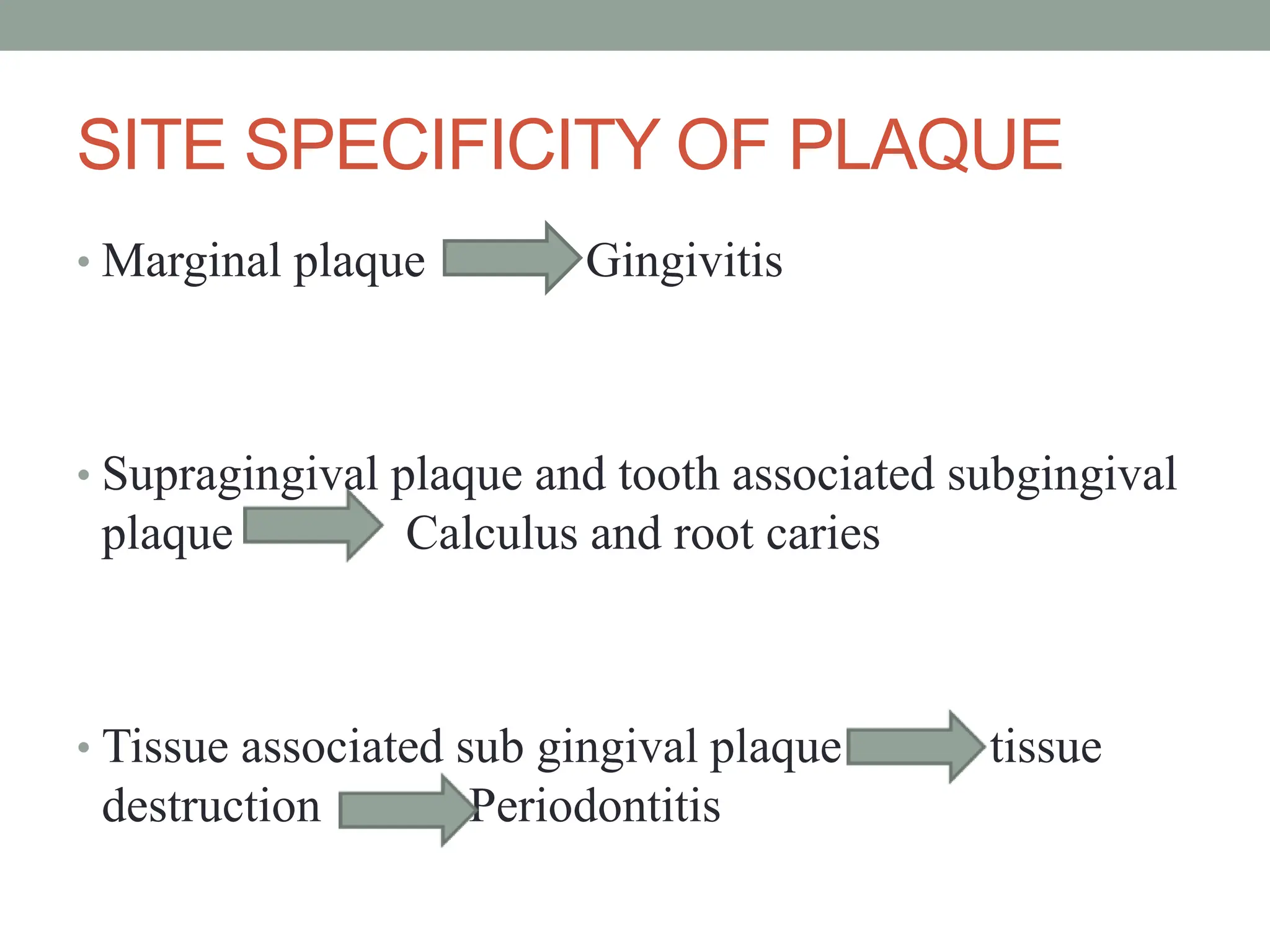
Dental plaque is a biofilm that forms on teeth and consists of bacteria embedded in an extracellular matrix. It develops in stages, beginning with the formation of a protein pellicle layer on the tooth surface within seconds of cleaning. Initial colonizers like streptococci then adhere to the pellicle. Secondary colonization involves more species adhering directly or co-aggregating with initial colonizers. Co-aggregation involves specific adhesins on bacteria binding together different species in complex biofilms. The plaque matures into distinct supragingival and subgingival biofilms as the environment changes below the gumline.
2. INTRODUCTION
Dental caries and periodontal diseases are the two most common
diseases of the oral cavity. Their prevalence is recorded along
with history of man after his appearance on earth. Experimental
and epidemiologic studies have demonstrated that these diseases
are dependent on the microorganisms present in plaque. It is the
build up of plaque that serves as an irritant to the gingiva.
3. ⢠Dental plaque is defined clinically as âa
structured resilient, yellow-grayish substance
that adheres tenaciously to the intra oral hard
surfaces, including removable and fixed
restorations.â â Bowen WH 1976
⢠Dental plaque- âhighly complex structural entity
which comprises of large species of
microorganisms embedded in a mucinous
matrixâ-American Academy of
Periodontology 1986
4. COMPOSITION OF DENTAL PLAQUE
⢠Composed of bacteria in a matrix of salivary glycoproteins and extra cellular
polysaccharides
⢠One gram of plaque - 1011 bacteria.(Schroeder etal 1970)
⢠Number of bacteria in supragingival plaque on a single tooth surface - > 109
⢠In a periodontal pocket - 103 to 108
⢠A single individual may harbor 150 or more different species.
⢠More than 500 distinct microbial species are found in dental plaque.(Moore etal
1994).
⢠Nonbacterial microorganisms found in plaque include
Mycoplasm species
yeasts
protozoa
viruses
Carranzaâs Clinical Periodontology 10th edition
5.
6. TEETH AS PORT OF ENTRY OF
PERIOPATHOGENS
ďŹ The unusual anatomic feature, that a mineralized structure, the
tooth, passes through the integument, so that part of it is
exposed to the external environment while part is within the
connective tissues.
ďŹ The tooth provides a surface for the colonization of a diverse
array of bacterial species.
ďŹ In contrast to the outer surfaces of most parts of the body,the
outer layers of the tooth do not shed, and thus microbial
colonization is facilitated.
ďŹ In addition the tooth provides sanctuaries in which organisms
can hide, persist at low levels during treatment and the re-
emerge to cause further problems.
⢠Clinical Periodontology & Implant Dentistry 5th edition
⢠Jan Lindhe
7. CLASSIFICATION
ďŹ Dental plaque is classified as
SUPRAGINGIVAL or
SUBGINGIVAL
ďŹ SUPRAGINGIVAL- found at or
above the gingival margin, when
in direct contact with the gingival
margin it is referred as marginal
plaque
ďŹ SUBINGIVAL- below the gingival margin, between the
tooth and the gingival pocket epithelium.
8. STRUCTURE OF SUPRAGINGIVAL PLAQUE
â˘Stratified organization of a multilayered accumulation of bacterial
morphocytes
â˘Gram positive cocci and short rods predominate at the tooth surface, where as
gram negative rods and filaments as well as spirochetes predominate in the
outer surface of the mature plaque mass
â˘1st bacteria to colonize are streptococci species andActinomyces. Veillonella
is also an early colonizer
â˘Plaque grows by cell division of adherent bacteria
â˘âCorncobâ structures (Listgarten et al. 1973)
â˘Feature of older plaque is the presence of dead, lysed cells. Provide nutrients
to the still viable bacteria in the neighbourhood. (Theilade and Theilade
1970)
10. STRUCTURE OF SUPRAGINGIVAL PLAQUE
â˘The material present between the bacterial cells- Intermicrobial
matrix
â˘3 sources contribute to it- the plaque microorganisms, the
saliva, gingival exudate
â˘Intermicrobial matrix varies in regions
- fibrillar component between gram +ve cocci
- granular or homogenous in other regions
- in the presence of gram âve organisms, vesicles seen. These
vesicles contain endotoxins and proteolytic enzymes and are
involved in adherence of bacteria (Hofstad et al. 1972, Grenier
& Mayrand 1987)
12. STRUCTURE OF SUBGINGIVAL PLAQUE
ďŹ Subgingival microbiata differs in
compostion from the supragingival plaque
primarily because:
a) Local availability of blood products.
b) Low oxidation-reduction (redox) potential
which characterizes the anaerobic
environment.
c) Gingival crevice or pocket is bathed by
the flow of crevicular fluid.
Carranzaâs Clinical Periodontology 10th edition
SUBGINGIVAL
PLAQE
13. STRUCTURE OF SUBGINGIVAL PLAQUE
⢠A relatively thin layer of adherent bacteria covers the tooth surface.
⢠Rods and filaments tend to be arranged in a palisading pattern, with the long
axis of the cells perpendicular to the tooth surface.
⢠Unique bacterial aggregates, resembling test-tube brushes, can be found
attached to the adhering plaque and extending into the space between the
bacterial layer and the adjacent soft tissue wall.
⢠The âbristlesâ of these test-tube brush formations are gram-negative
filamentous bacteria, some of which may be flagellated.
⢠The axial portion of the test-tube brush consists of a single or several long
filaments held together by an amorphous extracellular matrix.
⢠The bulk of the subgingival microbiota consists of a complex mixture of
predominantly anaerobic bacteria that surround and cover the test- tube brush
formations.
⢠The structure of Dental plaque- Max. Listgarten, Periodontology 2000,
Vol 5. 1994
14. STRUCTURE OF SUBGINGIVAL PLAQUE
The subgingival plaque has two
regions-
1). Tooth associated region of
subgingival plaque and
2). Tissue associated region of
subgingval plaque
Both morphologic and microbiologic
studies reveal distinction between
the tooth associated and tissue
associated regions of subgingival
plaque.(Listgarten etal 1970)
15. TOOTH ASSOCIATED PLAQUE
ďŹ Filamentous microorganisms dominate
ďŹ Increased number of gram positive rods and cocci are seen.
ďŹ In the deeper parts - filamentous organisms are fewer and in the
apical region - absent.
The apical border of the plaque mass is separated from the
junctional epithelium by a layer of host leukocytes, and the bacteria
of this apical tooth associated region show an increased number of
gram-negative rods.
ďŹ Tooth associated plaque- S.mitis, S.sanguis, A.viscosus, A.naeslundii,
Eubacterium species seen predominatly
Carranzaâs Clinical Periodontology 10th edition
16. TISSUE ASSOCIATED PLAQUE
⢠Primarily contain gram-negative rods and cocci, large numbers of filaments,
flagellated rods, and spirochetes.
⢠The multitude of spirochetes and flagellated organisms are motile bacteria and
there is no intermicrobial matrix between them. This outer part of the microbial
accumulation in the periodontal pocket adheres loosely to the soft-tissue pocket
wall. (Listgarten1976).
⢠Host tissue cells e.g. white blood cells and epithelial cells are also found.
⢠Soft tissue plaque- S.oralis, S.intermedius, P.gingivalis, P.intermedia, T.forsythia
and Fusobacterium Nucleatum. (Dzink etal 1989)
⢠Subgingivally located bacteria appear to have the capacity to invade dentinal
tubules, the openings of which have become exposed as a consequence of
inflammatory driven resorptions of the cementum (Adriaens et al. 1988). Such a
habitat might serve as the source for bacterial recolonization of the subgingival
space following treatment of periodontal disease.
⢠Clinical Periodontology & Implant Dentistry 5th edition
⢠Jan Lindhe
17. PERIIMPLANT PLAQUE
â˘Plaque forms on oral implants as well
â˘Similarities between peri-implant and subgingival microbial
deposits have been demonstrated in cross sectional studies
(Mombelli et al. 1987,1995) and longitudinal studies
(Mombelli et al. 1988; Pontoriero et al. 1994)
⢠Clinical Periodontology & Implant Dentistry 5th edition
⢠Jan Lindhe
18. SITE SPECIFICITY OF PLAQUE
⢠Marginal plaque Gingivitis
⢠Supragingival plaque and tooth associated subgingival
plaque Calculus and root caries
⢠Tissue associated sub gingival plaque tissue
destruction Periodontitis
19. FORMATION OF DENTAL PLAQUE
PLAQUE FORMATION
AT
ULTRASTRUCTURAL
LEVEL
1.Formation of the
pellicle on
the tooth surface
2.Initial colonization
by bacteria
3.Secondary colonization
and plaque maturation
20. FORMATION OF PELLICLE
â˘All surfaces of the oral cavity, hard and soft get coated with a pellicle
â˘Within nanoseconds after prophylaxis, saliva derived âacquired pellicleâ formed
â˘Composition- Glycoproteins, proline-rich proteins, phosphoproteins, histidine-rich
proteins, enzymes
â˘Studies indicate that bacteria can be part of the early deposit (Ronstrom A,
Edwardsson S, Atistrom R, 1977)
â˘Composition of pellicle differs from saliva indicating it forms by selective adsorption
of environmental macromolecules
â˘Forces involved are- Van der Waals, electrostatic and hydrophobic forces
â˘Functions as a protective barrier and also provides a substrate to which bacteria in the
environment attach
21. INITIAL COLONIZATION OF TOOTH
SURFACE
ďThe dental pellicle that is formed, alters the charge and free
energy of the surface which in turn increase the efficiency of
bacterial adhesion.
ďWithin few hrs. bacteria are found on the dental surface. In fact
with in 5min. 106 bacteria colonize per cm2 the tooth surface.
ďInitial bacteria that colonize the tooth surface are predominantly
gram +ve facultative micro organisms such as A. viscosus and
S. sangius.
ďAdhesion of bacteria is determined by the enviroment and the
physio-chemical surface properties of the bacterium and the
substratum.
24. SECONDARY COLONIZATION
AND PLAQUE MATURATION
ďThese include micro organisms that do not initially
colonize tooth surface and includes P.intermedia,
P.loesheii, Capnocytophaga, F. nucleatum and P.
gingivalis .
ďThe micro organism may interact with pellicle, bacterial
polysaccharide or there may be direct interaction between
bacterial cell surfaces.
ďThis last bacterial cell to cell interaction is termed as co-
aggregation and was first described by Gibbons and
Nygaard in 1970.
25. ďMc Intire, et al in 1978 described co-aggregation between A.
naeslundii and S. oralis. This interaction was between
proteinaceous molecule which acted as a lectin on A. naeslundii
and a carbohydrate receptor on S. oralis.
ďDirect cell to cell interaction were also noticed in early electron
microscopic studies of dental plaque. Clearly observed in these
studies were morphologic forms arising from the direct
association of different cell types.
ďThe interaction of filamentous cells with coccal cells were
particularly noticeable and these co-aggregated cells were
labeled âcorncobsâor âtest tube brushesâ or bristle brush due
to their appearance.
26. âcorncobsâ
ď The name corncobs was
coined by Jones in 1971.
ďIt was first described by
Vincentini in 1897 and
thought that the structures
were composed of a single
microbial species and
named them Letotrix
racemosa.
ďIt is now known that
corncob unit consists of a
central filamentous
bacterium covered by
coccal cells.
27. ďElectron micrographs of
cross section of corncobs
indicated that that
attachment of cocci to
the filament occurred via
hair like appendages that
are commonly found on
some species of oral
streptococci.(co-
aggregation)
ďThese fimbrae were
found to be located on
one pole rather than
uniformly distributed over
the cell surface as found
in other oral streptococci.
28. ďAnother feature of corncobs was the firmness of
attachment between component micro organism.
ďAttempt to separate component micro organism by
sonification failed.
ďIn 1997 Mouton, et al used a combination of
micromanipulation and culture to isolate both filamentous
organism and the attached streptococci.
29. ďLancy, et al 1980 developed a quantitative assay for
corncob formation.
ďUsing this assay and electron microscope it was
subsequently found that F. nucleatum also formed
corncobs with S.cristae. This was a very important finding
since F. nucleatum is a major inhabitant of sub gingival
plaque
ďThus formation of Fusobacterial corncobs could provided
a connective link between supra and sub gingival plaque
30. Multi generic co-aggregation
ďAnother important feature during secondary colonization
and plaque maturation includes the concept of multi
generic co-aggregation.
ďKolenbrander and Andersen 1986 showed that multi
generic aggregates are a composite of independent inter
generic co-aggregates.
31. ďBridging is also the property of co aggregating cells and
have important ecological implication. Bridging refers to
observation that two non-aggregating strains may
participate together in a multi generic if they recognize a
common partner by distinct mechanism.
ďE.g :- A. israelii does not co-aggregate with S. oralis.
However P. loescheii co-aggregates with both strain by
means of different adhesins. When the three were mixed,
all three cell types were found in aggregated form.
Kolenbrander 1985 .
32. ďAnother concept put forward by Kolenbrander, et al
in1990 that play a role in plaque formation is the
possibility that intra generic co-aggregation between
different streptococci.
ďOf all the bacteria that participated in intra generic co-
aggregation only Fusobacteria and streptococci were
capable of intra generic co-aggregation.
ď. F. nucleatum acts as a bridge between early and late
colonizers, which may partially explain why fusobacteria are so
numerous in samples from both healthy and diseased sites
33. ⢠In addition to interactions with oral bacteria and host cells, F. nucleatum
interacts with and binds host-derived molecules, such as plasminogen.
⢠F. nucleatum is generally nonproteolytic, but organisms that coexist with it,
such as P.gingivalis, are highly proteolytic and can activate fusobacterium-
bound plasminogen to form fusobacterium-bound plasmin,a plasma serine
⢠protease .
⢠Acquisition of proteolytic ability on its cell surface confers on the
fusobacteria a new
⢠metabolic property, the ability to process potential peptide signals in the
community.
⢠These peptides may be used as nutrients by fusobacteria or by other biofilm
residents. coaggregates with all the late colonizers.
⢠Coaggregation bridges are mechanisms of cooperation because they bring
together two species that are not coaggregation partners.
34. MICROBIAL COMPLEXES
⢠The association of bacteria within mixed biofilms is not
random, rather there are specific associations among bacterial
species
⢠Socransky et al.(1998) examined over 13,000 subgingival
plaque samples from 185 adult subjects and used cluster
analysis and community ordination techniques to demonstrate
the presence of specific microbial groups within dental plaque
⢠Six closely associated groups of bacterial species were
recognized
⢠Clinical Periodontology & Implant Dentistry 5th edition
⢠Jan Lindhe
35. ⢠These included the Actinomyces,
⢠A yellow complex consisting of members of genus Streptococcus
⢠A green complex consisting of Capnocytophaga species,
A.actinomycetemcomitans serotype a, E. corrodens and Campylobacter
concisus
⢠A purple complex consisting of V.parvula and Actinomyces odontolyticus
⢠These groups of species are early colonizers of the tooth surface whose
growth usually precedes the multiplication of the predominantly Gram-
negative orange and red complex
⢠The orange complex consists of Campylobacter gracilis, C. rectus, C.
showae, E. nodatum, F. nucleatum subspecies, F. periodonticum, P. micros,
P. intermedia, P. nigrescens and S. constellatus
⢠Clinical Periodontology & Implant Dentistry 5th edition
⢠Jan Lindhe
36. ⢠The red complex consists of B.forsythus, P. gingivalis and T.denticola (and
sometimes Eubacterium nodatum)
⢠The "red complex" was associated more commonly with clinical indicators of
periodontal diseases
⢠Red complex species increased strikingly in prevalence and numbers with increasing
pocket depth.
⢠The species of the red complex are also elevated at sites exhibiting gingival
inflammation, as measured by gingival redness, bleeding on probing and suppuration.
⢠Thus red complex species are not only related to periodontal disease status in a
subject, but to disease status at the periodontal site.
⢠Other species did not show this relationship
â˘
⢠Clinical Periodontology & Implant Dentistry 5th edition Jan Lindhe
39. ďThe term biofilm describes the relatively undefinable
microbial community associated with tooth surface or
any other hard, non-shedding material (Wilderer and
Charaklis 1989).
ďA biofilm is a well organized community of bacteria
that adheres to surfaces and is embedded in an
extracellular slime layer(Jill S.Nield-Gehrig).
41. NATURE OF BIOFILM
⢠Preferred method of growth for microorganisms
â Provides advantages for colonizing species
⢠Protection from
⢠Competing microorganisms
⢠Environmental factors, host defense
⢠Toxic substances, such as lethal chemicals, antibiotics
⢠Facilitate processing and uptake of nutrients, cross-
feeding,removal of harmful metabolic products
⢠Development of an appropriate physico-chemical environment.
⢠Clinical Periodontology & Implant Dentistry 5th edition
⢠Jan Lindhe
42. COMPOSITION OF BIOFILM
⢠Composed of micro colonies (15-20% by volume) distributed in a shaped matrix
or glycocalyx (75-80% volume)
⢠Presence of voids or water channels
o Permit the passage of nutrients and other agents, acting as âcirculatoryâ
⢠system
⢠Organic constituents include:
o Polysaccharides
o Proteins
o Glycoproteins
o Lipid material
o Albumin
⢠Inorganic components are mainly calcium and phosphorus with trace amounts of
other minerals including sodium, potassium and fluoride.
⢠Clinical Periodontology & Implant Dentistry 5th edition
⢠Jan Lindhe
43.
44.
45. Exopolysaccharides-backbone of the biofilm
⢠DRY material-exopolysaccharides,proteins,salts,cell material
⢠Exopolysaccharides-major component(50-95%)
⢠Plays major role in maintaining the integrity of biofilm
⢠Several different polysaccharides
⢠Some are neutral(mutans),some highly charged polyanionic
46. ⢠Protects microbial cells from dessication & attack by harmful
agents
⢠Creates a local nutritionally rich enviorment by binding to
essential nutrients
⢠Acts as a buffer
⢠Maintain biofilm structure-formation of networked cross linked
linear macromolecules
⢠Type and not the quantity of exopolysaccharide has an effect on
biofilm.
49. â˘Begins with pellicle formation
â˘Pellicle- thin coating of salivary proteins
â˘Acts as a double-sided adhesive tape
â˘Adhering to tooth surface on one side and providing a
sticky surface for bacterial attachment on the other side
⢠Bacteria connect to each other and pellicle by fimbrae,
fibrils
Attachment
50. â˘Once they stick,bacteria produce substances that stimulate
other free flowing bacteria to get attached
â˘2 days of no tooth cleaning tooth surface colonized by
gram +ve cocci(streptococci species)
â˘Attachment to a solid surface stimulates bacteria to
secrete extracellular slime layer that helps in anchoring
and protection for attached bacteria.
51. I-physical properties
⢠Increased surface roughness>increased surface
area>increase colonisation
II-chemical properties
⢠Chemical composition of surface;eg-brass,polyvinyl
chloride
⢠Cohesiveness of conditioning film(Bos R 1999)
⢠Surronding saliva and its flow rate
FACTORSAFFECTING
ATTACHMENT OF BIOFILMS
52. Formation of microcolonies
â˘Begins after tooth surface is covered with attached
bacteria
â˘Biofilm grows primarily through cell division of
adherent bacteria
â˘Bacteria begin to grow away from the tooth
â˘Plaque grows quickly in early development and slower
in more mature biofilms
â˘Bacterial blooms- specific species grow at rapid
accelerated rates
53. Secondary colonization and
biofilm maturation
â˘Prevotella intermedia, Prevotella loescheii, Capnocytophaga,
Fusobacterium nucleatum ,Porphyromonas gingivalis â Secondary
colonizers
â˘Adhere to cells of bacteria already attached
â˘Adhere to one another by coaggregation
â˘Bacteria cluster together to form sessile,mushroom-shaped micro
colonies that are attached to tooth surface at a narrow base
â˘Results in formation of a complex array of different bacteria linked
to another
54. Detachment
â˘Essential to allow colonization of new habitats
â˘Cells detach in different fashions-
-Erosion-detachment of single cells in a continuous
fashion
-Sloughing-sporadic detachment of large group of cells
-intermediate process where large pieces of biofilm are
shed
â˘Rate of detachment not clear (Watnick P, Kolter R,
2000)
55. ⢠Rate of growth in many biofilms is slow and detachment
is an uncommon event
â˘Cells in such biofilms are metabolically active and
capable of growth once released from the biofilm
â˘Detachment is active ongoing process
56.
57. Factors affecting biofilm
development and behaviour
â˘Shear stress-
-high shear-thinner and denser biofilms,colonies are elongated and
capable of rapid oscillation
-low shear-roughly circular cluster of cells separated by
voids,colonies are tower/mushroom shaped
â˘Hydrodynamics-
-Biofilms under laminar flow(low shear) and turbulent flow(high
shear) are different
â˘Changes in nutrient concentration
-addition of nutrients to a biofilm increased both mass and
structure
Stoodley et al 1999
58. Bacterial behavior within the biofilm
â˘Bacteria growing in microbial communities do not behave the
same as those growing in a planktonic state
â˘E.g. the resistance of bacteria to antibiotics is increased in the
biofilm about 1000-1500 times compared to those in their
planktonic state (Costerton JW 1999)
â˘Mechanism of increased resistance in biofilms differs from
species to species, antibiotic to antibiotic and biofilms growing in
different habitats
â˘Resistance of bacteria to antibiotics is affected by their
nutritional status, growth rate, temperature, pH and previous
exposure to sub effective concentrations of antimicrobials (Brown
MRW, Collier PJ, Gilbert P,1990)
59. â˘Slower rate of growth of bacteria within the biofilm also makes
them less susceptible to some antibiotics (Brooun A, Liu S, Lewis K,
2000)
â˘Matrix performs a âhomeostatic functionâ
-cells deep in the film and that at the periphery experience different
growing conditions or cells growing planktonically
-growth rates of the cells also differ
-slow growing cells(deeper cells)express non-specific defense
mechanisms i.e shock proteins and multi drug efflux mechanisms
and so increased exopolymer synthesis
-this exopolymer has certain properties that retards diffusion
Clinical Periodontology & Implant Dentistry 5th edition
Jan Lindhe
60. ⢠Eg-strongly charged or chemically highly reactive agents fail to
reach the deeper zones of biofilm as the biofilm acts as an ion
exchange resin removing such molecules from solution
⢠Extracellular enzymes get trapped and concentrated in the
extracellular matrix ,thus inactivating positively charged
hydrophilic antibiotics
⢠Hydrophobic antibiotics like macrolides though positively
charged are unaffected
⢠ability of extracellular matrix to act as a barrier depends on the
type of antibiotic,its binding to the matrix,levels of antibiotic
⢠As reaction between agent and matrix will reduce the level of
agent,a biofilm of greater bulk will deplete the agent more
⢠Alteration in genotype and phenotype of bacteria is also
important
⢠Clinical Periodontology & Implant Dentistry 5th edition
⢠Jan Lindhe
61. ⢠Recently a subpopulation of cells within a biofilm that are
âsuper-resistentâ was proposed
⢠Such cells explained the elevated levels of resistance to ceratin
antibiotic
⢠Brooun et al 2000 examined multi drug resistant pumps to
antibiotic resistance of organisms grown in biofilms
⢠These pumps extruded the chemically antimicrobial agents from
the cell
⢠Extrusion placed the antibiotics outside the cell
membrane,hence offering protection to the biofilm from the
antibiotics targeting the cell wall synthesis
⢠Clinical Periodontology & Implant Dentistry 5th edition
⢠Jan Lindhe
62. Quorum sensing
â˘Comes from the same term used in a committee when
enough members are present to legally take some action
â˘It was first observed in the marine bacterium Vibrio
fischeri, which can produce light after a sufficient
population of this bacterium has developed
â˘Is the ability of the bacteria and microcolonies to
communicate with each other in the biofilm
63. ⢠Involves the regulation of expression of specific
genes through the accumulation of signaling
compounds that mediate inter cellular
communication(Prosser 1999)
⢠depends on cell density
⢠At threshold level, (quorum cell density) gene
expression is activated
⢠Cell signaling appears to be mediated by an N-acyl
homoserine lactone encoded by a lux1 gene
64. Though planktonic cells secrete chemical signals (HSLs, for homoserine lactones), the
low concentration of signal molecules does not change genetic expression. Biofilm cells
are held together in dense populations, so the secreted HSLs attain higher
concentrations. HSL molecules then re-cross the cell membranes and trigger changes in
genetic activity
65. â˘Autoinducer-2 a universal signal molecule is recently
discovered in mixed species communities(Kolenbrander et
al 2006)
â˘Physiological properties of bacteria in a community
may be altered(Cooper et al 1995)
â˘Plays a role in
- antibiotic resistance at high cell densities
-encourages growth of beneficial species
-discourages growth of competitors
66. QUORUM SENSING
⢠Quorum sensing systems bacteria have been generally
divided into at least three classes:
⢠(1) LuxI/LuxRâtype quorum sensing in Gram-negative
bacteria, which use acyl-homoserine lactones (AHL) as
signal molecules. ( Lux- bacterial luciferase gene).
⢠(2) Oligopeptide-two-component-type quorum sensing in
Gram-positive bacteria, which use small peptides as
signal molecules.
⢠(3) luxS-encoded autoinducer 2 (AI-2) quorum sensing in
both Gram-negative and Gram-positive bacteria.
67. ⢠Autoinducer-2 a universal signal molecule is recently discovered in mixed
species communities(Kolenbrander et al 2006)
⢠AI-2 allows for inter-species communication, so it is called a âuniversal
languageâused for cross-species communication.
⢠AI-2 is produced from S-adenosylmethionine through several steps, including the
required enzymatic conversion of the intermediate S-ribosylhomocysteine by
LuxS to 4,5-dihydroxy-2,3-pentanedione, which is unstable and is predicted to
cyclize spontaneously (133, 134) into a variety of molecules called pro-AI-2
before forming a mature AI-2âLuxP complex.
⢠The luxS gene, encoding S-adenosylhomocysteinase (LuxS) is present in the
genome sequencesof many oral bacteria.
⢠The discovery of AI-2 that is produced and detected by a large number of diverse
bacteria implies that bacteria have a means to assess the cell density of other
species in a microbial community, facilitating interspecies communication and
social interactions among species in the community.
⢠Communication among Oral Bacteria (Paul E. Kolenbrander,* Roxanna N. Andersen, David S. Blehert, Paul G. Egland,
Jamie S. Foster, and Robert J. Palmer Jr)MICROBIOLOGY AND MOLECULAR BIOLOGY REVIEWS, Sept. 2002
68. DE NOVO SUPRAGINGIVAL PLAQUE
FORMATION: CLINICAL ASPECTS
⢠Plaque formation follows an exponential growth curve (Quirynen et al
1989)
⢠Negligible in the 1st 24 hours . Increases rapidly in the next 3 days and then
slows down
⢠There is a shift toward anaerobic and gram-negative flora
⢠Follows a typical topographic pattern. Initial growth along the gingival
margin and interdental space (areas protected by shear stress)
⢠Can also start from grooves, cracks, perikymata or pits
⢠Rough intra oral surfaces accumulate and retain more plaque (Quirynen and
Bollen)
⢠Plaque formation occurs much faster in the lower jaw, molar areas and
buccal tooth surfaces and interdental regions. (Lindhe etal 1992)
⢠More rapid on teeth with inflamed gingival margins (Sorensen etal 1986)
⢠No difference in de novo plaque formation with age (Fransson etal 1995,
Holm-Pedersen etal 1975, Winkel etal 1987)
69. DE NOVO SUBGINGIVALPLAQUE
FORMATION
⢠Technically impossible to record the dynamics of subgingival
plaque formation
⢠Studies show that tooth surfaces harbor plaque and calculus
after scaling. These remain the primary source for subgingival
recolonization
⢠Leknes et al in 1994 did a study on beagle dogs. Studied the
extent of colonization in 6mm pockets. Observed that smooth
surfaces harbored significantly less plaque
70. PHYSIOLOGIC PROPERTIES OF DENTAL
PLAQUE
⢠Transition from gram positive to gram negative is accompanied by
physiologic transition in the developing plaque
⢠Early colonizers lower the redox potential of the environment and
favour the growth of anaerobic species
⢠Lactate and formate, by products of metabolism of streptococci and
actinomycetes maybe used in the metabolism of other plaque
microorganisms
⢠Host is also an important source of nutrients
⢠Physiologic interactions occur between different microorganisms in
plaque and between the host and plaque microorganisms
71. ⢠Nutritional interdependencies are critical for growth and
survival of the microorganism in dental plaque and partly
explains the highly specific structural interactions among
bacteria in plaque
⢠Some researchers say the pathologic flora is due to âecological
plaque hypothesisâ
72. NONSPECIFIC PLAQUE HYPOTHESIS
(Theilade 1986)
Mid 1900âs- periodontal disease was thought to be due to:
⢠- accumulation of plaque over time
⢠- decreased host response
⢠- increased host susceptibility
⢠This hypothesis maintains that periodontal disease results from the
âelaboration of noxious products by the entire plaque floraâ.
⢠The theory maintains that control of periodontal disease depends on control of
the amount of plaque accumulation.
⢠Although discarded in favor of the Specific plaque hypothesis, clinical
treatment is still based on this theory.
⢠Carranzaâs Clinical Periodontology 10th edition
73. NONSPECIFIC PLAQUE HYPOTHESIS
⢠Contradictions of theory:
⢠Individuals with considerable amounts of plaque, calculus and
gingivitis did not develop destructive periodontitis.
⢠Individuals with periodontitis demonstrated site-specificity in
the disease pattern.
⢠Some sites were unaffected whereas adjacent sites were affected
by the disease.
⢠In the presence of uniform host response,these findings were
inconsistent with the concept that all plaque was equally
pathogenic
⢠Led to a renewed search for specific pathogens in periodontal disease.
Carranzaâs Clinical Periodontology 10th edition
74. SPECIFIC PLAQUE HYPOTHESIS
Sir Walter Loesche 1979
â˘States that only certain plaque is pathogenic
â˘Pathogenecity depends on presence or increase in specific
microorganisms
â˘Plaque which has specific bacterial pathogens results in periodontal
disease- these organisms destroy host tissues
â˘Association of specific bacterial species with disease originated in
1960
â˘Acceptance of specific plaque hypothesis came about by the
recognition of A. actinomycetemcomitans as a pathogen in localized
aggressive periodontitis.
Carranzaâs Clinical Periodontology 10th edition
75. ECOLOGICAL PLAQUE HYPOTHESIS
⢠Proposed by PD Marsh in 1991
⢠Says that a change in a key environmental factor will trigger a
shift in the balance of the resident plaque microflora which might
predispose a site to disease
⢠In health, these organisms are only weakly competitive and not
significant clinically. Microbial specificity in disease is because
only certain species are competitive under the new environmental
conditions
⢠It is a basic tenet of microbial ecology that a major change to an
ecosystem produces a corresponding disturbance to the stability
of the resident microbial community (Brock 1966; Alexander
1971; Fletcher et al. 1987)
77. CRITERIA FOR IDENTIFICATION OF
PERIODONTAL PATHOGENS
⢠In the 1870âs Robert Koch developed classic criteria by which a
microorganism can be judged to be a causative agent in human infections
⢠These criteria, known as KOCHâS POSTULATES, stipulate the following
for the causative agents:
⢠Must be routinely isolated from diseased individuals
⢠Must be grown in pure culture in the laboratory
⢠Must produce a similar disease when inoculated into susceptible laboratory
animals
⢠Must be recovered from lesions in a diseased laboratory animal
Carranzaâs Clinical Periodontology 10th edition
78. ⢠Difficulties exist in the application of these criteria in the case
of periodontitis as:
1. The inability to culture all the microorganisms that have been
associated with the disease.(spirochetes).
2. Difficulties inherent in defining and culturing sites of active
disease
3. Lack of good animal model system for study of periodontitis.
Carranzaâs Clinical Periodontology 10th edition
79. In 1992, Sigmund Socransky , a researcher at Forsyth Dental Institute at
Boston proposed criteria by which periodontal microorganisms maybe judged
to be potential pathogens
These criteria are:
â˘Must be increase in the number of organisms at diseased sites
â˘Must be decreased at sites that show improvement with treatment
â˘Must demonstrate a host response
â˘Capable of causing disease in experimental animals
â˘Must demonstrate virulence factors responsible for enabling the
microorganism to cause destruction of the periodontal tissues
Carranzaâs Clinical Periodontology 10th edition
80. Association of plaque microorganisms with
Periodontal disease
⢠3 factors determine occurrence of active periodontitis:
⢠Susceptible host
⢠Presence of pathogenic species
⢠Absence or a small proportion of âbeneficial bacteriaâ
⢠The clinical manifestations of periodontitis are due to an interplay
between specific pathogens in plaque and host tissues
Carranzaâs Clinical Periodontology 10th edition
81. SUSCEPTIBILITY OF HOST
⢠Partially hereditary. Maybe influenced by smoking, diabetes
stress
⢠Genetic mutations have been identified that alter host response
to bacteria and are associated with periodontal disease
⢠Grossi et al. 1998 found a direct relation between periodontitis
and the level of smoking
⢠Diabetics are at higher risk for periodontal destruction
⢠Severe stress conditions also aggravate periodontal destruction
Carranzaâs Clinical Periodontology 10th edition
82. Marsh PD, Devine DA. How is the development of dental biofilms influenced by the host? J
Clin Periodontol 2011;
83. PRESENCE OF PATHOGENS
⢠Presence of pathogens in sufficient numbers is essential
⢠Key pathogens- Aggregetibacter actinomycetemcomitans, Tannerella Forsythia
and Porphyromonas gingivalis
⢠Moderate evidence for etiology- Prevotella intermedia, Prevotella nigrescens,
Campylobacter rectus, Peptostreptococcus micros, Fusobacterium nucleatum,
Eubacterium nodatum (past a certain threshold level)
⢠Evidence based on epidemiologic data and results of animal innoculation
⢠Mere presence of pathogens is not enough. An elevation to a critical level is
required
⢠Periopathogens may be present in the gingival crevice, although in lower
numbers,as members of the normal resident flora.(Tanner etal 1991)
Carranzaâs Clinical Periodontology 10th edition
84. ROLE OF BENEFICIAL SPECIES
⢠Affect disease progression in the following ways:
- occupying a niche that might otherwise have pathogens
- limiting pathogens ability to adhere to appropriate tissue
surfaces
- affecting the growth of the pathogen
- affecting the ability to produce virulence factors
- degrading virulence factors produced by the pathogen
⢠e.g. S. Sanguis produces hydrogen peroxide that kills A.
actinomycetemcomitans. (Hillman etal 1985)
Carranzaâs Clinical Periodontology 10th edition
85. MICROBIAL SHIFT DURING DISEASE
⢠Gram positive to gram negative
⢠From cocci to rods (later to spirochetes)
⢠Non motile to motile organisms
⢠Facultative to obligate anaerobes
⢠Fermenting to proteolytic species
Carranzaâs Clinical Periodontology 10th edition
86. Microbial shifts during dental biofilm re-
development in the absence of oral hygiene in
periodontal health and disease
NaciyeG.Uzel1,â ,FlaviaR.Teles1,2,RicardoP
.Teles1,2, XiaogingQ.Song1,GayTorresyap1,âĄ, SigmundS.Socransky1,AnneD.Haffajee1
JournalofClinicalPeriodontology
Volume38,Issue7,pages612â620,July2011
⢠Abstract
⢠Aim: To monitor microbial shifts during dental biofilm re-development.
⢠Materials and methods: Supra- and subgingival plaque samples were taken separately from 28
teeth in 38 healthy and 17 periodontitis subjects at baseline and immediately after tooth cleaning.
Samples were taken again from seven teeth in randomly selected quadrants during 1, 2, 4 and 7
days of no oral hygiene. Samples were analysed using checkerboard DNAâDNA hybridization.
Species counts were averaged within subjects at each time point. Significant differences in the
counts between healthy and periodontitis subjects were determined using the MannâWhitney test.
⢠Results: The total supra- and subgingival counts were significantly higher in periodontitis
on entry and reached or exceeded the baseline values after day 2. Supragingival counts
of Veillonella parvula, Fusobacterium nucleatum ss vincentii and Neisseria
mucosa increased from 2 to 7 days. Subgingival counts were greater for Actinomyces,
green and orange complex species. Significant differences between groups in supragingival
counts occurred for 17 of 41 species at entry, 0 at day 7; for subgingival plaque, these
values were 39/41 taxa at entry, 17/41 at day 7.
⢠Conclusions: Supragingival plaque re-development was similar in periodontitis and health,
but subgingival species recolonization was more marked in periodontitis.
87. STRATEGIES TO PREVENT PERIODONTAL
DISEASES
⢠Conventional methods involve mechanical removal of subgingival
plaque along with antimicrobial therapy
⢠Ecologic approach- alter the environment of the pocket to prevent
growth of pathogens
⢠Can be done by:
⢠- Antimicrobial and anti-inflammatory agents
⢠- Oxygenating and redox agents
⢠Novel drugs that specifically target quorum sensing systems are
capable of attenuating bacterial infections in a manner that is less
likely to result in the development of resistant mutants.(eg Furanones)
1) Quorum Sensing and Bacterial Social Interactions in Biofilms(Yung-Hua Li 1,2,* and
And Xiaolin Tian )
Sensors 2012,12
88. OMNIGENE
⢠These are DNA probe systems for a number of
known periodontopathogen subgingival bacteria.
⢠A paper point sample of sub-gingival plaque is
placed in the container provided and mailed off
to the company for assay.
⢠Probes are available for the detection of A.
actinomycetemcomitans, P. gingivalis, P.
intermedia, F. nucleatum, C. rectus, T. denticola
and E. corrodens.
⢠Reports are provided within very short time
periods (few hours to few days).
89. EVALUSITE
Evalusite is a kit that employs a novel membrane-based enzyme
immunoassay for the detection of three putative periodontopathogens:
Aa, Pg and Pi.
A sub-gingival sample is collected using paper points and added to a
sample tube. The eluent is then added to the kit, which employs a
sandwich-type ELISA (enzyme-linked immunosorbent assay); a pink
spot is displayed if the test organism is present.
The main weaknesses of this test kit reside in
1) the assumption that the three detected organisms are causing
disease;
2) (2) it is a multistage test;
3) (3) it has a subjective calorimetric end point and
4) (4) there is no permanent record of the results [11].
90. PERIOSCAN
⢠Perioscan is a diagnostic test kit that utilizes the BANA (N-benzoyl-DL-arginine-
2-naphthylamide)-hydrolysis reaction, developed to detect bacterial trypsin-like
proteases in the dental plaque .
⢠A trypsin-like activity has been identified in strains of P. gingivalis, T. denticola,
T. forsythia and some Capnocytophagia strains. BANA is an example of a
substrate-conjugated beta-nepthylamine (p-NA), which is hydrolyzed by this
trypsin-like enzyme to release free p-NA. The latter is a chromophore and reacts
with a variety of dyes (e.g. Fast-Garnet GBC) to produce colored products.
Subgingival plaque is collected and placed on a BANA-containing strip, which is
then folded to contact a second strip containing the âFast-Blackâ dye reagent.
⢠The folded card is placed inside an oven for 15 min at 55°C and any blue-black
color that appears is scored positive for the above species.
â˘
One of the potential difficulties of this test is that it may be positive at clinically
healthy sites and might remain so after treatment.
91. CONCLUSION
⢠Dental plaque biofilm cannot be eliminated. However, the
pathogenic nature of the dental plaque biofilm can be reduced
by reducing the bioburden (total microbial load and different
pathogenic isolates within that dental plaque biofilm) and
maintaining a normal flora with appropriate oral hygiene
methods that include daily brushing, flossing and rinsing with
antimicrobial mouthrinses.This can result in the prevention or
management of the associated sequelae, including the
development of periodontal diseases and possibly the impact of
periodontal diseases on specific systemic disorder
92. REFERENCES
Carranzaâs Clinical Periodontology- 9th , 10th Edition
Clinical Periodontology and Implant Dentistry- Lindhe, 4th,5th Edition
The structure of Dental plaque- Max. Listgarten, Periodontology 2000, Vol 5.
1994
Microbial ecology of dental plaque and its significance in health and disease-
P.D. Marsh
Dental biofilms:difficult therapeutic targets- Sigmund Socransky and Anne D
Haffajee, Periodontology 2000, Vol 28. 2002
Dental plaque biofilms: communities, conflict and control.P. D. Marsh
Periodontology 2000 vol 55 2011