SlideShare a Scribd company logo
ANATOMY OF MANDIBLE AND ITS
IMPORTANCE IN IMPLANT
PLACEMENT
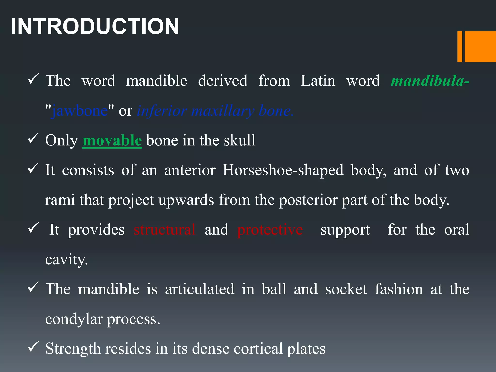
INTRODUCTION
 The word mandible derived from Latin word mandibula-
"jawbone" or inferior maxillary bone.
 Only movable bone in the skull
 It consists of an anterior Horseshoe-shaped body, and of two
rami that project upwards from the posterior part of the body.
 It provides structural and protective support for the oral
cavity.
 The mandible is articulated in ball and socket fashion at the
condylar process.
 Strength resides in its dense cortical plates
ANATOMY
OF
MANDIBLE
embryology
 Mandible is the second bone after clavicle to
ossify in the body.
 Parts that ossify in cartilage includes: incisive
part below the incisor teeth, coronoid and
condyloid processes
 Upper half of ramus above the level of the
mandibular foramen
 A single ossification centre for each half of the
mandible arises in the 6th week of I.U. life in
the region of bifurcation of inferior alveolar
nerve into mental and incisive branches. As the
ossification continues, the meckel’s cartilage
become surrounded and invaded by bone.
 It is partly membranous & partly cartilaginous in ossification.
• Incisive part below symphysis
menti
• Coronoid
• Condyloid process
• Upper half of ramus
Cartilage
• Whole of body except lower incisive
part
• Lower half of ramus upto mandibular
foramen
Membrane
OSTEOLOGY
BODY OF THE MANDIBLE
 Horseshoe-shaped BODY
 Has two surfaces EXTERNAL and INTERNAL surfaces ,
 Two borders UPPER and LOWER borders
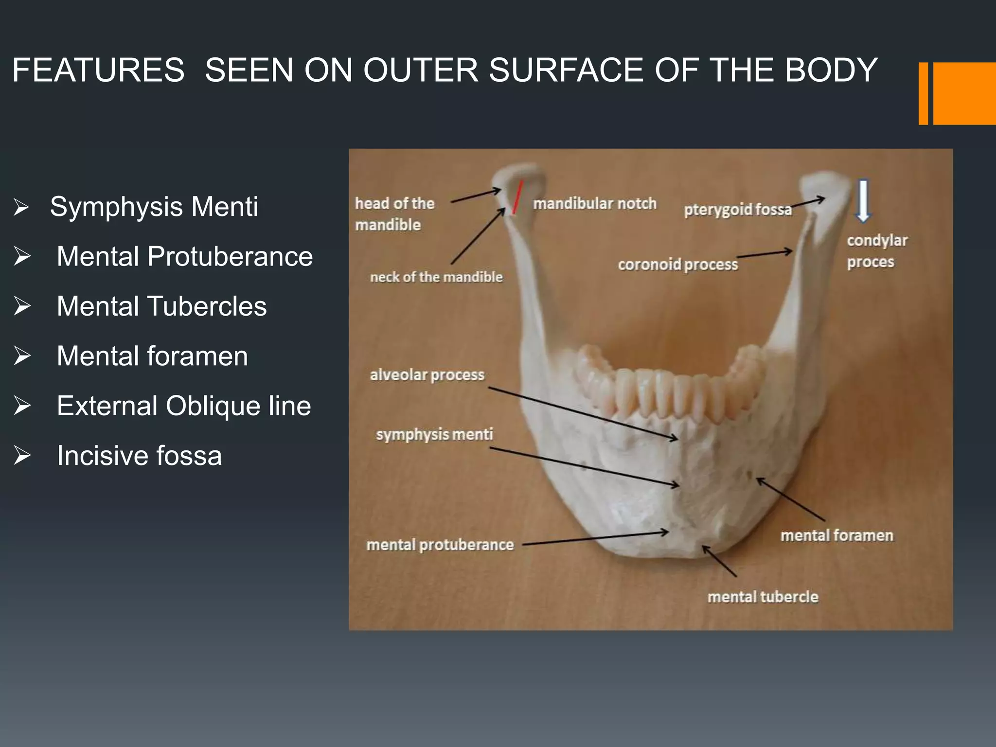
FEATURES SEEN ON OUTER SURFACE OF THE BODY
 Symphysis Menti
 Mental Protuberance
 Mental Tubercles
 Mental foramen
 External Oblique line
 Incisive fossa
FEATURES ON THE INNER SURFACE OF THE BODY
 Genial tubercles
 Mylohyoid line
 Submandibular fossae
 Sublingual fossae
 Mylohyoid groove
 Upper alveolar border
 Lower border base
 Digastric fossae
BORDERS OF THE MANDIBLE
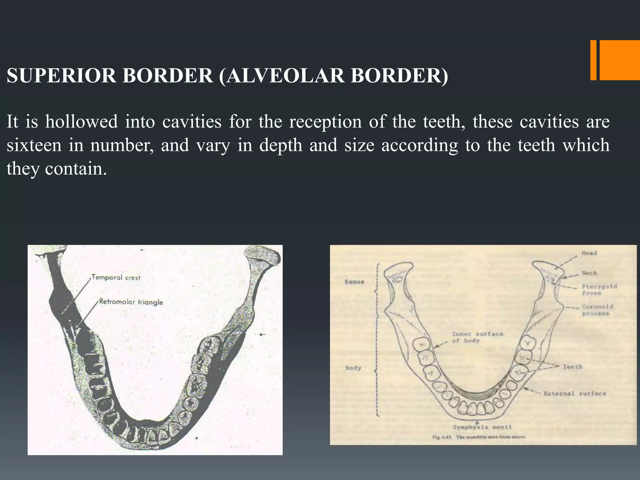
SUPERIOR BORDER (ALVEOLAR BORDER)
It is hollowed into cavities for the reception of the teeth, these cavities are
sixteen in number, and vary in depth and size according to the teeth which
they contain.
INFERIOR BORDER (base of mandible)
Is rounded, longer than the superior,
and thicker in front than behind.
RAMUS OF MANDIBLE
Is quadrilateral
2 surfaces
Lateral
Medial
4 borders
Superior
Inferior
Anterior
Posterior
2 processes
Coronoid
Condylar
PROCESSES
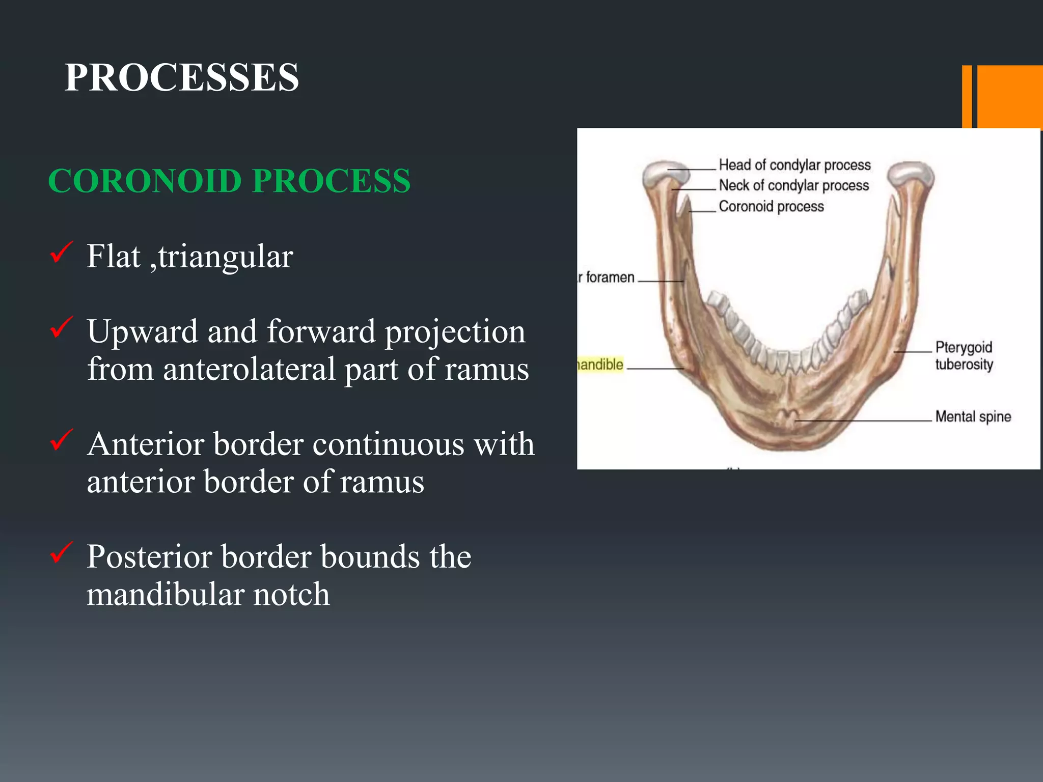
CORONOID PROCESS
 Flat ,triangular
 Upward and forward projection
from anterolateral part of ramus
 Anterior border continuous with
anterior border of ramus
 Posterior border bounds the
mandibular notch
CONDYLAR PROCESS
 Upward projection from postero
superior part of ramus
 Apically enlarged as head of
condyle.
 Articulates with temporal bone’s
mandibular fossa to form
temperomandibular joint
 Lateral aspect palpable in front
of tragus
 Pterygoid fovea anterior to neck
ATTACHMENTS AND RELATIONS
ON THE LATERAL
SURFACE:
1. From The Oblique line :
Buccinator, and depressor anguli
oris below the mental foramen
2. Incisive fossa:
gives origin to MENTALIS mental
slips of ORBICULARIS ORIS.
3. Whole of lateral surface of
ramus except posterosuperior part
provides insertion to MASSETER.
4.Posterosuperior part : covered
by PAROTID GLAND
5. Lateral surface of the neck
provides insertion to the
LATERAL LIGAMENT OF
TMJ.
6. Parts of both the inner and
outer surfaces just below the
alveolar margins are covered by
mucous membrane of the
mouth.
7. PLATYSMA is inserted into
the lower border.
8. The deep cervical fascia (
investing layer) is attached to
the whole length of the lower
border.
1. Digastric fossa: arises
ANTERIOR BELLY OF
DIGASTRIC
2. Genial tubercles: arises
GENIOGLOSSUS and
GENIOHYOID.
3. Mylohyoid line : arises
MYLOHYOID MUSCLE.
4. From an area above the
posterior end of mylohyoid line:
arises SUPERIOR
CONSTRICTOR OF PHARYNX.
5. Pterygomandibular raphe:
Attached immediately behind the
third molar tooth in continuation
with the origin of superior
constrictor
ON THE MEDIAL SURFACE
7.Below and behind the mylohyoid
groove: insertion of MEDIAL
PTERYGOID muscle .
8.At the apex of coronoid process :
TEMPORALIS is inserted ;extend
downwards on ant. Border of ramus.
9.Into the pterygoid fovea: insertion
of LATERAL PTERYGOID.
10.Sphenomandibular ligament : is
attached to the lingula.
BLOOD SUPPLY OF THE MANDIBLE
ARTERIAL SUPPLY
Mainly by Maxillary artery,
Branch of external carotid
artery
By its branches, mainly
through inferior alveolar
artery
Venous supply of mandible
 Drains into
Internal jugular vein and external
jugular vein through maxillary vein,
facial vein and pterygoid plexus
Nerve supply of mandible
Mainly through the trigeminal nerve -
V cranial nerve
MANDIBULAR NERVE
 Main trunk
 Anterior trunk
 Posterior trunk
FORAMINA AND OTHER RELATIONS
 Mental foramina - mental nerve and
vessels
 Mandibular notch - massetric nerve
and vessels
 Medial side of neck - auriculo
temporal nerve
 Mylohyoid groove - mylohyoid nerve
and vessels
 Mylohyoid groove in front of ramus -
lingual nerve
 Mandibular canal and foramina -
inferior alveolar nerve and vessels
POSTNATAL GROWTH OF
THE MANDIBLE
 According to the data from the vital staining experiments, the
posterior surface the ramus, the condyle and coronoid process are
principal sites of growth.
 Growth is quite general during the first year of life with all
surfaces showing bone apposition.
 Mandibular growth becomes more selective.
Postnatal Growth Of Mandible
The mandible can be divided into several sub-units like
 Chin
 Alveolar process
 Body
 Lingual tuberosity
 Ramus
 Angular process
 Coronoid process
 Condylar process
 Chin:
 1-2 years→ chin prominence is seen
 The mental protuberance forms by bone
deposition
 The change in the contour occurs by
following two mechanism.
1) The area just above the chin and
the base of the alveolar process,
is a resorptive area.
2) There is forward translation of
chin as mandible grows forward.
 Alveolar process:
 This develops in response to the developing tooth buds.
 Body: (corpus)
 The length of the body increases as the ramus
moves posteriorly
 Lingual tuberosity:
 It forms the boundary between
the ramus & body
 A combination of the resorption
and deposition accentuates its
prominence.
 Ramus:
 The ramus is seen to move posteriorly due to
deposition at its posterior border and resorption on
its anterior border
 Angle:
 The combined deposition and resorption causes flaring
of the angle of the mandible
 Coronoid process :
 Enlow’s enlarging “V” principle.
 Birth: Coronoid process is at
higher level than condylar
process.
 Childhood: Coronoid & condylar
processes are at same level.
 Adult: Condylar process is at
higher level.
 Condyle:
 Condylar growth rate increases at
puberty and reaches its peak by
12-14 years.
 The growth ceases at around 20
years
 Role of condyle:
o Primary displacement
APPLIED ANATOMY OF MANDIBLE
 Parasymphysis region lateral to the mental prominence is a
naturally weak area susceptible for parasymphyseal fracture.
This is because of the presence of incisive fossa and mental
foramen
 The body of the mandible is considerably thicker than the
ramus and the junction between these two portions
constitutes a line of structural weakness.
 Strength of the lower jaw varies with the presence or absence
of teeth. The presence of impacted lower third molars or
excessive long roots of canines make the area more
vulnerable for fracture.
IMPORTANCE IN IMPLANT PLACEMENT
Mandibular Foramen
 The location of the mandibular foramen may vary based on race and
ethnicity
 Among adult cadaveric mandibles, the foramen was found inferior to the
occlusal plane, at its level, or above it 75%, 22.5%, and 2.5% of the time,
respectively
 Therefore, according to these investigations, 2.5% to 23.5% of block
injections given at the level of occlusion would be ineffective.
 Advisable to inject patients 6 to10mmsuperior to the occlusal plane, which
usually accounts for anatomic variations
 Short needles can be used to attain anesthesia in the mandible
Inferior Alveolar Canal
 The inferior alveolar canal houses the IAN nerve , artery, vein and
lymphatics
 The canal is; 3.4mm wide, and the nerve is ;2.2mm thick
 Therefore, during implant placement it is possible to in advertently
penetrate into the mandibular canal and induce neurologic damage
without provoking hemorrhaging and vice versa.
 When developing an osteotomy over the mandibular canal, cortical
bone is penetrated first, and the preparation terminates within softer
cancellous bone
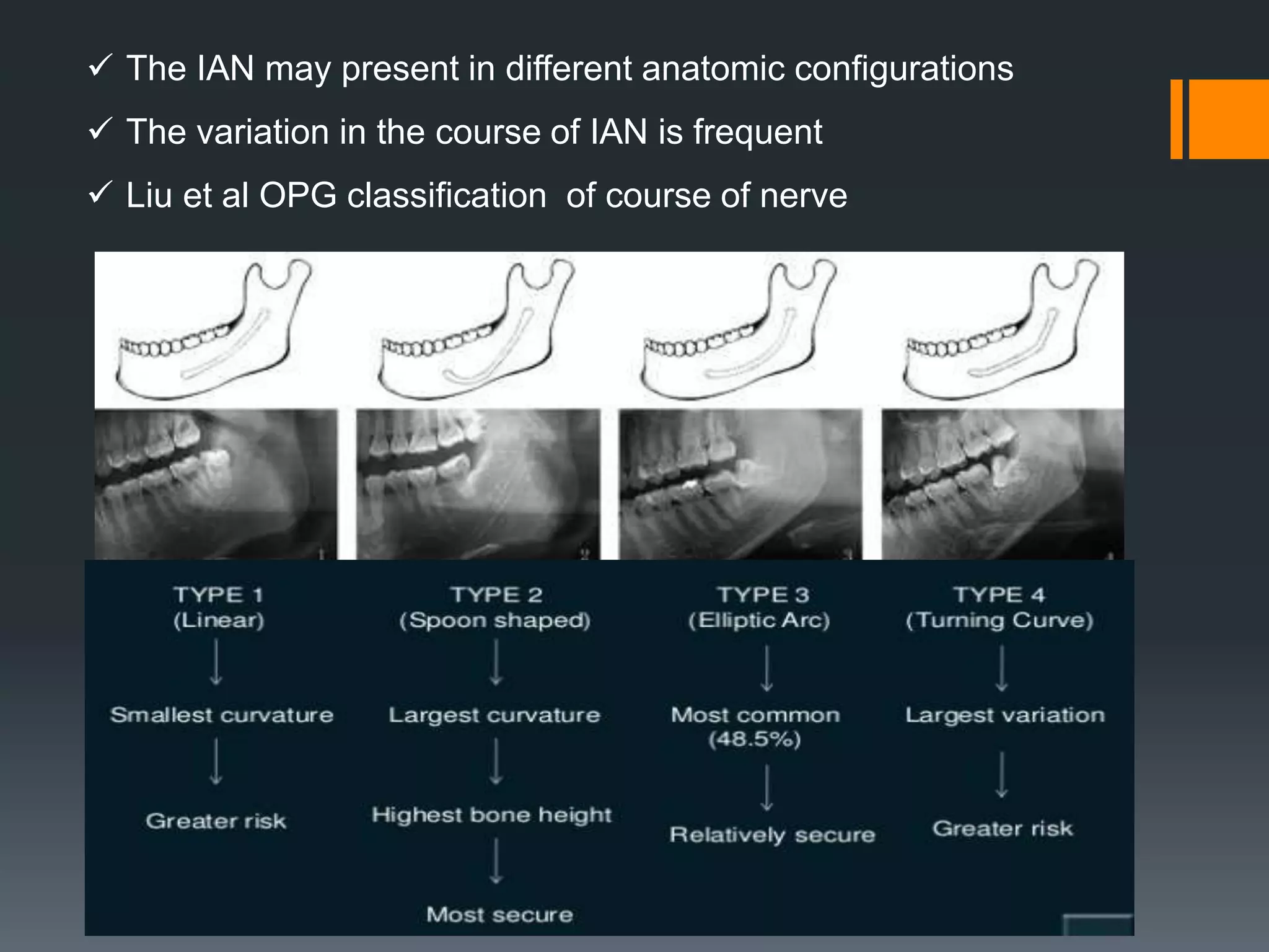
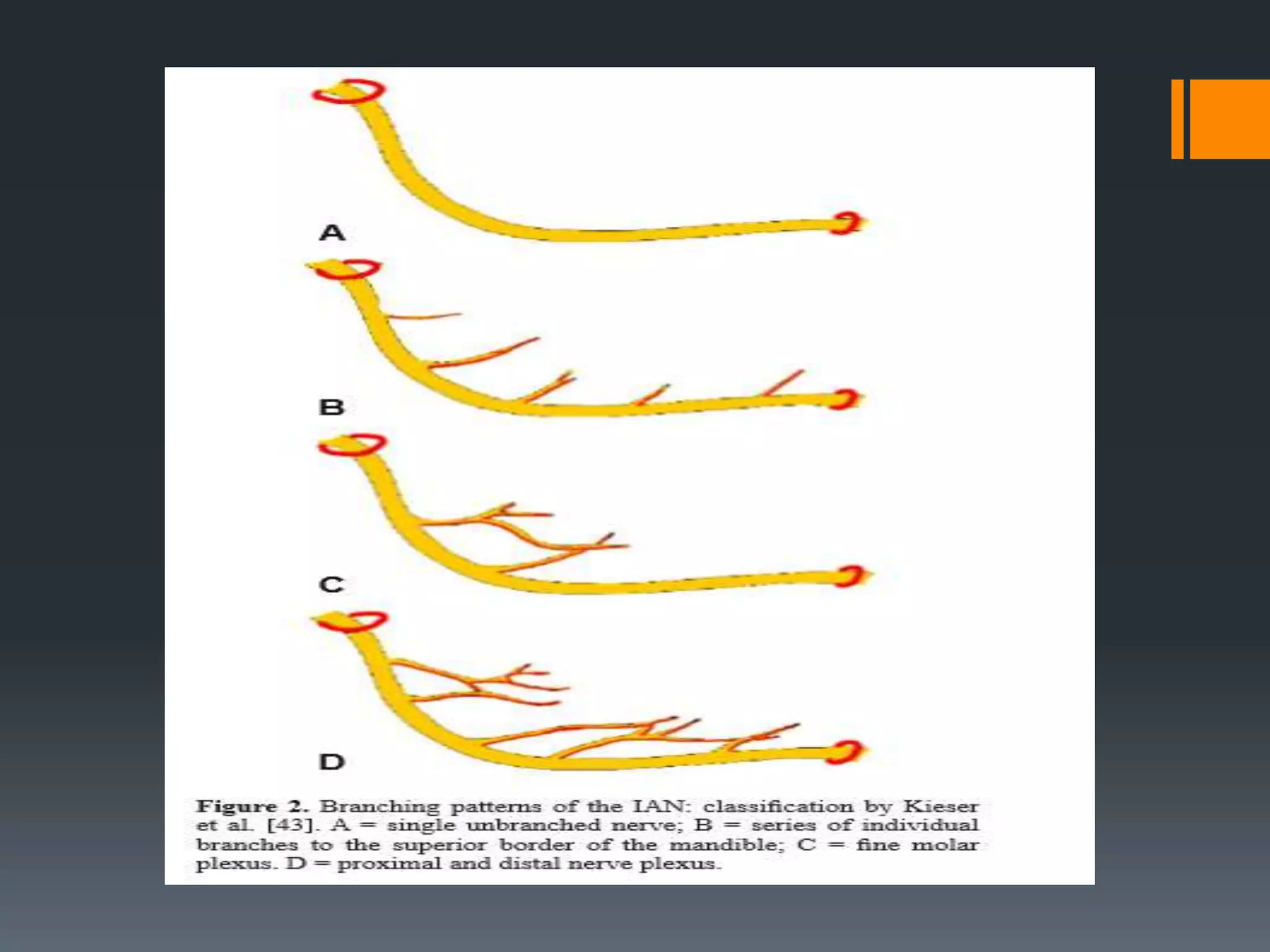
 The IAN may present in different anatomic configurations
 The variation in the course of IAN is frequent
 Liu et al OPG classification of course of nerve
Mental Foramen and Nerve
 Commonly, three nerve branches of the mental nerve emerge from the
mental foramen (each ;1 mm in diameter).
 Location- Differs in horizontal and vertical plane
 Atypically near canine or molar.
The anterior loop of the mental foramen refers to the IAN when it
courses inferiorly and anteriorly to the foramen and then loops back to
emerge from the foramen
 The dimensions of anterior loops in panoramic radiographs varied
from 0.5 to 3 mm, and cadaver specimens manifested anterior
loops that ranged from 0.11to3.31mm
 Choose an implant length a safety margin of 2mm
 It is prudent to place the distal aspect of the implant 6 mm anterior
to the mental foramen to avoid damaging the loop when drilling the
osteotomy
Mandibular Incisive Canal
 Numerous investigations reported that there is ‘‘true’’ incisive canal
mesial to the mental foramen, which is a continuation of the mandibular
canal.
 The incisive canal is typically found in the middle third of the mandible
(in 86%of cases).It usually narrows as it approaches the midline and
only reaches the midline 18%of the time
 Terminates apical to lateral or central incisor
 OPG -15% CT-93%
 Only large sized canal posed problems
Lingual Foramen and Lateral Canals
 The lingual foramen was detected in 99% of the mandibles when
evaluating skull dissections, but only 45% in radiographs
 The lingual foramen harbors an artery that corresponds to an
anastomosis of the right and left sublingual arteries.
 Risk of haemorrhage if canal size > 1mm
Submental and Sublingual Arteries
 The submental artery (2-mm average diameter) is derived from the
facial artery, and the sublingual artery (2-mm average diameter) is a
branch of the lingual artery
 If resected can cause haematoma in submandibular and sublingual
space, leading to swelling and airway obstruction
Submandibular and Sublingual Fossae
 The submandibular and sublingual Fossae must be palpated prior to
osteotomy development; if there is a large undercut, the lingual
bony plate can be perforated in advertently, resulting in
hemorrhaging.
 If there is a large undercut, an instrument Can be placed into and
parallel to the undercut to visualize and measure the extent of the
depression
 A CT scan with radiopaque markers provides the most accurate
information.
The Lingual and Mylohyoid Nerves
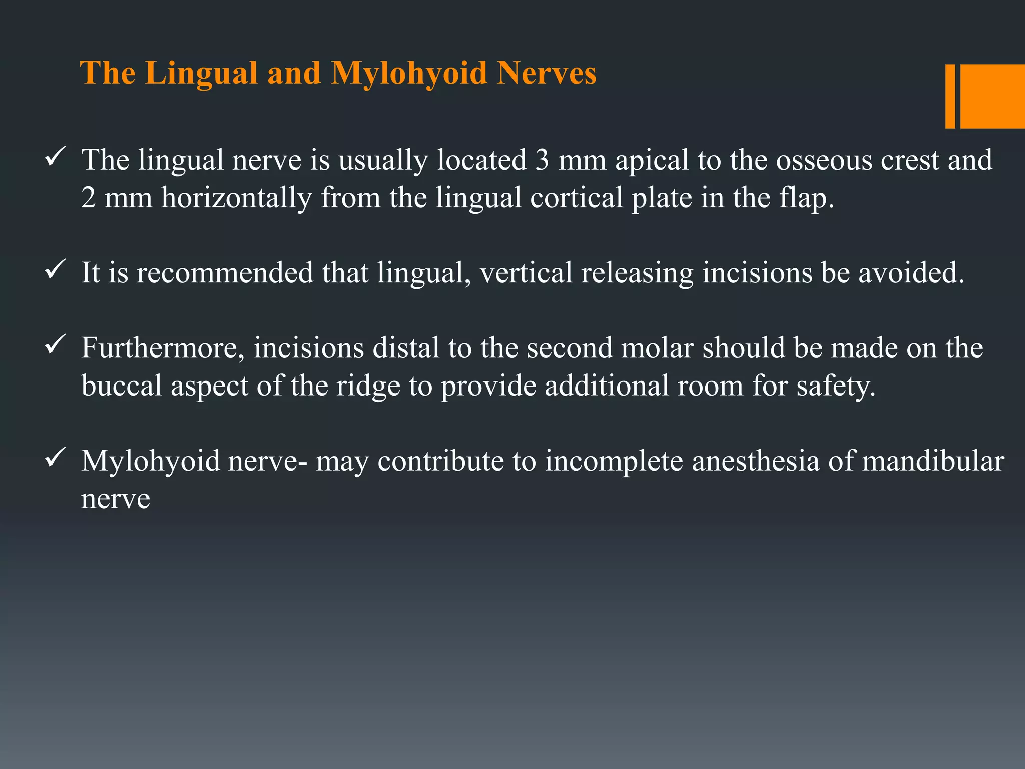
 The lingual nerve is usually located 3 mm apical to the osseous crest and
2 mm horizontally from the lingual cortical plate in the flap.
 It is recommended that lingual, vertical releasing incisions be avoided.
 Furthermore, incisions distal to the second molar should be made on the
buccal aspect of the ridge to provide additional room for safety.
 Mylohyoid nerve- may contribute to incomplete anesthesia of mandibular
nerve
Mandibular zone of safety
Given by Carl E Misch
Determined radiograpically or clinically during surgery
CONCLUSION
 Familiarity with the anatomic structures pertaining to dental
implantology is critically important.
 Preplanning and review of anatomy before surgical procedures can
help to avoid problems.
 Also many of the shortcomings of two-dimensional radiography for
treatment planning can be eliminated with the use of three-
dimensional imaging.
 And an understanding of basic surgical principle is necessary to
insure a successful implant.
REFERENCES:
B.D CHAURASIA’S HUMAN ANATOMY – 6TH EDITION
TEXTBOOK OF ANATOMY BY INDERBIR SINGH- 5TH EDITION •
GRAY’S ANATOMY – 2ND EDITION
Greenstein G1, Cavallaro J, Tarnow D. J Periodontol. 2008 Oct;79(10):1833-46
J Oral Maxillofac Res 2010 (Jan-Mar) | vol. 1 | No 1 | e2 | p.1 -5

More Related Content

What's hot

Applied anatomy of tmj
Applied anatomy of tmjApplied anatomy of tmj
Applied anatomy of tmj
Dr. Samarth Johari
 
Anatomy of temporomandibular joint
Anatomy of temporomandibular jointAnatomy of temporomandibular joint
Anatomy of temporomandibular joint
DrGayatriMehrotra
 
Muscles of mastication
Muscles of masticationMuscles of mastication
Muscles of mastication
Dr Khushboo Sinhmar
 
External carotid artery, branches and ligation
External carotid artery, branches and ligationExternal carotid artery, branches and ligation
External carotid artery, branches and ligation
benjamin Emmanuel
 
Anatomy of TMJ
Anatomy of TMJAnatomy of TMJ
Anatomy of TMJ
Dr Laxman Khanal
 
Muscles of Mastication
Muscles of MasticationMuscles of Mastication
Muscles of Mastication
Dr. Anshul Sahu
 
Maxillary sinus
Maxillary sinusMaxillary sinus
Maxillary sinus
Amrit Jaishi
 
TMJ and its relation to periodontics
TMJ and its relation to periodonticsTMJ and its relation to periodontics
TMJ and its relation to periodontics
Chittoor Deals
 
Development & Growth of Maxilla
Development & Growth of Maxilla  Development & Growth of Maxilla
Development & Growth of Maxilla
Menatalla Elhindawy
 
Growth of maxilla
Growth of maxillaGrowth of maxilla
Growth of maxilla
Sushant Pandey
 
Temporomandibular joint development and applied aspects
Temporomandibular joint development and applied aspectsTemporomandibular joint development and applied aspects
Temporomandibular joint development and applied aspects
Ravi banavathu
 
TMJ
TMJ TMJ
Maxilla
MaxillaMaxilla
Maxilla
dviya jain
 
pre natal &; post-natal growth of maxilla & palate
 pre natal &; post-natal growth of maxilla & palate  pre natal &; post-natal growth of maxilla & palate
pre natal &; post-natal growth of maxilla & palate
mahesh kumar
 
Development,anatomy and applied anatomy of Maxilla
Development,anatomy and applied anatomy of MaxillaDevelopment,anatomy and applied anatomy of Maxilla
Development,anatomy and applied anatomy of Maxilla
Soni Bista
 
Muscles of mastication
Muscles of masticationMuscles of mastication
Muscles of mastication
Dr. SHRUTI SUDARSANAN
 
Anatomy of temporomandibular joint(tmj)
Anatomy of temporomandibular joint(tmj)Anatomy of temporomandibular joint(tmj)
Anatomy of temporomandibular joint(tmj)
oorvi
 
Growth & development of maxilla and mandible
Growth & development of maxilla and mandibleGrowth & development of maxilla and mandible
Growth & development of maxilla and mandibleRajesh Bariker
 
Maxilla anatomy, development & surgical anatomy
Maxilla  anatomy, development & surgical anatomyMaxilla  anatomy, development & surgical anatomy
Maxilla anatomy, development & surgical anatomyDr. SHEETAL KAPSE
 

What's hot (20)

Applied anatomy of tmj
Applied anatomy of tmjApplied anatomy of tmj
Applied anatomy of tmj
 
Anatomy of temporomandibular joint
Anatomy of temporomandibular jointAnatomy of temporomandibular joint
Anatomy of temporomandibular joint
 
Nerve supply of head and neck
Nerve supply of head and neck Nerve supply of head and neck
Nerve supply of head and neck
 
Muscles of mastication
Muscles of masticationMuscles of mastication
Muscles of mastication
 
External carotid artery, branches and ligation
External carotid artery, branches and ligationExternal carotid artery, branches and ligation
External carotid artery, branches and ligation
 
Anatomy of TMJ
Anatomy of TMJAnatomy of TMJ
Anatomy of TMJ
 
Muscles of Mastication
Muscles of MasticationMuscles of Mastication
Muscles of Mastication
 
Maxillary sinus
Maxillary sinusMaxillary sinus
Maxillary sinus
 
TMJ and its relation to periodontics
TMJ and its relation to periodonticsTMJ and its relation to periodontics
TMJ and its relation to periodontics
 
Development & Growth of Maxilla
Development & Growth of Maxilla  Development & Growth of Maxilla
Development & Growth of Maxilla
 
Growth of maxilla
Growth of maxillaGrowth of maxilla
Growth of maxilla
 
Temporomandibular joint development and applied aspects
Temporomandibular joint development and applied aspectsTemporomandibular joint development and applied aspects
Temporomandibular joint development and applied aspects
 
TMJ
TMJ TMJ
TMJ
 
Maxilla
MaxillaMaxilla
Maxilla
 
pre natal &; post-natal growth of maxilla & palate
 pre natal &; post-natal growth of maxilla & palate  pre natal &; post-natal growth of maxilla & palate
pre natal &; post-natal growth of maxilla & palate
 
Development,anatomy and applied anatomy of Maxilla
Development,anatomy and applied anatomy of MaxillaDevelopment,anatomy and applied anatomy of Maxilla
Development,anatomy and applied anatomy of Maxilla
 
Muscles of mastication
Muscles of masticationMuscles of mastication
Muscles of mastication
 
Anatomy of temporomandibular joint(tmj)
Anatomy of temporomandibular joint(tmj)Anatomy of temporomandibular joint(tmj)
Anatomy of temporomandibular joint(tmj)
 
Growth & development of maxilla and mandible
Growth & development of maxilla and mandibleGrowth & development of maxilla and mandible
Growth & development of maxilla and mandible
 
Maxilla anatomy, development & surgical anatomy
Maxilla  anatomy, development & surgical anatomyMaxilla  anatomy, development & surgical anatomy
Maxilla anatomy, development & surgical anatomy
 

Similar to Anatomy of mandible and its importance in implant placement

Mandible
MandibleMandible
ANATOMY,DEVELOPMENT AND CLINICAL CONSIDERATION OF Mandible
ANATOMY,DEVELOPMENT AND CLINICAL CONSIDERATION OF MandibleANATOMY,DEVELOPMENT AND CLINICAL CONSIDERATION OF Mandible
ANATOMY,DEVELOPMENT AND CLINICAL CONSIDERATION OF Mandible
Soni Bista
 
1mandiblelaxmi 150502145137-conversion-gate01
1mandiblelaxmi 150502145137-conversion-gate011mandiblelaxmi 150502145137-conversion-gate01
1mandiblelaxmi 150502145137-conversion-gate01
AKRAMTARIQ2
 
mandible
 mandible  mandible
mandible
Mehul Shinde
 
mandible
mandiblemandible
mandible
KevinMungasia
 
SURGICAL ANATOMY OF MANDIBLE.pptx
SURGICAL ANATOMY OF MANDIBLE.pptxSURGICAL ANATOMY OF MANDIBLE.pptx
SURGICAL ANATOMY OF MANDIBLE.pptx
Priyanka Pai
 
surgicalanatomyofmandible-190510072105 (1).pptx
surgicalanatomyofmandible-190510072105 (1).pptxsurgicalanatomyofmandible-190510072105 (1).pptx
surgicalanatomyofmandible-190510072105 (1).pptx
Sakshi617058
 
SURGICAL ANATOMY OF MANDIBLE.pptx
SURGICAL ANATOMY OF MANDIBLE.pptxSURGICAL ANATOMY OF MANDIBLE.pptx
SURGICAL ANATOMY OF MANDIBLE.pptx
shalini sampreethi
 
Anatomy and Development of Mandible
Anatomy and Development of MandibleAnatomy and Development of Mandible
Anatomy and Development of Mandible
nikhilreddyg63
 
Osteology of head & neck ii
Osteology of head & neck iiOsteology of head & neck ii
Osteology of head & neck ii
ShadowFighter1
 
osteology.pptx
osteology.pptxosteology.pptx
osteology.pptx
sucrathajohn1
 
Anatomy of oropharynx maxilla mandible neck nodes
Anatomy of oropharynx maxilla mandible neck nodesAnatomy of oropharynx maxilla mandible neck nodes
Anatomy of oropharynx maxilla mandible neck nodes
Samik Sharma
 
Development of mandible
Development of mandibleDevelopment of mandible
Development of mandible
Jignesh Tate
 
Maxilla, Mandible & Hyoid Bone & Clinical Correlations by Dr. RIG.pptx
Maxilla, Mandible & Hyoid Bone & Clinical Correlations by Dr. RIG.pptxMaxilla, Mandible & Hyoid Bone & Clinical Correlations by Dr. RIG.pptx
Maxilla, Mandible & Hyoid Bone & Clinical Correlations by Dr. RIG.pptx
Dr. Rabia Inam Gandapore
 
growth of_mandible__orthodontics human.ppt
growth of_mandible__orthodontics human.pptgrowth of_mandible__orthodontics human.ppt
growth of_mandible__orthodontics human.ppt
PseudoPocket
 
Prenatal and postnatal growth of mandible
Prenatal and postnatal growth of mandiblePrenatal and postnatal growth of mandible
Prenatal and postnatal growth of mandibleshayonisen2012
 
Anatomy of orbit
Anatomy of orbitAnatomy of orbit
Anatomy of orbit
ChrisPius
 
Mandible
MandibleMandible
Mandible
shaimaaf12
 
Ear discharge and otalgia
Ear discharge and otalgiaEar discharge and otalgia
Ear discharge and otalgia
Dennis Lee
 

Similar to Anatomy of mandible and its importance in implant placement (20)

Mandible
MandibleMandible
Mandible
 
ANATOMY,DEVELOPMENT AND CLINICAL CONSIDERATION OF Mandible
ANATOMY,DEVELOPMENT AND CLINICAL CONSIDERATION OF MandibleANATOMY,DEVELOPMENT AND CLINICAL CONSIDERATION OF Mandible
ANATOMY,DEVELOPMENT AND CLINICAL CONSIDERATION OF Mandible
 
1mandiblelaxmi 150502145137-conversion-gate01
1mandiblelaxmi 150502145137-conversion-gate011mandiblelaxmi 150502145137-conversion-gate01
1mandiblelaxmi 150502145137-conversion-gate01
 
mandible
 mandible  mandible
mandible
 
mandible
mandiblemandible
mandible
 
SURGICAL ANATOMY OF MANDIBLE.pptx
SURGICAL ANATOMY OF MANDIBLE.pptxSURGICAL ANATOMY OF MANDIBLE.pptx
SURGICAL ANATOMY OF MANDIBLE.pptx
 
surgicalanatomyofmandible-190510072105 (1).pptx
surgicalanatomyofmandible-190510072105 (1).pptxsurgicalanatomyofmandible-190510072105 (1).pptx
surgicalanatomyofmandible-190510072105 (1).pptx
 
SURGICAL ANATOMY OF MANDIBLE.pptx
SURGICAL ANATOMY OF MANDIBLE.pptxSURGICAL ANATOMY OF MANDIBLE.pptx
SURGICAL ANATOMY OF MANDIBLE.pptx
 
Anatomy and Development of Mandible
Anatomy and Development of MandibleAnatomy and Development of Mandible
Anatomy and Development of Mandible
 
Osteology of head & neck ii
Osteology of head & neck iiOsteology of head & neck ii
Osteology of head & neck ii
 
osteology.pptx
osteology.pptxosteology.pptx
osteology.pptx
 
Anatomy of oropharynx maxilla mandible neck nodes
Anatomy of oropharynx maxilla mandible neck nodesAnatomy of oropharynx maxilla mandible neck nodes
Anatomy of oropharynx maxilla mandible neck nodes
 
Development of mandible
Development of mandibleDevelopment of mandible
Development of mandible
 
Maxilla, Mandible & Hyoid Bone & Clinical Correlations by Dr. RIG.pptx
Maxilla, Mandible & Hyoid Bone & Clinical Correlations by Dr. RIG.pptxMaxilla, Mandible & Hyoid Bone & Clinical Correlations by Dr. RIG.pptx
Maxilla, Mandible & Hyoid Bone & Clinical Correlations by Dr. RIG.pptx
 
growth of_mandible__orthodontics human.ppt
growth of_mandible__orthodontics human.pptgrowth of_mandible__orthodontics human.ppt
growth of_mandible__orthodontics human.ppt
 
Prenatal and postnatal growth of mandible
Prenatal and postnatal growth of mandiblePrenatal and postnatal growth of mandible
Prenatal and postnatal growth of mandible
 
Anatomy of orbit
Anatomy of orbitAnatomy of orbit
Anatomy of orbit
 
Growth of mandible
Growth of mandibleGrowth of mandible
Growth of mandible
 
Mandible
MandibleMandible
Mandible
 
Ear discharge and otalgia
Ear discharge and otalgiaEar discharge and otalgia
Ear discharge and otalgia
 

Recently uploaded

Evaluation of antidepressant activity of clitoris ternatea in animals
Evaluation of antidepressant activity of clitoris ternatea in animalsEvaluation of antidepressant activity of clitoris ternatea in animals
Evaluation of antidepressant activity of clitoris ternatea in animals
Shweta
 
Triangles of Neck and Clinical Correlation by Dr. RIG.pptx
Triangles of Neck and Clinical Correlation by Dr. RIG.pptxTriangles of Neck and Clinical Correlation by Dr. RIG.pptx
Triangles of Neck and Clinical Correlation by Dr. RIG.pptx
Dr. Rabia Inam Gandapore
 
Pulmonary Thromboembolism - etilogy, types, medical- Surgical and nursing man...
Pulmonary Thromboembolism - etilogy, types, medical- Surgical and nursing man...Pulmonary Thromboembolism - etilogy, types, medical- Surgical and nursing man...
Pulmonary Thromboembolism - etilogy, types, medical- Surgical and nursing man...
VarunMahajani
 
Surat @ℂall @Girls ꧁❤8527049040❤꧂@ℂall @Girls Service Vip Top Model Safe
Surat @ℂall @Girls ꧁❤8527049040❤꧂@ℂall @Girls Service Vip Top Model SafeSurat @ℂall @Girls ꧁❤8527049040❤꧂@ℂall @Girls Service Vip Top Model Safe
Surat @ℂall @Girls ꧁❤8527049040❤꧂@ℂall @Girls Service Vip Top Model Safe
Savita Shen $i11
 
Cervical & Brachial Plexus By Dr. RIG.pptx
Cervical & Brachial Plexus By Dr. RIG.pptxCervical & Brachial Plexus By Dr. RIG.pptx
Cervical & Brachial Plexus By Dr. RIG.pptx
Dr. Rabia Inam Gandapore
 
Prix Galien International 2024 Forum Program
Prix Galien International 2024 Forum ProgramPrix Galien International 2024 Forum Program
Prix Galien International 2024 Forum Program
Levi Shapiro
 
ARTIFICIAL INTELLIGENCE IN HEALTHCARE.pdf
ARTIFICIAL INTELLIGENCE IN  HEALTHCARE.pdfARTIFICIAL INTELLIGENCE IN  HEALTHCARE.pdf
ARTIFICIAL INTELLIGENCE IN HEALTHCARE.pdf
Anujkumaranit
 
24 Upakrama.pptx class ppt useful in all
24 Upakrama.pptx class ppt useful in all24 Upakrama.pptx class ppt useful in all
24 Upakrama.pptx class ppt useful in all
DrSathishMS1
 
Charaka Samhita Sutra sthana Chapter 15 Upakalpaniyaadhyaya
Charaka Samhita Sutra sthana Chapter 15 UpakalpaniyaadhyayaCharaka Samhita Sutra sthana Chapter 15 Upakalpaniyaadhyaya
Charaka Samhita Sutra sthana Chapter 15 Upakalpaniyaadhyaya
Dr KHALID B.M
 
Couples presenting to the infertility clinic- Do they really have infertility...
Couples presenting to the infertility clinic- Do they really have infertility...Couples presenting to the infertility clinic- Do they really have infertility...
Couples presenting to the infertility clinic- Do they really have infertility...
Sujoy Dasgupta
 
The Normal Electrocardiogram - Part I of II
The Normal Electrocardiogram - Part I of IIThe Normal Electrocardiogram - Part I of II
The Normal Electrocardiogram - Part I of II
MedicoseAcademics
 
Surgical Site Infections, pathophysiology, and prevention.pptx
Surgical Site Infections, pathophysiology, and prevention.pptxSurgical Site Infections, pathophysiology, and prevention.pptx
Surgical Site Infections, pathophysiology, and prevention.pptx
jval Landero
 
THOA 2.ppt Human Organ Transplantation Act
THOA 2.ppt Human Organ Transplantation ActTHOA 2.ppt Human Organ Transplantation Act
THOA 2.ppt Human Organ Transplantation Act
DrSathishMS1
 
Tom Selleck Health: A Comprehensive Look at the Iconic Actor’s Wellness Journey
Tom Selleck Health: A Comprehensive Look at the Iconic Actor’s Wellness JourneyTom Selleck Health: A Comprehensive Look at the Iconic Actor’s Wellness Journey
Tom Selleck Health: A Comprehensive Look at the Iconic Actor’s Wellness Journey
greendigital
 
The hemodynamic and autonomic determinants of elevated blood pressure in obes...
The hemodynamic and autonomic determinants of elevated blood pressure in obes...The hemodynamic and autonomic determinants of elevated blood pressure in obes...
The hemodynamic and autonomic determinants of elevated blood pressure in obes...
Catherine Liao
 
basicmodesofventilation2022-220313203758.pdf
basicmodesofventilation2022-220313203758.pdfbasicmodesofventilation2022-220313203758.pdf
basicmodesofventilation2022-220313203758.pdf
aljamhori teaching hospital
 
Physiology of Chemical Sensation of smell.pdf
Physiology of Chemical Sensation of smell.pdfPhysiology of Chemical Sensation of smell.pdf
Physiology of Chemical Sensation of smell.pdf
MedicoseAcademics
 
BRACHYTHERAPY OVERVIEW AND APPLICATORS
BRACHYTHERAPY OVERVIEW  AND  APPLICATORSBRACHYTHERAPY OVERVIEW  AND  APPLICATORS
BRACHYTHERAPY OVERVIEW AND APPLICATORS
Krishan Murari
 
Alcohol_Dr. Jeenal Mistry MD Pharmacology.pdf
Alcohol_Dr. Jeenal Mistry MD Pharmacology.pdfAlcohol_Dr. Jeenal Mistry MD Pharmacology.pdf
Alcohol_Dr. Jeenal Mistry MD Pharmacology.pdf
Dr Jeenal Mistry
 
Non-respiratory Functions of the Lungs.pdf
Non-respiratory Functions of the Lungs.pdfNon-respiratory Functions of the Lungs.pdf
Non-respiratory Functions of the Lungs.pdf
MedicoseAcademics
 

Recently uploaded (20)

Evaluation of antidepressant activity of clitoris ternatea in animals
Evaluation of antidepressant activity of clitoris ternatea in animalsEvaluation of antidepressant activity of clitoris ternatea in animals
Evaluation of antidepressant activity of clitoris ternatea in animals
 
Triangles of Neck and Clinical Correlation by Dr. RIG.pptx
Triangles of Neck and Clinical Correlation by Dr. RIG.pptxTriangles of Neck and Clinical Correlation by Dr. RIG.pptx
Triangles of Neck and Clinical Correlation by Dr. RIG.pptx
 
Pulmonary Thromboembolism - etilogy, types, medical- Surgical and nursing man...
Pulmonary Thromboembolism - etilogy, types, medical- Surgical and nursing man...Pulmonary Thromboembolism - etilogy, types, medical- Surgical and nursing man...
Pulmonary Thromboembolism - etilogy, types, medical- Surgical and nursing man...
 
Surat @ℂall @Girls ꧁❤8527049040❤꧂@ℂall @Girls Service Vip Top Model Safe
Surat @ℂall @Girls ꧁❤8527049040❤꧂@ℂall @Girls Service Vip Top Model SafeSurat @ℂall @Girls ꧁❤8527049040❤꧂@ℂall @Girls Service Vip Top Model Safe
Surat @ℂall @Girls ꧁❤8527049040❤꧂@ℂall @Girls Service Vip Top Model Safe
 
Cervical & Brachial Plexus By Dr. RIG.pptx
Cervical & Brachial Plexus By Dr. RIG.pptxCervical & Brachial Plexus By Dr. RIG.pptx
Cervical & Brachial Plexus By Dr. RIG.pptx
 
Prix Galien International 2024 Forum Program
Prix Galien International 2024 Forum ProgramPrix Galien International 2024 Forum Program
Prix Galien International 2024 Forum Program
 
ARTIFICIAL INTELLIGENCE IN HEALTHCARE.pdf
ARTIFICIAL INTELLIGENCE IN  HEALTHCARE.pdfARTIFICIAL INTELLIGENCE IN  HEALTHCARE.pdf
ARTIFICIAL INTELLIGENCE IN HEALTHCARE.pdf
 
24 Upakrama.pptx class ppt useful in all
24 Upakrama.pptx class ppt useful in all24 Upakrama.pptx class ppt useful in all
24 Upakrama.pptx class ppt useful in all
 
Charaka Samhita Sutra sthana Chapter 15 Upakalpaniyaadhyaya
Charaka Samhita Sutra sthana Chapter 15 UpakalpaniyaadhyayaCharaka Samhita Sutra sthana Chapter 15 Upakalpaniyaadhyaya
Charaka Samhita Sutra sthana Chapter 15 Upakalpaniyaadhyaya
 
Couples presenting to the infertility clinic- Do they really have infertility...
Couples presenting to the infertility clinic- Do they really have infertility...Couples presenting to the infertility clinic- Do they really have infertility...
Couples presenting to the infertility clinic- Do they really have infertility...
 
The Normal Electrocardiogram - Part I of II
The Normal Electrocardiogram - Part I of IIThe Normal Electrocardiogram - Part I of II
The Normal Electrocardiogram - Part I of II
 
Surgical Site Infections, pathophysiology, and prevention.pptx
Surgical Site Infections, pathophysiology, and prevention.pptxSurgical Site Infections, pathophysiology, and prevention.pptx
Surgical Site Infections, pathophysiology, and prevention.pptx
 
THOA 2.ppt Human Organ Transplantation Act
THOA 2.ppt Human Organ Transplantation ActTHOA 2.ppt Human Organ Transplantation Act
THOA 2.ppt Human Organ Transplantation Act
 
Tom Selleck Health: A Comprehensive Look at the Iconic Actor’s Wellness Journey
Tom Selleck Health: A Comprehensive Look at the Iconic Actor’s Wellness JourneyTom Selleck Health: A Comprehensive Look at the Iconic Actor’s Wellness Journey
Tom Selleck Health: A Comprehensive Look at the Iconic Actor’s Wellness Journey
 
The hemodynamic and autonomic determinants of elevated blood pressure in obes...
The hemodynamic and autonomic determinants of elevated blood pressure in obes...The hemodynamic and autonomic determinants of elevated blood pressure in obes...
The hemodynamic and autonomic determinants of elevated blood pressure in obes...
 
basicmodesofventilation2022-220313203758.pdf
basicmodesofventilation2022-220313203758.pdfbasicmodesofventilation2022-220313203758.pdf
basicmodesofventilation2022-220313203758.pdf
 
Physiology of Chemical Sensation of smell.pdf
Physiology of Chemical Sensation of smell.pdfPhysiology of Chemical Sensation of smell.pdf
Physiology of Chemical Sensation of smell.pdf
 
BRACHYTHERAPY OVERVIEW AND APPLICATORS
BRACHYTHERAPY OVERVIEW  AND  APPLICATORSBRACHYTHERAPY OVERVIEW  AND  APPLICATORS
BRACHYTHERAPY OVERVIEW AND APPLICATORS
 
Alcohol_Dr. Jeenal Mistry MD Pharmacology.pdf
Alcohol_Dr. Jeenal Mistry MD Pharmacology.pdfAlcohol_Dr. Jeenal Mistry MD Pharmacology.pdf
Alcohol_Dr. Jeenal Mistry MD Pharmacology.pdf
 
Non-respiratory Functions of the Lungs.pdf
Non-respiratory Functions of the Lungs.pdfNon-respiratory Functions of the Lungs.pdf
Non-respiratory Functions of the Lungs.pdf
 

Anatomy of mandible and its importance in implant placement

  • 1. ANATOMY OF MANDIBLE AND ITS IMPORTANCE IN IMPLANT PLACEMENT
  • 2. INTRODUCTION  The word mandible derived from Latin word mandibula- "jawbone" or inferior maxillary bone.  Only movable bone in the skull  It consists of an anterior Horseshoe-shaped body, and of two rami that project upwards from the posterior part of the body.  It provides structural and protective support for the oral cavity.  The mandible is articulated in ball and socket fashion at the condylar process.  Strength resides in its dense cortical plates
  • 4. embryology  Mandible is the second bone after clavicle to ossify in the body.  Parts that ossify in cartilage includes: incisive part below the incisor teeth, coronoid and condyloid processes  Upper half of ramus above the level of the mandibular foramen  A single ossification centre for each half of the mandible arises in the 6th week of I.U. life in the region of bifurcation of inferior alveolar nerve into mental and incisive branches. As the ossification continues, the meckel’s cartilage become surrounded and invaded by bone.
  • 5.  It is partly membranous & partly cartilaginous in ossification. • Incisive part below symphysis menti • Coronoid • Condyloid process • Upper half of ramus Cartilage • Whole of body except lower incisive part • Lower half of ramus upto mandibular foramen Membrane
  • 6. OSTEOLOGY BODY OF THE MANDIBLE  Horseshoe-shaped BODY  Has two surfaces EXTERNAL and INTERNAL surfaces ,  Two borders UPPER and LOWER borders
  • 7. FEATURES SEEN ON OUTER SURFACE OF THE BODY  Symphysis Menti  Mental Protuberance  Mental Tubercles  Mental foramen  External Oblique line  Incisive fossa
  • 8. FEATURES ON THE INNER SURFACE OF THE BODY  Genial tubercles  Mylohyoid line  Submandibular fossae  Sublingual fossae  Mylohyoid groove  Upper alveolar border  Lower border base  Digastric fossae
  • 9. BORDERS OF THE MANDIBLE
  • 10. SUPERIOR BORDER (ALVEOLAR BORDER) It is hollowed into cavities for the reception of the teeth, these cavities are sixteen in number, and vary in depth and size according to the teeth which they contain.
  • 11. INFERIOR BORDER (base of mandible) Is rounded, longer than the superior, and thicker in front than behind.
  • 12. RAMUS OF MANDIBLE Is quadrilateral 2 surfaces Lateral Medial 4 borders Superior Inferior Anterior Posterior 2 processes Coronoid Condylar
  • 13. PROCESSES CORONOID PROCESS  Flat ,triangular  Upward and forward projection from anterolateral part of ramus  Anterior border continuous with anterior border of ramus  Posterior border bounds the mandibular notch
  • 14. CONDYLAR PROCESS  Upward projection from postero superior part of ramus  Apically enlarged as head of condyle.  Articulates with temporal bone’s mandibular fossa to form temperomandibular joint  Lateral aspect palpable in front of tragus  Pterygoid fovea anterior to neck
  • 16. ON THE LATERAL SURFACE: 1. From The Oblique line : Buccinator, and depressor anguli oris below the mental foramen 2. Incisive fossa: gives origin to MENTALIS mental slips of ORBICULARIS ORIS. 3. Whole of lateral surface of ramus except posterosuperior part provides insertion to MASSETER. 4.Posterosuperior part : covered by PAROTID GLAND
  • 17. 5. Lateral surface of the neck provides insertion to the LATERAL LIGAMENT OF TMJ. 6. Parts of both the inner and outer surfaces just below the alveolar margins are covered by mucous membrane of the mouth. 7. PLATYSMA is inserted into the lower border. 8. The deep cervical fascia ( investing layer) is attached to the whole length of the lower border.
  • 18. 1. Digastric fossa: arises ANTERIOR BELLY OF DIGASTRIC 2. Genial tubercles: arises GENIOGLOSSUS and GENIOHYOID. 3. Mylohyoid line : arises MYLOHYOID MUSCLE. 4. From an area above the posterior end of mylohyoid line: arises SUPERIOR CONSTRICTOR OF PHARYNX. 5. Pterygomandibular raphe: Attached immediately behind the third molar tooth in continuation with the origin of superior constrictor ON THE MEDIAL SURFACE
  • 19. 7.Below and behind the mylohyoid groove: insertion of MEDIAL PTERYGOID muscle . 8.At the apex of coronoid process : TEMPORALIS is inserted ;extend downwards on ant. Border of ramus. 9.Into the pterygoid fovea: insertion of LATERAL PTERYGOID. 10.Sphenomandibular ligament : is attached to the lingula.
  • 20. BLOOD SUPPLY OF THE MANDIBLE ARTERIAL SUPPLY Mainly by Maxillary artery, Branch of external carotid artery By its branches, mainly through inferior alveolar artery
  • 21. Venous supply of mandible  Drains into Internal jugular vein and external jugular vein through maxillary vein, facial vein and pterygoid plexus
  • 22. Nerve supply of mandible Mainly through the trigeminal nerve - V cranial nerve MANDIBULAR NERVE  Main trunk  Anterior trunk  Posterior trunk
  • 23. FORAMINA AND OTHER RELATIONS  Mental foramina - mental nerve and vessels  Mandibular notch - massetric nerve and vessels  Medial side of neck - auriculo temporal nerve  Mylohyoid groove - mylohyoid nerve and vessels  Mylohyoid groove in front of ramus - lingual nerve  Mandibular canal and foramina - inferior alveolar nerve and vessels
  • 25.  According to the data from the vital staining experiments, the posterior surface the ramus, the condyle and coronoid process are principal sites of growth.  Growth is quite general during the first year of life with all surfaces showing bone apposition.  Mandibular growth becomes more selective. Postnatal Growth Of Mandible
  • 26. The mandible can be divided into several sub-units like  Chin  Alveolar process  Body  Lingual tuberosity  Ramus  Angular process  Coronoid process  Condylar process
  • 27.  Chin:  1-2 years→ chin prominence is seen  The mental protuberance forms by bone deposition  The change in the contour occurs by following two mechanism. 1) The area just above the chin and the base of the alveolar process, is a resorptive area. 2) There is forward translation of chin as mandible grows forward.
  • 28.  Alveolar process:  This develops in response to the developing tooth buds.
  • 29.  Body: (corpus)  The length of the body increases as the ramus moves posteriorly
  • 30.  Lingual tuberosity:  It forms the boundary between the ramus & body  A combination of the resorption and deposition accentuates its prominence.
  • 31.  Ramus:  The ramus is seen to move posteriorly due to deposition at its posterior border and resorption on its anterior border
  • 32.  Angle:  The combined deposition and resorption causes flaring of the angle of the mandible
  • 33.  Coronoid process :  Enlow’s enlarging “V” principle.  Birth: Coronoid process is at higher level than condylar process.  Childhood: Coronoid & condylar processes are at same level.  Adult: Condylar process is at higher level.
  • 34.  Condyle:  Condylar growth rate increases at puberty and reaches its peak by 12-14 years.  The growth ceases at around 20 years  Role of condyle: o Primary displacement
  • 35.
  • 36. APPLIED ANATOMY OF MANDIBLE  Parasymphysis region lateral to the mental prominence is a naturally weak area susceptible for parasymphyseal fracture. This is because of the presence of incisive fossa and mental foramen  The body of the mandible is considerably thicker than the ramus and the junction between these two portions constitutes a line of structural weakness.  Strength of the lower jaw varies with the presence or absence of teeth. The presence of impacted lower third molars or excessive long roots of canines make the area more vulnerable for fracture.
  • 38. Mandibular Foramen  The location of the mandibular foramen may vary based on race and ethnicity  Among adult cadaveric mandibles, the foramen was found inferior to the occlusal plane, at its level, or above it 75%, 22.5%, and 2.5% of the time, respectively  Therefore, according to these investigations, 2.5% to 23.5% of block injections given at the level of occlusion would be ineffective.  Advisable to inject patients 6 to10mmsuperior to the occlusal plane, which usually accounts for anatomic variations  Short needles can be used to attain anesthesia in the mandible
  • 39. Inferior Alveolar Canal  The inferior alveolar canal houses the IAN nerve , artery, vein and lymphatics  The canal is; 3.4mm wide, and the nerve is ;2.2mm thick  Therefore, during implant placement it is possible to in advertently penetrate into the mandibular canal and induce neurologic damage without provoking hemorrhaging and vice versa.  When developing an osteotomy over the mandibular canal, cortical bone is penetrated first, and the preparation terminates within softer cancellous bone  The IAN may present in different anatomic configurations
  • 40.  The IAN may present in different anatomic configurations  The variation in the course of IAN is frequent  Liu et al OPG classification of course of nerve
  • 41.
  • 42. Mental Foramen and Nerve  Commonly, three nerve branches of the mental nerve emerge from the mental foramen (each ;1 mm in diameter).  Location- Differs in horizontal and vertical plane  Atypically near canine or molar.
  • 43. The anterior loop of the mental foramen refers to the IAN when it courses inferiorly and anteriorly to the foramen and then loops back to emerge from the foramen  The dimensions of anterior loops in panoramic radiographs varied from 0.5 to 3 mm, and cadaver specimens manifested anterior loops that ranged from 0.11to3.31mm  Choose an implant length a safety margin of 2mm  It is prudent to place the distal aspect of the implant 6 mm anterior to the mental foramen to avoid damaging the loop when drilling the osteotomy
  • 44.
  • 45. Mandibular Incisive Canal  Numerous investigations reported that there is ‘‘true’’ incisive canal mesial to the mental foramen, which is a continuation of the mandibular canal.  The incisive canal is typically found in the middle third of the mandible (in 86%of cases).It usually narrows as it approaches the midline and only reaches the midline 18%of the time  Terminates apical to lateral or central incisor  OPG -15% CT-93%  Only large sized canal posed problems
  • 46. Lingual Foramen and Lateral Canals  The lingual foramen was detected in 99% of the mandibles when evaluating skull dissections, but only 45% in radiographs  The lingual foramen harbors an artery that corresponds to an anastomosis of the right and left sublingual arteries.  Risk of haemorrhage if canal size > 1mm Submental and Sublingual Arteries  The submental artery (2-mm average diameter) is derived from the facial artery, and the sublingual artery (2-mm average diameter) is a branch of the lingual artery  If resected can cause haematoma in submandibular and sublingual space, leading to swelling and airway obstruction
  • 47. Submandibular and Sublingual Fossae  The submandibular and sublingual Fossae must be palpated prior to osteotomy development; if there is a large undercut, the lingual bony plate can be perforated in advertently, resulting in hemorrhaging.  If there is a large undercut, an instrument Can be placed into and parallel to the undercut to visualize and measure the extent of the depression  A CT scan with radiopaque markers provides the most accurate information.
  • 48. The Lingual and Mylohyoid Nerves  The lingual nerve is usually located 3 mm apical to the osseous crest and 2 mm horizontally from the lingual cortical plate in the flap.  It is recommended that lingual, vertical releasing incisions be avoided.  Furthermore, incisions distal to the second molar should be made on the buccal aspect of the ridge to provide additional room for safety.  Mylohyoid nerve- may contribute to incomplete anesthesia of mandibular nerve
  • 49. Mandibular zone of safety Given by Carl E Misch Determined radiograpically or clinically during surgery
  • 50. CONCLUSION  Familiarity with the anatomic structures pertaining to dental implantology is critically important.  Preplanning and review of anatomy before surgical procedures can help to avoid problems.  Also many of the shortcomings of two-dimensional radiography for treatment planning can be eliminated with the use of three- dimensional imaging.  And an understanding of basic surgical principle is necessary to insure a successful implant.
  • 51. REFERENCES: B.D CHAURASIA’S HUMAN ANATOMY – 6TH EDITION TEXTBOOK OF ANATOMY BY INDERBIR SINGH- 5TH EDITION • GRAY’S ANATOMY – 2ND EDITION Greenstein G1, Cavallaro J, Tarnow D. J Periodontol. 2008 Oct;79(10):1833-46 J Oral Maxillofac Res 2010 (Jan-Mar) | vol. 1 | No 1 | e2 | p.1 -5

Editor's Notes

  1. by four small elevations called the superior and inferior tubercle
  2. Lateral surface – flat with oblique ridges Medial surface -. MANDIBULAR FORAMEN (centre of ramus at the level of occlusal plane. It leadsmandibular canal. ) LINGULA : Anterior margin of foramen marked by tongue shaped projection MYLOHYOID GROOVE , UPPER BORXER , LOWER BORDER
  3. MAIN – meningeal and nerve to medial ptretgoid Anterior- buccal, masseteric , deeep temporal and lateral ptyerygoid
  4. Mainly sensory fibres
  5. Occasionally, the mental nerve emerges from the buccal plateof bone and reenters thealveolarboneto provide innervation for the incisor teeth.
  6. Surgical dissection furnished the best evidence for validating thepresence of the anteriorloop ofthe mental foramen Thus, when there is concern with respect to the location of the mental nerve, it should be exposed to identify its position before implant insertion. First, determineontheradiographwherethementalforamenis located