SlideShare a Scribd company logo
1 of 36
Download to read offline
ACNS Standardized Critical
Care EEG Terminology 2021
EEG and Legends
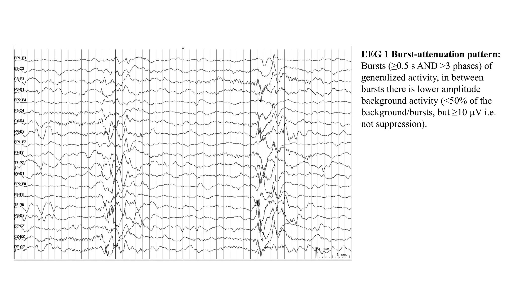
EEG 1 Burst-attenuation pattern:
Bursts (≥0.5 s AND >3 phases) of
generalized activity, in between
bursts there is lower amplitude
background activity (<50% of the
background/bursts, but ≥10 µV i.e.
not suppression).
EEG 2 Burst-suppression pattern:
Bursts (≥0.5 s AND >3 phases) of
generalized activity on a suppressed
(<10 µV) background.
EEG 3 Identical Highly-
Epileptiform Bursts: The pattern is
burst suppression. The first 0.5 s of
each burst appears visually similar in
all channels, qualifying as identical.
Each burst also contains 2 or more
epileptiform discharges occurring at
an average of 1 Hz or faster within a
single burst, qualifying as highly
epileptiform.
EEG 4 Identical Non-Highly-
Epileptiform Bursts: Burst
suppression pattern, with each burst
approximately 1 s (≥0.5 s) and
containing 4 – 6 phases each (>3
phases). The first 0.5 seconds of
each burst is visually similar in all
channels, qualifying as identical
bursts. Although there is an
epileptiform discharge in each burst,
there are not consistently 2 or more
in each burst and not occurring at
greater than 1-Hz frequency;
therefore, this is not highly
epileptiform.
EEG 5 Generalized Periodic
Discharges (GPDs): 1 Hz sharp
GPDs.
EEG 6 Generalized Rhythmic
Delta Activity (GRDA):1.5 Hz
frontally predominant GRDA. If the
lower amplitude faster (alpha range)
frequencies are not present in the
background when the GRDA is not
present, then this would qualify as
GRDA+F.
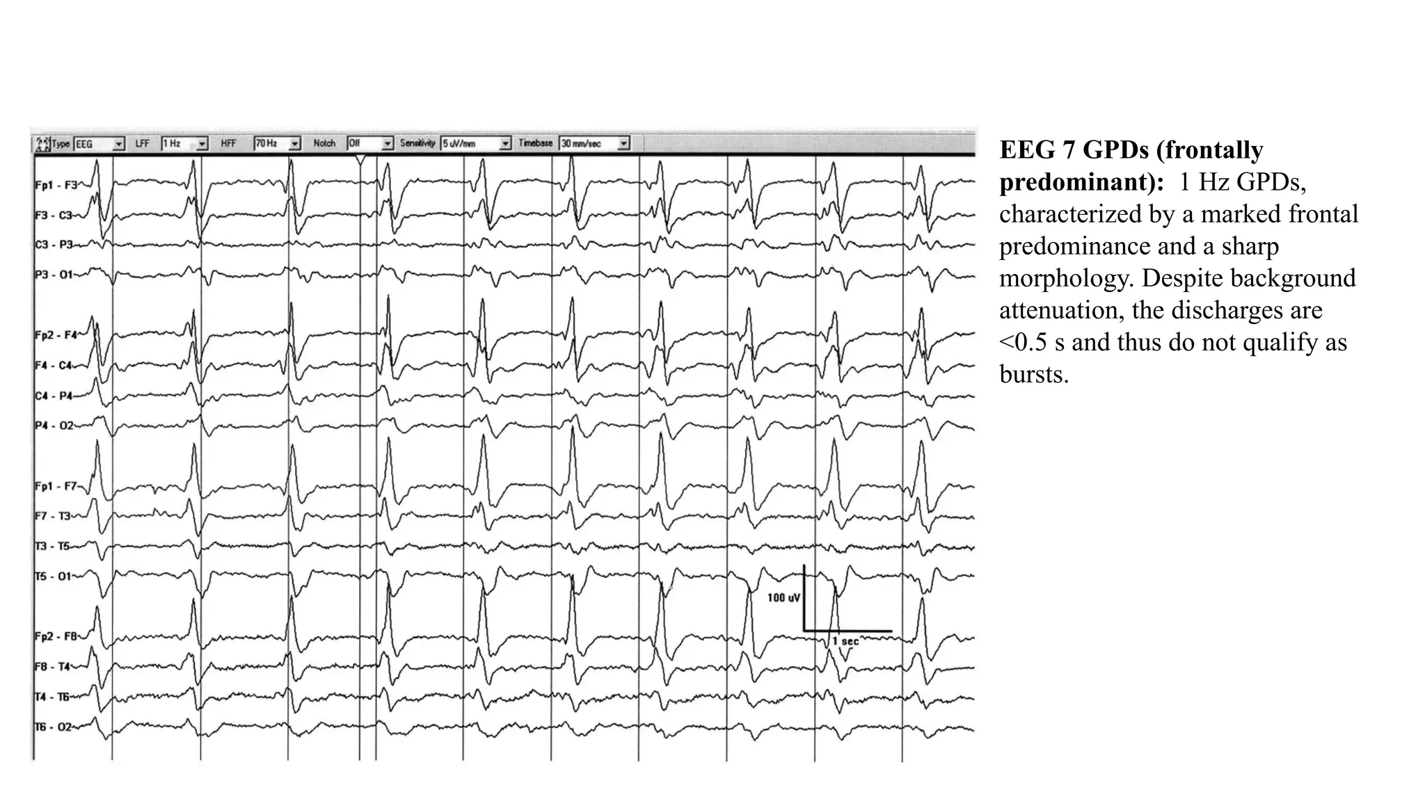
EEG 7 GPDs (frontally
predominant): 1 Hz GPDs,
characterized by a marked frontal
predominance and a sharp
morphology. Despite background
attenuation, the discharges are
<0.5 s and thus do not qualify as
bursts.
EEG 7 Lateralized Periodic
Discharges (LPDs): 0.5-1 Hz spiky
LPDs. Despite their spike-and-wave
morphology, the discharges are
periodic (as there is a quantifiable
inter-discharge interval between
consecutive waveforms and
recurrence of the waveform at nearly
regular intervals).
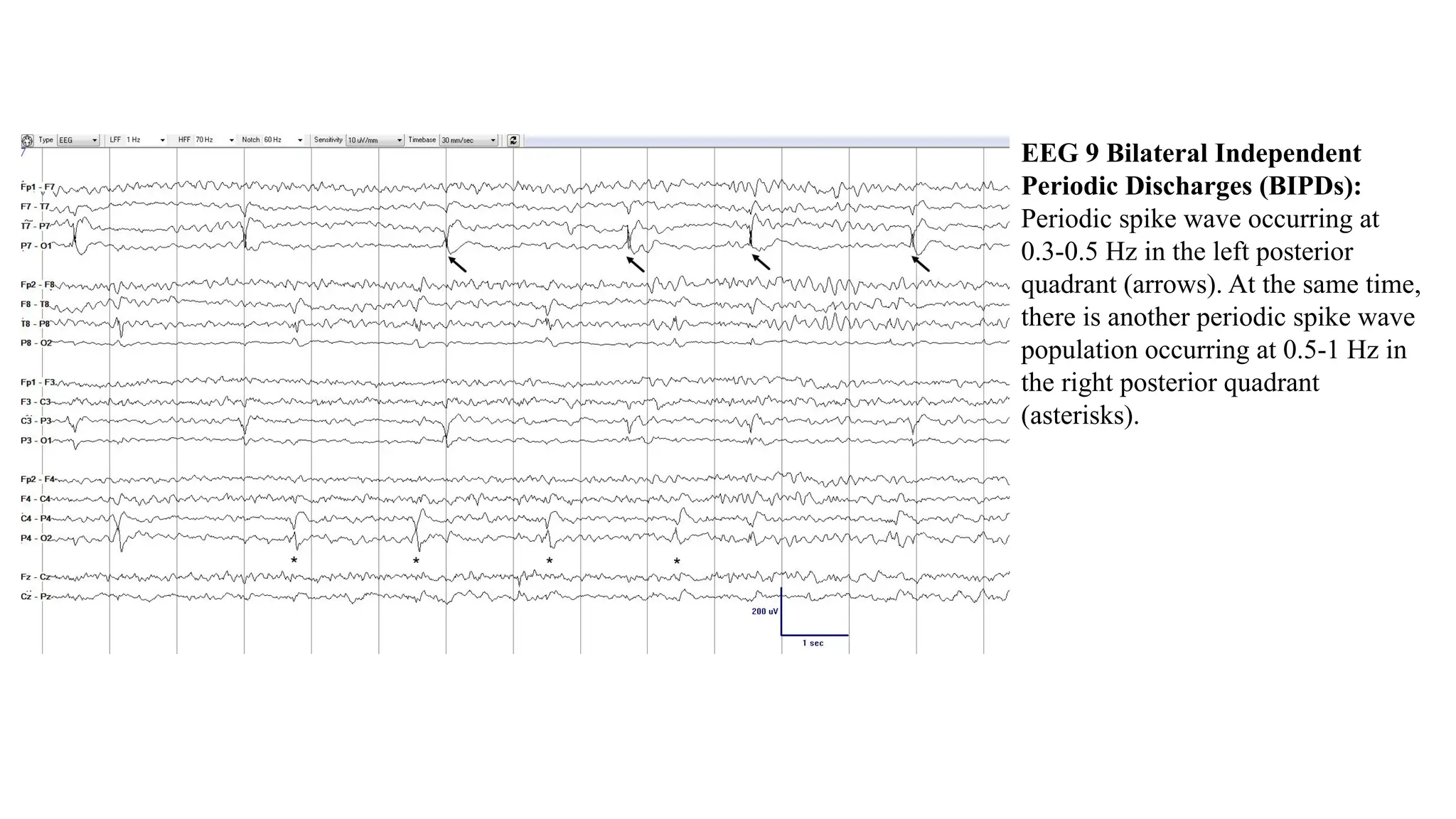
EEG 9 Bilateral Independent
Periodic Discharges (BIPDs):
Periodic spike wave occurring at
0.3-0.5 Hz in the left posterior
quadrant (arrows). At the same time,
there is another periodic spike wave
population occurring at 0.5-1 Hz in
the right posterior quadrant
(asterisks).
EEG 10 Unilateral
Independent Periodic
Discharges (UIPDs): 1-1.5 Hz
Periodic Discharges (PDs)
maximal at F3. At the same time
there are independent 0.5-Hz
PDs in the central region. The
two PD patterns in the same
hemisphere qualify as UIPDs.
NOTE a focal midline pattern
may still be classified in the
same hemisphere (unilateral) as
an independent pattern in either
the right or left hemisphere.
Courtesy of Dr. Jong Woo Lee.
EEG 11 Unilateral Independent
Rhythmic Delta Activity
(UIRDA): 1.5-Hz Rhythmic Delta
Activity (RDA) in the right frontal
region (solid box). At the same time,
there is 1-Hz RDA in the right
temporal region (dashed box). The
patterns are independent from each
other but are both in the same
hemisphere (so unilateral).
EEG 12 Multifocal Periodic
Discharges (MfPDs): Three
independent lateralized periodic
patterns occurring at the same time,
with at least one in each hemisphere
(2 on the right and 1 on the left).
Blunt PDs left fronto-central at 0.75
Hz (boxes), sharp PDs at F4 at 0.33
Hz (arrows) and spiky PDs at P4 at
0.2-0.25 Hz (asterisks). Courtesy of
Dr. Luis Octavio Caboclo.
EEG 13 LPDs (bilateral
asymmetric): Sharply contoured
LPDs. In this case they are clearly
lateralized to the left hemisphere
but can be seen in both hemispheres
(for example at F4). These are still
classified as a lateralized pattern
(LPDs) but are bilateral
asymmetric.
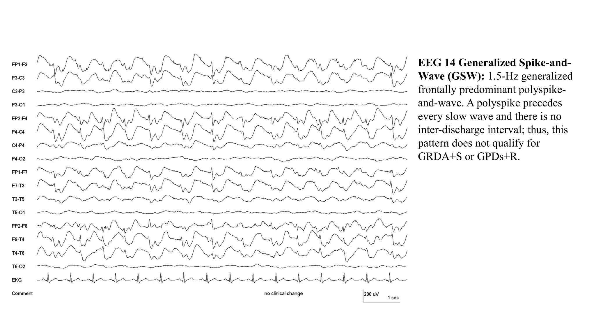
EEG 14 Generalized Spike-and-
Wave (GSW): 1.5-Hz generalized
frontally predominant polyspike-
and-wave. A polyspike precedes
every slow wave and there is no
inter-discharge interval; thus, this
pattern does not qualify for
GRDA+S or GPDs+R.
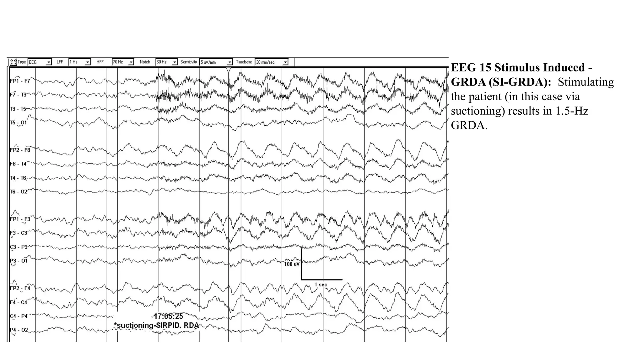
EEG 15 Stimulus Induced -
GRDA (SI-GRDA): Stimulating
the patient (in this case via
suctioning) results in 1.5-Hz
GRDA.
EEG 16 LRDA with evolution:
LRDA that evolves in morphology
and frequency. It begins as low
voltage sharply contoured 1.5-Hz
delta in the left parasagittal region,
evolves to 3-Hz rhythmic delta, then
again slows.
NOTE: The pattern only lasts 9 s and
therefore does not meet the ≥10 s cut
off for an electrographic seizure. It
also only reaches a maximum of 3
Hz. If the rhythmic activity were >4
Hz with evolution this would be
classified as definite BIRDs.
EEG 17 LPDs with
fluctuation: LPDs
that fluctuate in
frequency between
0.5 and 1 Hz.
EEG 18 GPDs+F: 1-1.25 Hz sharp
GPDs with superimposed low
amplitude quasi-rhythmic fast
activity (highlighted in boxes).
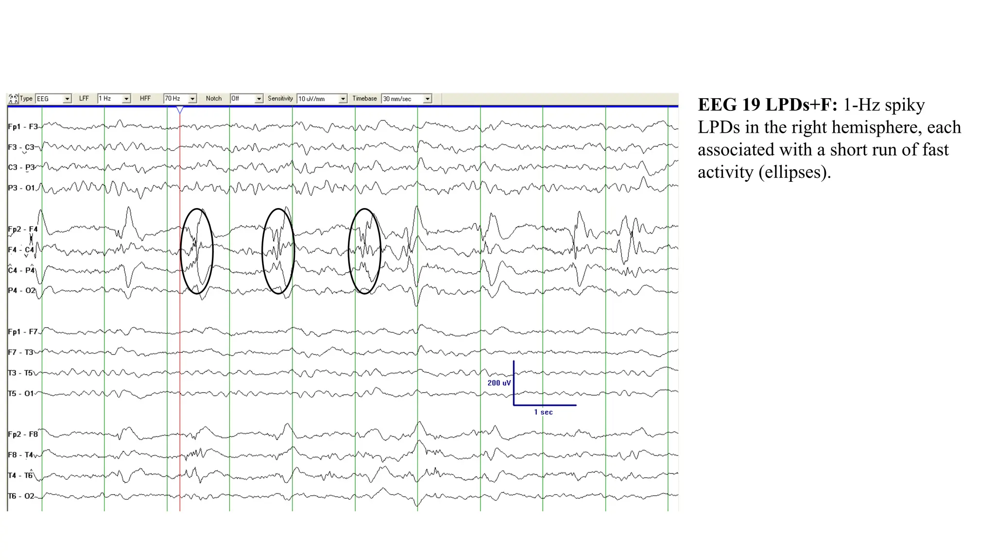
EEG 19 LPDs+F: 1-Hz spiky
LPDs in the right hemisphere, each
associated with a short run of fast
activity (ellipses).
EEG 20 LRDA+S: 2-Hz LRDA with
superimposed repetitive sharp waves
(several marked with asterisks)
(LRDA+S). The superimposed low
amplitude fast activity is also present
on the right hemisphere and should
not be recorded as +F.
EEG 21 BIPDs+F: Bilateral
independent periodic discharges at
0.5-1 Hz, most prominent
centroparietally on both sides. The
periodic discharges have a sharp
morphology and are associated with
low amplitude sharply contoured
quasi-rhythmic fast activity,
especially posteriorly, and more
prominent on the right where the
fast activity is nearly continuous.
EEG 22 Extreme Delta Brush
(EDB: 1-Hz periodic delta brush
pattern (i.e., there is a clear interval
between each consecutive delta brush
waveform). Using the current
terminology, this would be best
characterized as 1-Hz GPDs+F of
blunt morphology, where each
discharge is a delta wave. Fast
activity occurs in a stereotyped
relation with each delta wave (in this
case at the crest and on the
downslope [ellipses]). This is better
seen in the blown-up section of the
EEG in the box. If this pattern were
abundant or continuous it would be
definite EDB, but if only occasional
or frequent it would be possible EDB.
Courtesy of Dr. Nicolas Gaspard.
EEG 23 GPDs with triphasic
morphology and A-P lag: GPDs at
just under 1.5 Hz. In this case there
is also triphasic morphology and an
anterior-posterior lag, highlighted
with the diagonal line in the upper
right of the figure.
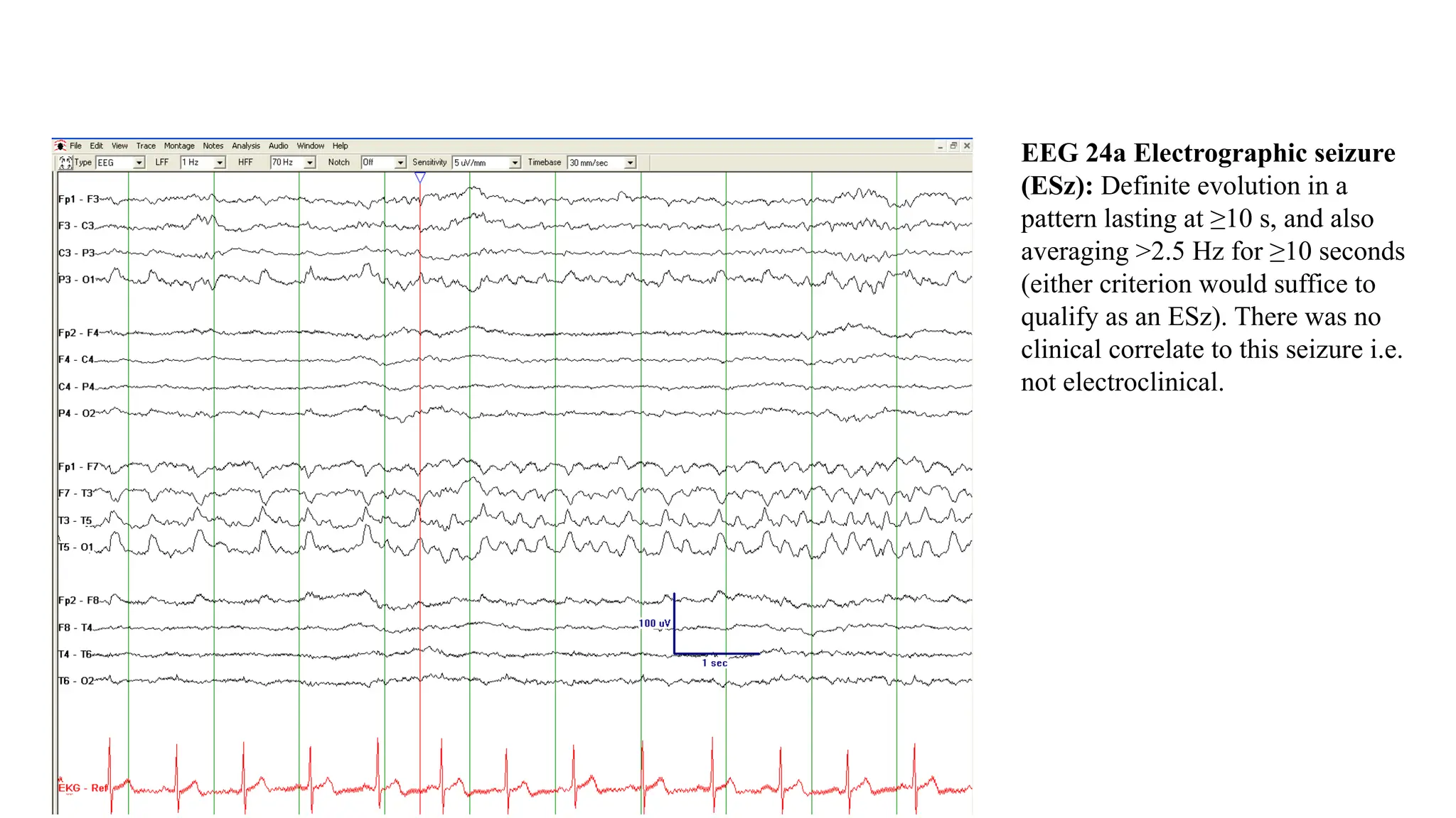
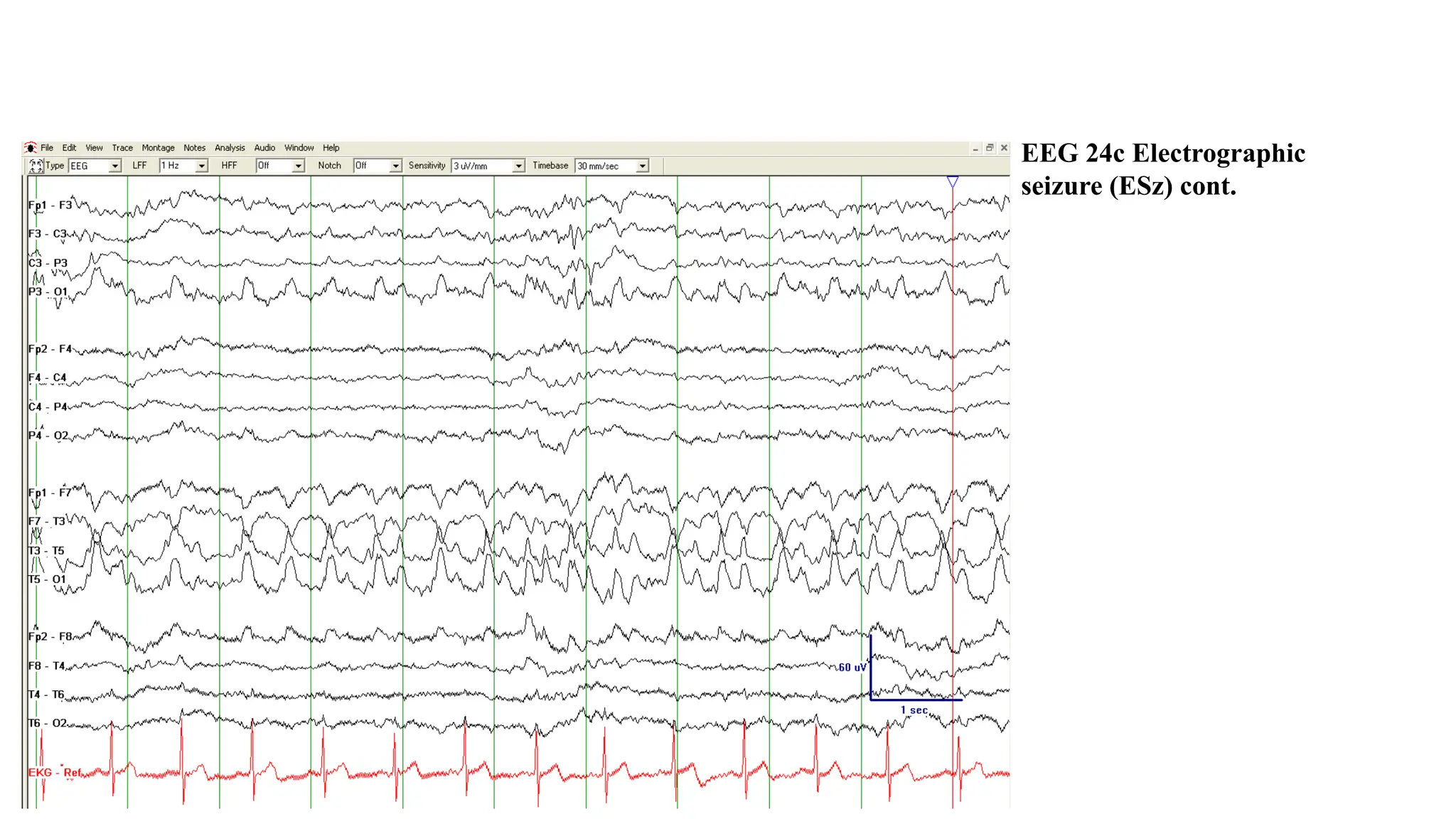
EEG 24a Electrographic seizure
(ESz): Definite evolution in a
pattern lasting at ≥10 s, and also
averaging >2.5 Hz for ≥10 seconds
(either criterion would suffice to
qualify as an ESz). There was no
clinical correlate to this seizure i.e.
not electroclinical.
EEG 24b Electrographic
seizure (ESz) cont.
EEG 24c Electrographic
seizure (ESz) cont.
EEG 25 Electroclinical seizure
(ECSz): The spike and polyspike
pattern at C3 is associated with right
hand twitching (EMG trace at the
bottom of the page). [From Hirsch
LJ, Brenner RP. Atlas of EEG in
Critical Care. London: Wiley, 2010.
With permission.]
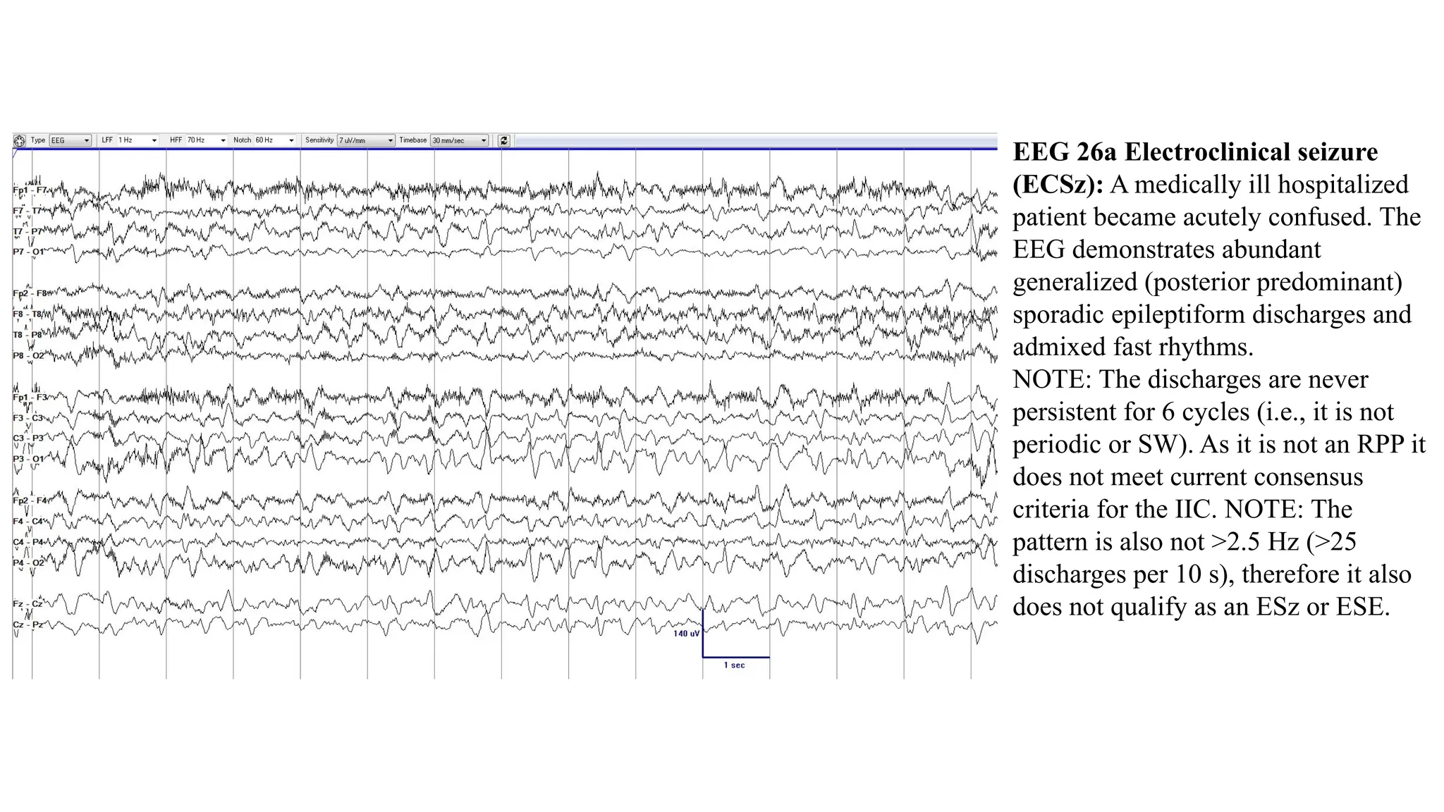
EEG 26a Electroclinical seizure
(ECSz): A medically ill hospitalized
patient became acutely confused. The
EEG demonstrates abundant
generalized (posterior predominant)
sporadic epileptiform discharges and
admixed fast rhythms.
NOTE: The discharges are never
persistent for 6 cycles (i.e., it is not
periodic or SW). As it is not an RPP it
does not meet current consensus
criteria for the IIC. NOTE: The
pattern is also not >2.5 Hz (>25
discharges per 10 s), therefore it also
does not qualify as an ESz or ESE.
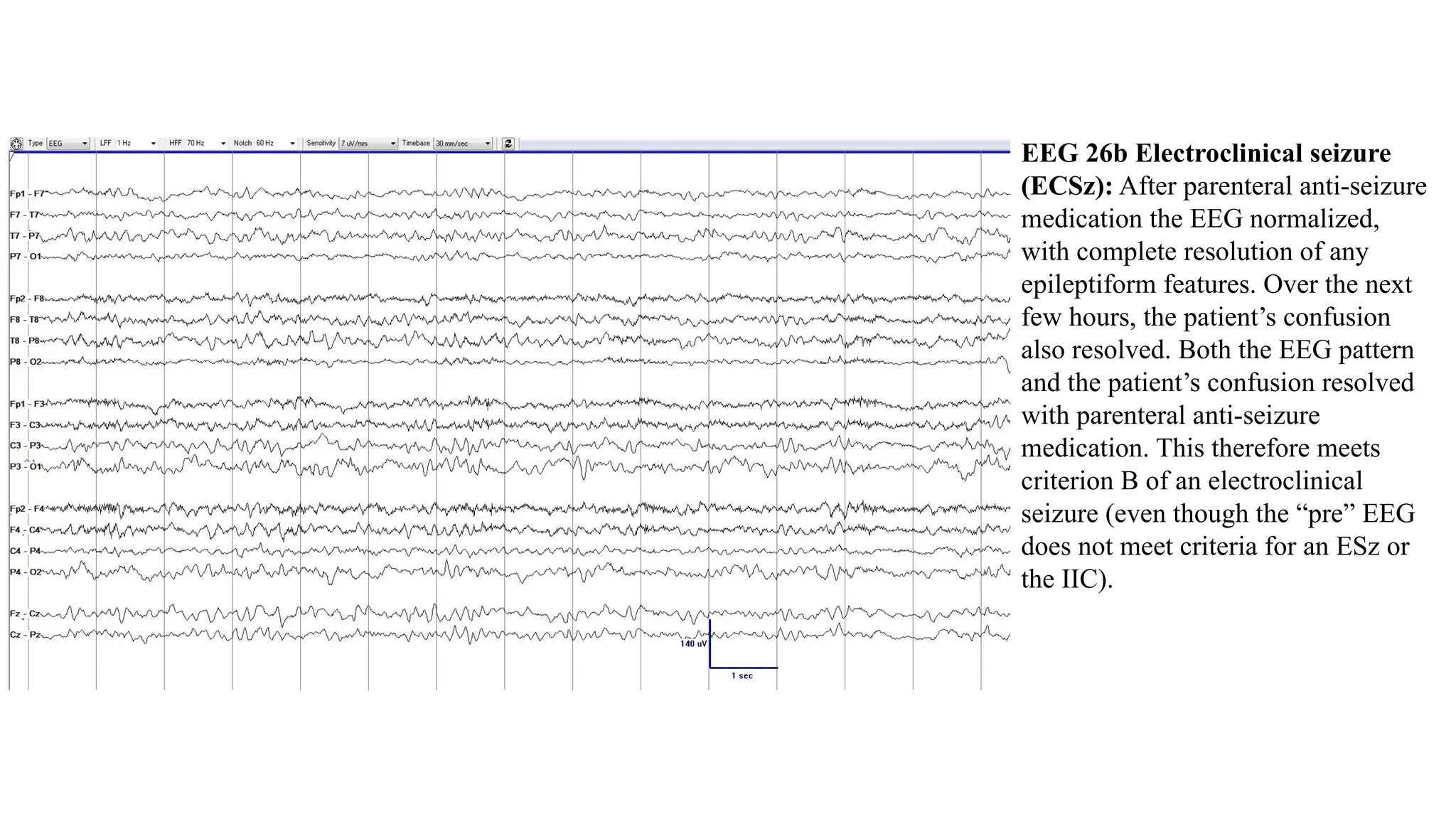
EEG 26b Electroclinical seizure
(ECSz): After parenteral anti-seizure
medication the EEG normalized,
with complete resolution of any
epileptiform features. Over the next
few hours, the patient’s confusion
also resolved. Both the EEG pattern
and the patient’s confusion resolved
with parenteral anti-seizure
medication. This therefore meets
criterion B of an electroclinical
seizure (even though the “pre” EEG
does not meet criteria for an ESz or
the IIC).
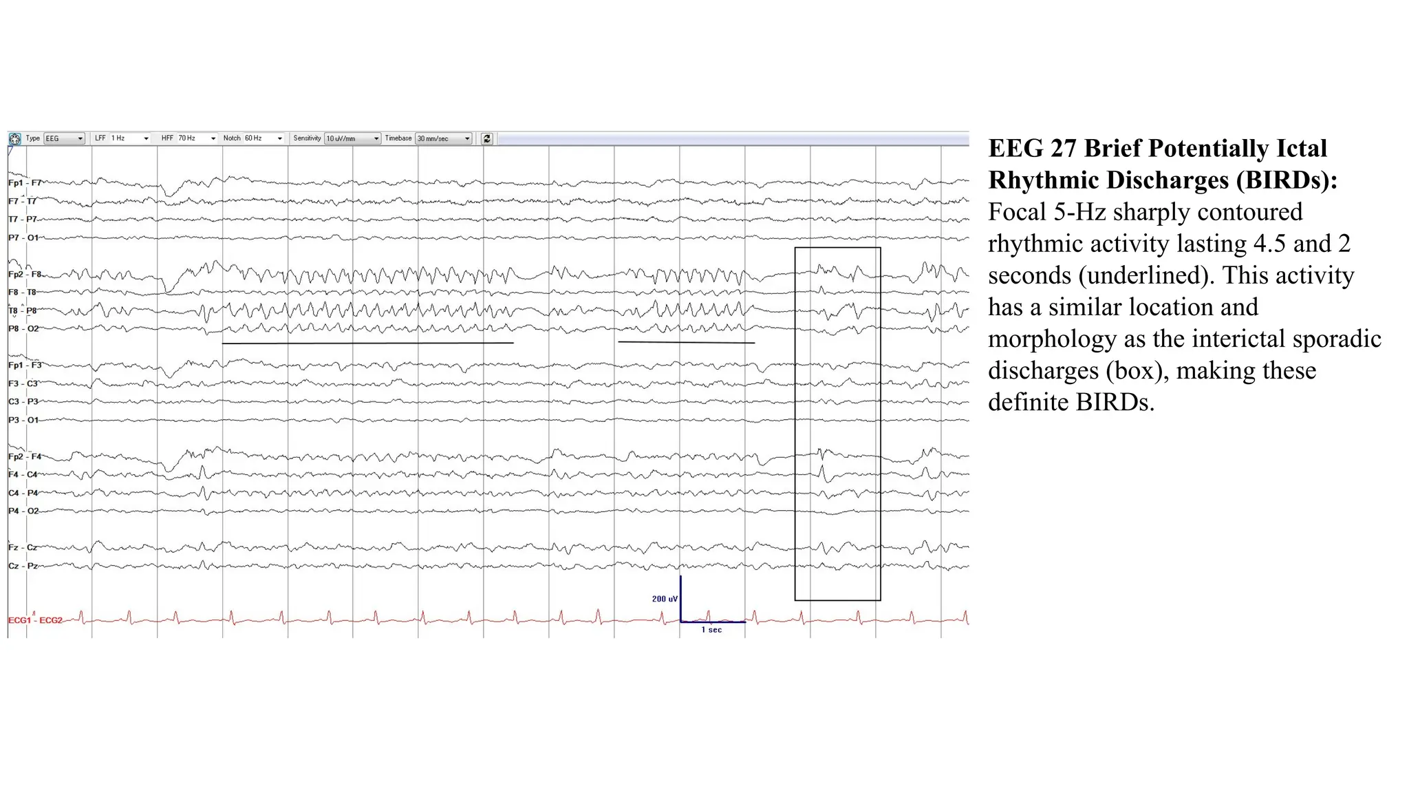
EEG 27 Brief Potentially Ictal
Rhythmic Discharges (BIRDs):
Focal 5-Hz sharply contoured
rhythmic activity lasting 4.5 and 2
seconds (underlined). This activity
has a similar location and
morphology as the interictal sporadic
discharges (box), making these
definite BIRDs.
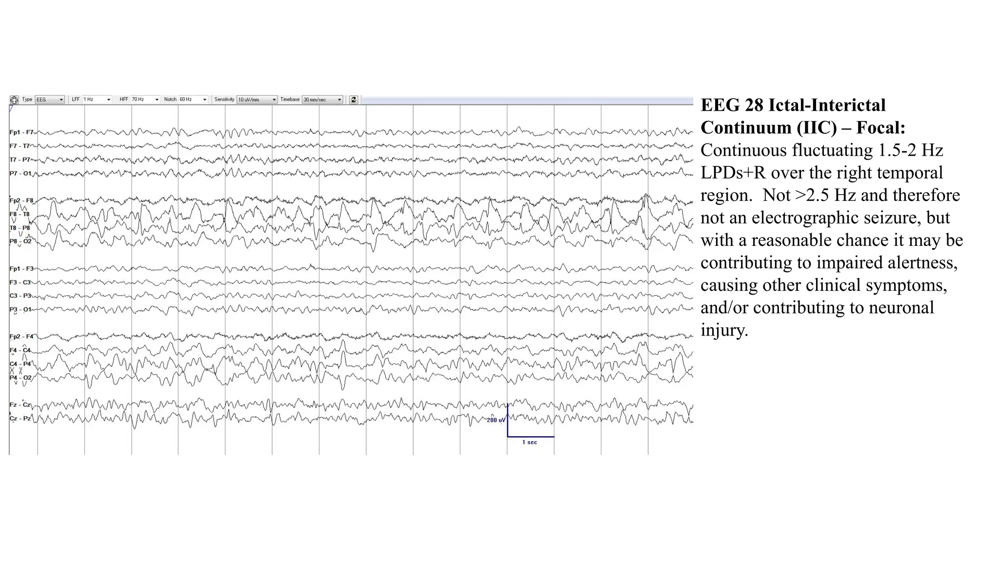
EEG 28 Ictal-Interictal
Continuum (IIC) – Focal:
Continuous fluctuating 1.5-2 Hz
LPDs+R over the right temporal
region. Not >2.5 Hz and therefore
not an electrographic seizure, but
with a reasonable chance it may be
contributing to impaired alertness,
causing other clinical symptoms,
and/or contributing to neuronal
injury.
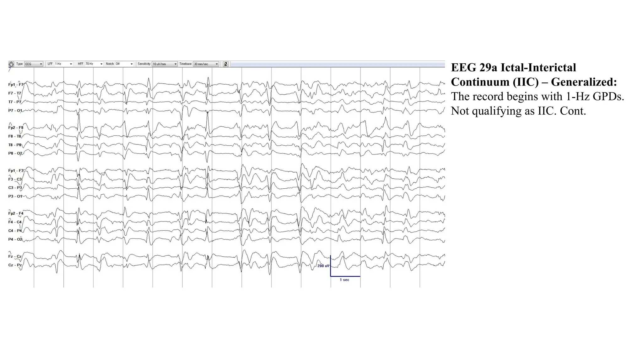
EEG 29a Ictal-Interictal
Continuum (IIC) – Generalized:
The record begins with 1-Hz GPDs.
Not qualifying as IIC. Cont.
EEG 29b Ictal-Interictal
Continuum (IIC) – Generalized:
As the recording continues the EEG
begins to change, with GPDs
fluctuating between 1 Hz and very
briefly up to 1.5 Hz. Now qualifying
as a pattern on the IIC. Cont.
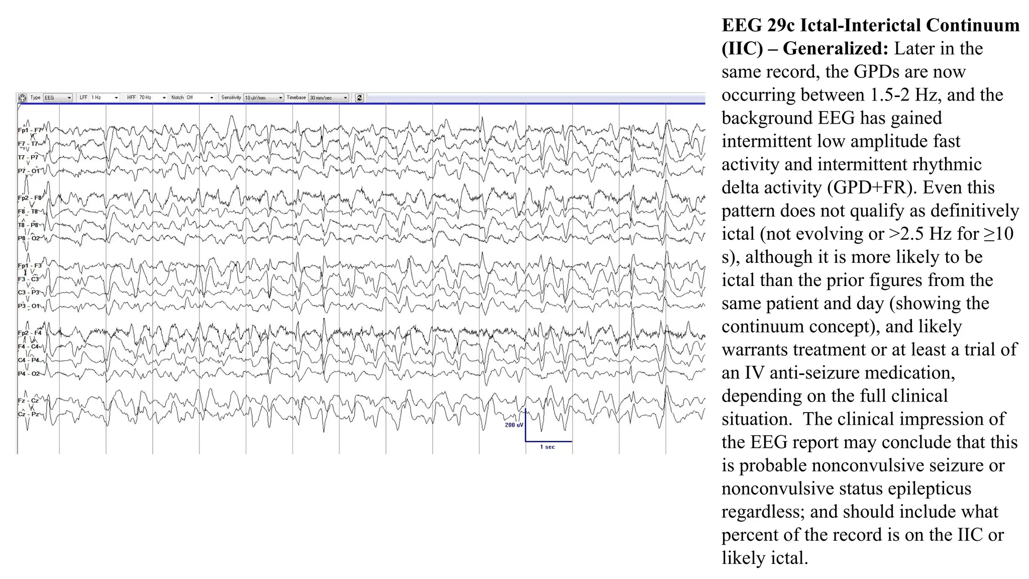
EEG 29c Ictal-Interictal Continuum
(IIC) – Generalized: Later in the
same record, the GPDs are now
occurring between 1.5-2 Hz, and the
background EEG has gained
intermittent low amplitude fast
activity and intermittent rhythmic
delta activity (GPD+FR). Even this
pattern does not qualify as definitively
ictal (not evolving or >2.5 Hz for ≥10
s), although it is more likely to be
ictal than the prior figures from the
same patient and day (showing the
continuum concept), and likely
warrants treatment or at least a trial of
an IV anti-seizure medication,
depending on the full clinical
situation. The clinical impression of
the EEG report may conclude that this
is probable nonconvulsive seizure or
nonconvulsive status epilepticus
regardless; and should include what
percent of the record is on the IIC or
likely ictal.
EEG 30 Ictal-Interictal Continuum (IIC) with Quantitative
EEG (QEEG). The figure demonstrates the concept of the IIC.
Panels A and B show the Color Density Spectral Array (CSA)
for the left hemisphere (A) and the right hemisphere (B). The
CSA displays EEG power by frequency band. The y axis is
frequency (from 0 to 30 Hz), the x axis is time (in this case
showing a 12-hour trend). The amount of power at each
frequency is demonstrated by the intensity of the color on a Z
scale. If there is no power the QEEG is black, through to high
power, which demonstrates intense red then pink and white
colors. The QEEG demonstrates that over the 12 hours of the
recording the power in each hemisphere is slowly and gradually
reduced across all frequencies. Panel C shows the EEG at the
respective time points (arrows). Near the beginning there are 1-
1.5 Hz posterior predominant GPDs with fast and rhythmic
activity (GPD+FR), high amplitude (note the 15 uV/mm
sensitivity), a pattern on the IIC, not qualifying as definitively
ictal, but interpreted (clinical impression) as probable
nonconvulsive status epilepticus. By the end of the recording
the periodic pattern, fast activity and rhythmicity have resolved,
now only demonstrating diffuse dysfunction with abundant
sporadic epileptiform discharges (clearly not ictal, and not on
the IIC). The middle panel shows a state in between the two.
The cutoff point where the highly epileptiform patten becomes
“interictal” is not easily defined. This demonstrates the concept
of the IIC, a spectrum of EEG findings from interictal to
potentially ictal, at times progressing into definite
electrographic seizures and status epilepticus. There is no
abrupt transition between ictal and non-ictal, but rather a
gradual continuum. [Adapted from Hirsch LJ, Brenner RP.
Atlas of EEG in Critical Care. London: Wiley, 2010. With
permission.]

More Related Content

Similar to ACNS Standardized Critical Care EEG Terminology 2021

EEG artifacts 2
EEG artifacts  2EEG artifacts  2
EEG artifacts 2
DGIST
 
Diagrams-Eeg-Epilepsy.ppt
Diagrams-Eeg-Epilepsy.pptDiagrams-Eeg-Epilepsy.ppt
Diagrams-Eeg-Epilepsy.ppt
PANFRAGGER
 
west syndrome ppt hypsarrhythmia infantile.pptx
west syndrome  ppt hypsarrhythmia infantile.pptxwest syndrome  ppt hypsarrhythmia infantile.pptx
west syndrome ppt hypsarrhythmia infantile.pptx
priya99148
 
Fundamentals of nerve conduction study
Fundamentals of nerve conduction studyFundamentals of nerve conduction study
Fundamentals of nerve conduction study
NeurologyKota
 

Similar to ACNS Standardized Critical Care EEG Terminology 2021 (20)

Neurophysiological investigations
Neurophysiological investigationsNeurophysiological investigations
Neurophysiological investigations
 
Polysomnogram interpretetion
Polysomnogram interpretetionPolysomnogram interpretetion
Polysomnogram interpretetion
 
EEG basic to practice 1
EEG basic to practice 1EEG basic to practice 1
EEG basic to practice 1
 
Basics of EEG - A brief review of the EEG.ppt
Basics of EEG - A brief review of the EEG.pptBasics of EEG - A brief review of the EEG.ppt
Basics of EEG - A brief review of the EEG.ppt
 
چرخه های زیستی و خواب
چرخه های  زیستی  و خوابچرخه های  زیستی  و خواب
چرخه های زیستی و خواب
 
normal eeg
 normal eeg  normal eeg
normal eeg
 
Eeg in encephalopathy
Eeg in encephalopathyEeg in encephalopathy
Eeg in encephalopathy
 
Approaching the eeg
Approaching the eegApproaching the eeg
Approaching the eeg
 
EEG
EEGEEG
EEG
 
Eeg examples
Eeg examplesEeg examples
Eeg examples
 
EEG artifacts 2
EEG artifacts  2EEG artifacts  2
EEG artifacts 2
 
Ncs
NcsNcs
Ncs
 
Abnormal EEG patterns
Abnormal EEG patternsAbnormal EEG patterns
Abnormal EEG patterns
 
Diagrams-Eeg-Epilepsy.ppt
Diagrams-Eeg-Epilepsy.pptDiagrams-Eeg-Epilepsy.ppt
Diagrams-Eeg-Epilepsy.ppt
 
پاور چدید آموزش نوروفیدبک درمورد خواب و تشنج.ppt
پاور چدید آموزش نوروفیدبک درمورد خواب و تشنج.pptپاور چدید آموزش نوروفیدبک درمورد خواب و تشنج.ppt
پاور چدید آموزش نوروفیدبک درمورد خواب و تشنج.ppt
 
Erg and eog
Erg and eogErg and eog
Erg and eog
 
west syndrome ppt hypsarrhythmia infantile.pptx
west syndrome  ppt hypsarrhythmia infantile.pptxwest syndrome  ppt hypsarrhythmia infantile.pptx
west syndrome ppt hypsarrhythmia infantile.pptx
 
Basics of eeg signal
Basics of eeg signalBasics of eeg signal
Basics of eeg signal
 
Electrodiadnostic tests ERG and VEP
Electrodiadnostic tests ERG and VEPElectrodiadnostic tests ERG and VEP
Electrodiadnostic tests ERG and VEP
 
Fundamentals of nerve conduction study
Fundamentals of nerve conduction studyFundamentals of nerve conduction study
Fundamentals of nerve conduction study
 

Recently uploaded

Difference Between Skeletal Smooth and Cardiac Muscles
Difference Between Skeletal Smooth and Cardiac MusclesDifference Between Skeletal Smooth and Cardiac Muscles
Difference Between Skeletal Smooth and Cardiac Muscles
MedicoseAcademics
 
Russian Call Girls In Pune 👉 Just CALL ME: 9352988975 ✅❤️💯low cost unlimited ...
Russian Call Girls In Pune 👉 Just CALL ME: 9352988975 ✅❤️💯low cost unlimited ...Russian Call Girls In Pune 👉 Just CALL ME: 9352988975 ✅❤️💯low cost unlimited ...
Russian Call Girls In Pune 👉 Just CALL ME: 9352988975 ✅❤️💯low cost unlimited ...
chanderprakash5506
 
❤️ Chandigarh Call Girls☎️98151-579OO☎️ Call Girl service in Chandigarh ☎️ Ch...
❤️ Chandigarh Call Girls☎️98151-579OO☎️ Call Girl service in Chandigarh ☎️ Ch...❤️ Chandigarh Call Girls☎️98151-579OO☎️ Call Girl service in Chandigarh ☎️ Ch...
❤️ Chandigarh Call Girls☎️98151-579OO☎️ Call Girl service in Chandigarh ☎️ Ch...
Rashmi Entertainment
 
Call Girl in Chennai | Whatsapp No 📞 7427069034 📞 VIP Escorts Service Availab...
Call Girl in Chennai | Whatsapp No 📞 7427069034 📞 VIP Escorts Service Availab...Call Girl in Chennai | Whatsapp No 📞 7427069034 📞 VIP Escorts Service Availab...
Call Girl in Chennai | Whatsapp No 📞 7427069034 📞 VIP Escorts Service Availab...
amritaverma53
 
Cara Menggugurkan Kandungan Dengan Cepat Selesai Dalam 24 Jam Secara Alami Bu...
Cara Menggugurkan Kandungan Dengan Cepat Selesai Dalam 24 Jam Secara Alami Bu...Cara Menggugurkan Kandungan Dengan Cepat Selesai Dalam 24 Jam Secara Alami Bu...
Cara Menggugurkan Kandungan Dengan Cepat Selesai Dalam 24 Jam Secara Alami Bu...
Cara Menggugurkan Kandungan 087776558899
 

Recently uploaded (20)

Chennai ❣️ Call Girl 6378878445 Call Girls in Chennai Escort service book now
Chennai ❣️ Call Girl 6378878445 Call Girls in Chennai Escort service book nowChennai ❣️ Call Girl 6378878445 Call Girls in Chennai Escort service book now
Chennai ❣️ Call Girl 6378878445 Call Girls in Chennai Escort service book now
 
💞 Safe And Secure Call Girls Coimbatore🧿 6378878445 🧿 High Class Coimbatore C...
💞 Safe And Secure Call Girls Coimbatore🧿 6378878445 🧿 High Class Coimbatore C...💞 Safe And Secure Call Girls Coimbatore🧿 6378878445 🧿 High Class Coimbatore C...
💞 Safe And Secure Call Girls Coimbatore🧿 6378878445 🧿 High Class Coimbatore C...
 
Call Girls Mussoorie Just Call 8854095900 Top Class Call Girl Service Available
Call Girls Mussoorie Just Call 8854095900 Top Class Call Girl Service AvailableCall Girls Mussoorie Just Call 8854095900 Top Class Call Girl Service Available
Call Girls Mussoorie Just Call 8854095900 Top Class Call Girl Service Available
 
Cardiac Output, Venous Return, and Their Regulation
Cardiac Output, Venous Return, and Their RegulationCardiac Output, Venous Return, and Their Regulation
Cardiac Output, Venous Return, and Their Regulation
 
ANATOMY AND PHYSIOLOGY OF RESPIRATORY SYSTEM.pptx
ANATOMY AND PHYSIOLOGY OF RESPIRATORY SYSTEM.pptxANATOMY AND PHYSIOLOGY OF RESPIRATORY SYSTEM.pptx
ANATOMY AND PHYSIOLOGY OF RESPIRATORY SYSTEM.pptx
 
7 steps How to prevent Thalassemia : Dr Sharda Jain & Vandana Gupta
7 steps How to prevent Thalassemia : Dr Sharda Jain & Vandana Gupta7 steps How to prevent Thalassemia : Dr Sharda Jain & Vandana Gupta
7 steps How to prevent Thalassemia : Dr Sharda Jain & Vandana Gupta
 
Call Girls in Lucknow Just Call 👉👉8630512678 Top Class Call Girl Service Avai...
Call Girls in Lucknow Just Call 👉👉8630512678 Top Class Call Girl Service Avai...Call Girls in Lucknow Just Call 👉👉8630512678 Top Class Call Girl Service Avai...
Call Girls in Lucknow Just Call 👉👉8630512678 Top Class Call Girl Service Avai...
 
ANATOMY AND PHYSIOLOGY OF REPRODUCTIVE SYSTEM.pptx
ANATOMY AND PHYSIOLOGY OF REPRODUCTIVE SYSTEM.pptxANATOMY AND PHYSIOLOGY OF REPRODUCTIVE SYSTEM.pptx
ANATOMY AND PHYSIOLOGY OF REPRODUCTIVE SYSTEM.pptx
 
Difference Between Skeletal Smooth and Cardiac Muscles
Difference Between Skeletal Smooth and Cardiac MusclesDifference Between Skeletal Smooth and Cardiac Muscles
Difference Between Skeletal Smooth and Cardiac Muscles
 
Russian Call Girls In Pune 👉 Just CALL ME: 9352988975 ✅❤️💯low cost unlimited ...
Russian Call Girls In Pune 👉 Just CALL ME: 9352988975 ✅❤️💯low cost unlimited ...Russian Call Girls In Pune 👉 Just CALL ME: 9352988975 ✅❤️💯low cost unlimited ...
Russian Call Girls In Pune 👉 Just CALL ME: 9352988975 ✅❤️💯low cost unlimited ...
 
Call Girls Service Jaipur {9521753030 } ❤️VVIP BHAWNA Call Girl in Jaipur Raj...
Call Girls Service Jaipur {9521753030 } ❤️VVIP BHAWNA Call Girl in Jaipur Raj...Call Girls Service Jaipur {9521753030 } ❤️VVIP BHAWNA Call Girl in Jaipur Raj...
Call Girls Service Jaipur {9521753030 } ❤️VVIP BHAWNA Call Girl in Jaipur Raj...
 
💰Call Girl In Bangalore☎️63788-78445💰 Call Girl service in Bangalore☎️Bangalo...
💰Call Girl In Bangalore☎️63788-78445💰 Call Girl service in Bangalore☎️Bangalo...💰Call Girl In Bangalore☎️63788-78445💰 Call Girl service in Bangalore☎️Bangalo...
💰Call Girl In Bangalore☎️63788-78445💰 Call Girl service in Bangalore☎️Bangalo...
 
Call girls Service Phullen / 9332606886 Genuine Call girls with real Photos a...
Call girls Service Phullen / 9332606886 Genuine Call girls with real Photos a...Call girls Service Phullen / 9332606886 Genuine Call girls with real Photos a...
Call girls Service Phullen / 9332606886 Genuine Call girls with real Photos a...
 
Lucknow Call Girls Service { 9984666624 } ❤️VVIP ROCKY Call Girl in Lucknow U...
Lucknow Call Girls Service { 9984666624 } ❤️VVIP ROCKY Call Girl in Lucknow U...Lucknow Call Girls Service { 9984666624 } ❤️VVIP ROCKY Call Girl in Lucknow U...
Lucknow Call Girls Service { 9984666624 } ❤️VVIP ROCKY Call Girl in Lucknow U...
 
Call Girls Kathua Just Call 8250077686 Top Class Call Girl Service Available
Call Girls Kathua Just Call 8250077686 Top Class Call Girl Service AvailableCall Girls Kathua Just Call 8250077686 Top Class Call Girl Service Available
Call Girls Kathua Just Call 8250077686 Top Class Call Girl Service Available
 
Call Girls Bangalore - 450+ Call Girl Cash Payment 💯Call Us 🔝 6378878445 🔝 💃 ...
Call Girls Bangalore - 450+ Call Girl Cash Payment 💯Call Us 🔝 6378878445 🔝 💃 ...Call Girls Bangalore - 450+ Call Girl Cash Payment 💯Call Us 🔝 6378878445 🔝 💃 ...
Call Girls Bangalore - 450+ Call Girl Cash Payment 💯Call Us 🔝 6378878445 🔝 💃 ...
 
❤️ Chandigarh Call Girls☎️98151-579OO☎️ Call Girl service in Chandigarh ☎️ Ch...
❤️ Chandigarh Call Girls☎️98151-579OO☎️ Call Girl service in Chandigarh ☎️ Ch...❤️ Chandigarh Call Girls☎️98151-579OO☎️ Call Girl service in Chandigarh ☎️ Ch...
❤️ Chandigarh Call Girls☎️98151-579OO☎️ Call Girl service in Chandigarh ☎️ Ch...
 
Call Girl in Chennai | Whatsapp No 📞 7427069034 📞 VIP Escorts Service Availab...
Call Girl in Chennai | Whatsapp No 📞 7427069034 📞 VIP Escorts Service Availab...Call Girl in Chennai | Whatsapp No 📞 7427069034 📞 VIP Escorts Service Availab...
Call Girl in Chennai | Whatsapp No 📞 7427069034 📞 VIP Escorts Service Availab...
 
Cara Menggugurkan Kandungan Dengan Cepat Selesai Dalam 24 Jam Secara Alami Bu...
Cara Menggugurkan Kandungan Dengan Cepat Selesai Dalam 24 Jam Secara Alami Bu...Cara Menggugurkan Kandungan Dengan Cepat Selesai Dalam 24 Jam Secara Alami Bu...
Cara Menggugurkan Kandungan Dengan Cepat Selesai Dalam 24 Jam Secara Alami Bu...
 
Lucknow Call Girls Just Call 👉👉8630512678 Top Class Call Girl Service Available
Lucknow Call Girls Just Call 👉👉8630512678 Top Class Call Girl Service AvailableLucknow Call Girls Just Call 👉👉8630512678 Top Class Call Girl Service Available
Lucknow Call Girls Just Call 👉👉8630512678 Top Class Call Girl Service Available
 

ACNS Standardized Critical Care EEG Terminology 2021

  • 1. ACNS Standardized Critical Care EEG Terminology 2021 EEG and Legends
  • 2. EEG 1 Burst-attenuation pattern: Bursts (≥0.5 s AND >3 phases) of generalized activity, in between bursts there is lower amplitude background activity (<50% of the background/bursts, but ≥10 µV i.e. not suppression).
  • 3. EEG 2 Burst-suppression pattern: Bursts (≥0.5 s AND >3 phases) of generalized activity on a suppressed (<10 µV) background.
  • 4. EEG 3 Identical Highly- Epileptiform Bursts: The pattern is burst suppression. The first 0.5 s of each burst appears visually similar in all channels, qualifying as identical. Each burst also contains 2 or more epileptiform discharges occurring at an average of 1 Hz or faster within a single burst, qualifying as highly epileptiform.
  • 5. EEG 4 Identical Non-Highly- Epileptiform Bursts: Burst suppression pattern, with each burst approximately 1 s (≥0.5 s) and containing 4 – 6 phases each (>3 phases). The first 0.5 seconds of each burst is visually similar in all channels, qualifying as identical bursts. Although there is an epileptiform discharge in each burst, there are not consistently 2 or more in each burst and not occurring at greater than 1-Hz frequency; therefore, this is not highly epileptiform.
  • 6. EEG 5 Generalized Periodic Discharges (GPDs): 1 Hz sharp GPDs.
  • 7. EEG 6 Generalized Rhythmic Delta Activity (GRDA):1.5 Hz frontally predominant GRDA. If the lower amplitude faster (alpha range) frequencies are not present in the background when the GRDA is not present, then this would qualify as GRDA+F.
  • 8. EEG 7 GPDs (frontally predominant): 1 Hz GPDs, characterized by a marked frontal predominance and a sharp morphology. Despite background attenuation, the discharges are <0.5 s and thus do not qualify as bursts.
  • 9. EEG 7 Lateralized Periodic Discharges (LPDs): 0.5-1 Hz spiky LPDs. Despite their spike-and-wave morphology, the discharges are periodic (as there is a quantifiable inter-discharge interval between consecutive waveforms and recurrence of the waveform at nearly regular intervals).
  • 10. EEG 9 Bilateral Independent Periodic Discharges (BIPDs): Periodic spike wave occurring at 0.3-0.5 Hz in the left posterior quadrant (arrows). At the same time, there is another periodic spike wave population occurring at 0.5-1 Hz in the right posterior quadrant (asterisks).
  • 11. EEG 10 Unilateral Independent Periodic Discharges (UIPDs): 1-1.5 Hz Periodic Discharges (PDs) maximal at F3. At the same time there are independent 0.5-Hz PDs in the central region. The two PD patterns in the same hemisphere qualify as UIPDs. NOTE a focal midline pattern may still be classified in the same hemisphere (unilateral) as an independent pattern in either the right or left hemisphere. Courtesy of Dr. Jong Woo Lee.
  • 12. EEG 11 Unilateral Independent Rhythmic Delta Activity (UIRDA): 1.5-Hz Rhythmic Delta Activity (RDA) in the right frontal region (solid box). At the same time, there is 1-Hz RDA in the right temporal region (dashed box). The patterns are independent from each other but are both in the same hemisphere (so unilateral).
  • 13. EEG 12 Multifocal Periodic Discharges (MfPDs): Three independent lateralized periodic patterns occurring at the same time, with at least one in each hemisphere (2 on the right and 1 on the left). Blunt PDs left fronto-central at 0.75 Hz (boxes), sharp PDs at F4 at 0.33 Hz (arrows) and spiky PDs at P4 at 0.2-0.25 Hz (asterisks). Courtesy of Dr. Luis Octavio Caboclo.
  • 14. EEG 13 LPDs (bilateral asymmetric): Sharply contoured LPDs. In this case they are clearly lateralized to the left hemisphere but can be seen in both hemispheres (for example at F4). These are still classified as a lateralized pattern (LPDs) but are bilateral asymmetric.
  • 15. EEG 14 Generalized Spike-and- Wave (GSW): 1.5-Hz generalized frontally predominant polyspike- and-wave. A polyspike precedes every slow wave and there is no inter-discharge interval; thus, this pattern does not qualify for GRDA+S or GPDs+R.
  • 16. EEG 15 Stimulus Induced - GRDA (SI-GRDA): Stimulating the patient (in this case via suctioning) results in 1.5-Hz GRDA.
  • 17. EEG 16 LRDA with evolution: LRDA that evolves in morphology and frequency. It begins as low voltage sharply contoured 1.5-Hz delta in the left parasagittal region, evolves to 3-Hz rhythmic delta, then again slows. NOTE: The pattern only lasts 9 s and therefore does not meet the ≥10 s cut off for an electrographic seizure. It also only reaches a maximum of 3 Hz. If the rhythmic activity were >4 Hz with evolution this would be classified as definite BIRDs.
  • 18. EEG 17 LPDs with fluctuation: LPDs that fluctuate in frequency between 0.5 and 1 Hz.
  • 19. EEG 18 GPDs+F: 1-1.25 Hz sharp GPDs with superimposed low amplitude quasi-rhythmic fast activity (highlighted in boxes).
  • 20. EEG 19 LPDs+F: 1-Hz spiky LPDs in the right hemisphere, each associated with a short run of fast activity (ellipses).
  • 21. EEG 20 LRDA+S: 2-Hz LRDA with superimposed repetitive sharp waves (several marked with asterisks) (LRDA+S). The superimposed low amplitude fast activity is also present on the right hemisphere and should not be recorded as +F.
  • 22. EEG 21 BIPDs+F: Bilateral independent periodic discharges at 0.5-1 Hz, most prominent centroparietally on both sides. The periodic discharges have a sharp morphology and are associated with low amplitude sharply contoured quasi-rhythmic fast activity, especially posteriorly, and more prominent on the right where the fast activity is nearly continuous.
  • 23. EEG 22 Extreme Delta Brush (EDB: 1-Hz periodic delta brush pattern (i.e., there is a clear interval between each consecutive delta brush waveform). Using the current terminology, this would be best characterized as 1-Hz GPDs+F of blunt morphology, where each discharge is a delta wave. Fast activity occurs in a stereotyped relation with each delta wave (in this case at the crest and on the downslope [ellipses]). This is better seen in the blown-up section of the EEG in the box. If this pattern were abundant or continuous it would be definite EDB, but if only occasional or frequent it would be possible EDB. Courtesy of Dr. Nicolas Gaspard.
  • 24. EEG 23 GPDs with triphasic morphology and A-P lag: GPDs at just under 1.5 Hz. In this case there is also triphasic morphology and an anterior-posterior lag, highlighted with the diagonal line in the upper right of the figure.
  • 25. EEG 24a Electrographic seizure (ESz): Definite evolution in a pattern lasting at ≥10 s, and also averaging >2.5 Hz for ≥10 seconds (either criterion would suffice to qualify as an ESz). There was no clinical correlate to this seizure i.e. not electroclinical.
  • 28. EEG 25 Electroclinical seizure (ECSz): The spike and polyspike pattern at C3 is associated with right hand twitching (EMG trace at the bottom of the page). [From Hirsch LJ, Brenner RP. Atlas of EEG in Critical Care. London: Wiley, 2010. With permission.]
  • 29. EEG 26a Electroclinical seizure (ECSz): A medically ill hospitalized patient became acutely confused. The EEG demonstrates abundant generalized (posterior predominant) sporadic epileptiform discharges and admixed fast rhythms. NOTE: The discharges are never persistent for 6 cycles (i.e., it is not periodic or SW). As it is not an RPP it does not meet current consensus criteria for the IIC. NOTE: The pattern is also not >2.5 Hz (>25 discharges per 10 s), therefore it also does not qualify as an ESz or ESE.
  • 30. EEG 26b Electroclinical seizure (ECSz): After parenteral anti-seizure medication the EEG normalized, with complete resolution of any epileptiform features. Over the next few hours, the patient’s confusion also resolved. Both the EEG pattern and the patient’s confusion resolved with parenteral anti-seizure medication. This therefore meets criterion B of an electroclinical seizure (even though the “pre” EEG does not meet criteria for an ESz or the IIC).
  • 31. EEG 27 Brief Potentially Ictal Rhythmic Discharges (BIRDs): Focal 5-Hz sharply contoured rhythmic activity lasting 4.5 and 2 seconds (underlined). This activity has a similar location and morphology as the interictal sporadic discharges (box), making these definite BIRDs.
  • 32. EEG 28 Ictal-Interictal Continuum (IIC) – Focal: Continuous fluctuating 1.5-2 Hz LPDs+R over the right temporal region. Not >2.5 Hz and therefore not an electrographic seizure, but with a reasonable chance it may be contributing to impaired alertness, causing other clinical symptoms, and/or contributing to neuronal injury.
  • 33. EEG 29a Ictal-Interictal Continuum (IIC) – Generalized: The record begins with 1-Hz GPDs. Not qualifying as IIC. Cont.
  • 34. EEG 29b Ictal-Interictal Continuum (IIC) – Generalized: As the recording continues the EEG begins to change, with GPDs fluctuating between 1 Hz and very briefly up to 1.5 Hz. Now qualifying as a pattern on the IIC. Cont.
  • 35. EEG 29c Ictal-Interictal Continuum (IIC) – Generalized: Later in the same record, the GPDs are now occurring between 1.5-2 Hz, and the background EEG has gained intermittent low amplitude fast activity and intermittent rhythmic delta activity (GPD+FR). Even this pattern does not qualify as definitively ictal (not evolving or >2.5 Hz for ≥10 s), although it is more likely to be ictal than the prior figures from the same patient and day (showing the continuum concept), and likely warrants treatment or at least a trial of an IV anti-seizure medication, depending on the full clinical situation. The clinical impression of the EEG report may conclude that this is probable nonconvulsive seizure or nonconvulsive status epilepticus regardless; and should include what percent of the record is on the IIC or likely ictal.
  • 36. EEG 30 Ictal-Interictal Continuum (IIC) with Quantitative EEG (QEEG). The figure demonstrates the concept of the IIC. Panels A and B show the Color Density Spectral Array (CSA) for the left hemisphere (A) and the right hemisphere (B). The CSA displays EEG power by frequency band. The y axis is frequency (from 0 to 30 Hz), the x axis is time (in this case showing a 12-hour trend). The amount of power at each frequency is demonstrated by the intensity of the color on a Z scale. If there is no power the QEEG is black, through to high power, which demonstrates intense red then pink and white colors. The QEEG demonstrates that over the 12 hours of the recording the power in each hemisphere is slowly and gradually reduced across all frequencies. Panel C shows the EEG at the respective time points (arrows). Near the beginning there are 1- 1.5 Hz posterior predominant GPDs with fast and rhythmic activity (GPD+FR), high amplitude (note the 15 uV/mm sensitivity), a pattern on the IIC, not qualifying as definitively ictal, but interpreted (clinical impression) as probable nonconvulsive status epilepticus. By the end of the recording the periodic pattern, fast activity and rhythmicity have resolved, now only demonstrating diffuse dysfunction with abundant sporadic epileptiform discharges (clearly not ictal, and not on the IIC). The middle panel shows a state in between the two. The cutoff point where the highly epileptiform patten becomes “interictal” is not easily defined. This demonstrates the concept of the IIC, a spectrum of EEG findings from interictal to potentially ictal, at times progressing into definite electrographic seizures and status epilepticus. There is no abrupt transition between ictal and non-ictal, but rather a gradual continuum. [Adapted from Hirsch LJ, Brenner RP. Atlas of EEG in Critical Care. London: Wiley, 2010. With permission.]