Percutaneous nephrolithotomy surgery india
•Download as PPTX, PDF•
2 likes•3,258 views

Percutaneous nephrolithotripsy (PCNL or PNL) is a minimally invasive endoscopic treatment for removing large kidney stones called staghorn stones or large or multiple stones impacted at the upper ureter.
Report
Share
Report
Share

Recommended
Percutaneous Nephrolithotomy
This study evaluated the safety and efficacy of percutaneous nephrolithotomy (PCNL) guided solely by ultrasonography in over 700 cases over 5 years. Access to the pelvicalyceal system was successful in all cases using ultrasonography. The overall stone-free rate was 87.4% and complications were minor, with a low 16% rate. The study demonstrated that PCNL can be performed safely and effectively using only ultrasonography guidance, avoiding the risks of radiation exposure from fluoroscopy.
Percutaneous Nephrolithotomy PCNL by Dr. Majid Kakakhel IKD, Peshawar.
The document describes the procedure and techniques for percutaneous nephrolithotomy (PCNL). PCNL is used to remove kidney stones through a small incision in the skin and involves four main steps: 1) opacification of the collecting system, 2) puncture of the system, 3) dilation of the tract, and 4) stone fragmentation and removal. Key techniques for puncture include the bull's eye, triangulation, and gradual descent methods. Potential complications include hemorrhage, injury to surrounding organs, failed access, pneumothorax, and sepsis. The document outlines the indications, positioning, surgical approach, and complications of PCNL.
PCNL - the Perfect Puncture
Dr Ho Siew Hong shared his experience on how to perform the ideal puncture for PCNL in a lecture to Asian urologists during the Advanced Urology Course 2008 in Singapore
TURP TECHNIQUE
How to perform TURP Technique
More info please visit https://senseheal.blogspot.com/2019/05/turp-technique.html
Extracorporeal shock wave lithotripsy (eswl)
Use focusing Shock Waves to breakdown
a stone into small pieces.
Shock waves are acoustic pulses.
Pass through better in water and solid but
not in air.
Introduce in 1980 by Dornier which is a supersonic aircraft company
Urethral & bladder injury
Urethral and bladder injuries can occur from pelvic fractures or direct trauma. Posterior urethral injuries commonly occur from shearing forces in pelvic fractures and require initial suprapubic cystostomy with delayed repair to avoid complications. Anterior urethral injuries from straddle injuries may be contusions or lacerations, treated with catheterization or cystostomy depending on severity. Bladder injuries are often extraperitoneal from pelvic fractures and present as hematuria, diagnosed by cystography or CT cystography and treated with catheter drainage. Intraperitoneal bladder injuries require surgery.
PCNL Advances and updates
This document discusses the challenges facing endourologists performing percutaneous nephrolithotomy (PCNL). It outlines several challenges including difficult patient populations, complex kidney stones, congenital kidney anomalies, and technical difficulties. It also describes advances in imaging technologies like multimodal imaging and stone morphometry analyses that help surgical planning. Advances in patient positioning like prone, supine, and flank positions and new instruments for lithotripsy, retrieval, and hemostasis are discussed. The document emphasizes the importance of training and experience to successfully perform the complicated PCNL procedure.
Nephrectomy : Operative Technique
A nephrectomy is a surgical procedure to remove a kidney. There are several types including simple, partial, and radical nephrectomies. A surgeon must have knowledge of renal anatomy and vasculature. Approaches can be open, laparoscopic, or robotic. Key steps include mobilizing the kidney, isolating and ligating the renal vessels, and closing fascial layers. Complications include bleeding, fistula, and loss of renal function.
Flexible Uretero-renoscopy or RIRS
This document discusses flexible ureterorenoscopy (RIRS) for treating conditions of the kidney and urinary tract. RIRS uses flexible instruments introduced through the ureter to access the kidney in a minimally invasive manner. It has advantages over rigid ureteroscopy like shorter hospital stays and recovery time. The document outlines the history, indications, instrumentation, technique and complications of RIRS. Emerging technologies discussed include digital flexible ureteroscopy, flexible robotic assistance and virtual reconstruction of ureteroscopic views.
Ln in ca penis
The document discusses different procedures for inguinal lymph node dissection, including standard, modified, and radical dissection. It describes key aspects of modified inguinal lymphadenectomy such as a shorter skin incision and preservation of structures like the saphenous vein. Complications of inguinal node dissection are also outlined, ranging from minor issues like lymphocele and wound infection to major complications including debilitating lymphedema, flap necrosis, and blood clots. The document provides details on surgical techniques, postoperative care, and risks associated with dissection of lymph nodes in the groin area.
COMPLICATIONS OF PCNL
This document discusses complications of percutaneous nephrolithotomy (PCNL). It describes the most common complications as acute hemorrhage from the renal parenchyma or collecting system. Delayed hemorrhage can also occur due to arteriovenous fistulas or pseudoaneurysms. Collecting system injuries like tears or perforations need drainage with stents or nephrostomy tubes. Rare but serious complications include visceral injuries to nearby organs, pleural injuries, metabolic disturbances, and neurological issues from positioning. Management involves drainage, angioembolization, or open surgery depending on the complication. The document also reviews drainage techniques after PCNL including tubeless procedures with just ureteral stents or
Partial nephrectomy
This document provides details on partial nephrectomy, including its history, definition, surgical technique considerations, and approaches. It discusses renal vascular anatomy, tolerance of warm ischemia, and techniques for tumor resection including polar segmental nephrectomy, wedge resection, and transverse resection. Factors for surgical planning like nephrometry score and imaging are also covered. The document aims to inform surgeons on performing partial nephrectomy while maximizing preservation of renal function.
Hypospadias
This document discusses hypospadias, a congenital abnormality where the opening of the urethra is on the underside of the penis instead of at the tip. It begins with definitions and classifications of hypospadias. It then discusses the incidence, associated anomalies like cryptorchidism and inguinal hernia, role of imaging, timing of repair, anesthesia techniques, hemostasis methods, suture techniques, and dressings used postoperatively. It outlines indications for repair and the use of preoperative hormonal stimulation. It provides an intraoperative algorithm and details techniques for repair of distal, middle, and proximal hypospadias. Finally, it discusses potential complications like bleeding, meatal stenosis
Foot fractures -meta tarsal fractures
The document discusses fractures of the bones in the foot, including the metatarsals and phalanges. It describes the anatomy of the foot bones and their divisions. It discusses the causes, presentations, findings and treatments for different types of metatarsal fractures, including stress fractures, shaft fractures, neck fractures, and fractures of the fifth metatarsal. It also covers phalangeal fractures of the toes. Treatment options discussed include closed reduction, percutaneous pinning, open reduction and internal fixation with plates or screws.
Lap pyeloplasty
This document discusses urinary tract obstruction, specifically ureteropelvic junction obstruction (UPJO). It covers the causes, evaluation, and surgical treatment options for UPJO, with a focus on laparoscopic pyeloplasty. Key points include that UPJO can be congenital or acquired, and indications for intervention include symptoms, impaired renal function, stones or infection. Laparoscopic pyeloplasty is a less invasive alternative to open surgery that provides comparable success rates while reducing morbidity. The procedure involves mobilizing the colon, dissecting the ureter, and performing a dismembered pyeloplasty reconstruction.
Ectopic ureter & ureterocoele
This document discusses ectopic ureters and ureteroceles. Some key points:
1. Ectopic ureters and ureteroceles are congenital abnormalities that occur due to abnormal development of the ureter and urinary tract.
2. Clinical presentations can include urinary tract infections, incontinence, pain, and obstruction. Evaluation involves ultrasound, voiding cystourethrogram, nuclear scans, and possibly MRI.
3. Management depends on factors like obstruction, reflux, and renal function. Options include observation, acute decompression, definitive surgery like reimplantation, and in some cases total reconstruction or upper pole nephrectomy. Complications
Open prostatectomy tray
This document discusses prostatectomy procedures including simple and radical prostatectomy. Simple prostatectomy involves removing part of the prostate for benign conditions, while radical prostatectomy removes the entire prostate and surrounding tissues for prostate cancer. The document describes different approaches for radical prostatectomy including radical perineal, supra pubic, and retro pubic. Key instruments used in prostatectomy are also listed such as retractors, forceps, scissors, and hemoclip appliers.
Turp techniques
- TURP (transurethral resection of the prostate) is a minimally invasive surgical procedure that remains the standard treatment for obstructive prostatic hypertrophy.
- The prostate anatomy is described, noting areas of thin tissue anteriorly and abundant blood vessels just anterior to the prostatic capsule that can cause bleeding if damaged.
- Different types of resectoscopes and electrosurgical techniques are discussed for safely and effectively performing a TURP, including irrigation solutions to use.
- Various resection techniques are outlined such as Nesbit, Milner, and Barnes' methods, emphasizing establishing landmarks before removing tissue in stages.
Radical cystectomy
This document describes the procedure for radical cystectomy. It provides details on:
- The indications for radical cystectomy including muscle-invasive bladder cancer and refractory non-muscle invasive disease.
- The surgical technique for radical cystectomy in males, including lymph node dissection, division of the bladder pedicles, and removal of the bladder and prostate.
- The surgical technique for radical cystectomy in females, including ligation of the round ligaments, division of the ureters, and removal of the bladder, uterus, and vaginal cuff.
- Key steps like mobilization of the bowel, identification and preservation of the ureters, and closure are discussed.
ureterocele
This document discusses ureteroceles, which are cystic dilations of the terminal ureter. It describes classifications of ureteroceles and their embryology. Diagnosis can be made through prenatal ultrasound or MRI showing hydronephrosis and the intravesical cyst. Evaluation involves ultrasound, intravenous pyelography, voiding cystourethrography, and nuclear scans. Management is individualized and may include prenatal decompression or postnatal surgical procedures to preserve renal function, eliminate infection/obstruction/reflux, and maintain continence. Treatment aims to minimize morbidity while meeting these goals.
Urodynamic studies
This document provides an overview of urodynamics, including the physiology of micturition, urodynamics equipment, cystometry, uroflowmetry, pressure flow studies, and abnormal findings. Key points include:
1. Urodynamics evaluates bladder filling, storage, and emptying through tests like cystometry, uroflowmetry, and pressure flow studies.
2. Cystometry measures bladder pressure during filling and identifies abnormalities like impaired compliance, detrusor overactivity, and high leak point pressures.
3. Pressure flow studies quantify voiding and help differentiate obstruction from poor contractility.
4. Abnormal findings on urodynamics like high pressures, impaired compliance,
Principles of laparotomy for trauma
Academic presentationduring Junior residency rotation at general surgery unit, surgery department, AKTH by
Dr Kabiru SALISU
Plates-form and function
The document discusses the form and function of plates used in orthopedic surgery. It describes how plate design has evolved over time to provide more stable fixation and minimize complications. Some of the plate systems discussed include the dynamic compression plate (DCP), less invasive compression plate (LC-DCP), locking compression plate (LCP), and less invasive stabilization system (LISS). The functions of plates, such as neutralization, compression, buttress, and bridge plating are also outlined.
Management of pelviureteric junction obstruction onyeze copy
This document provides an outline and overview of pelviureteric junction obstruction. It discusses the epidemiology, relevant anatomy, etiology, pathophysiology, clinical features, investigations, and management including surgical and non-surgical treatment options. The standard surgical procedure for repair is the Anderson-Hynes dismembered pyeloplasty technique, which involves excision of the narrowed segment and anastomosis of the renal pelvis to the ureter. Other approaches include endoscopic techniques, laparoscopic pyeloplasty, and robotic-assisted surgery. Proper pre-operative evaluation and post-operative care are important for optimal outcomes.
LAPAROSCOPIC UROLOGICAL SURGERY
Every upcoming surgeon practising minimal access surgery should know the basics of urology , so that he or she can put his or her,s capabilities as a surgeon
Tips and tricks semirigid urs final
This document provides tips and tricks for performing semirigid ureteroscopy. It discusses preoperative preparation including reviewing imaging and planning the procedure. Patient positioning and access to the ureter are important considerations. Negotiating the ureteric orifice may require dilatation for difficult cases. Advancing the scope can be challenging in tortuous ureters and kinks may need to be straightened. Stone manipulation involves techniques to fragment and extract stones while preventing migration. Completing the procedure involves ensuring drainage with stenting when needed. Troubleshooting tips address problems that may arise like false passages or difficult baskets. Safety and recognizing complications early are emphasized.
Flexible ureteroscopy and RIRS
This document provides an overview of flexible ureteroscopy (URS) and retrograde intrarenal surgery (RIRS) for treating conditions of the urinary tract. It discusses the history and rising trends of URS, types of flexible ureteroscopes, instrumentation used in RIRS including guidewires, dilators, and laser lithotrites. Indications for diagnostic and therapeutic RIRS are outlined. The document details techniques for flexible ureteroscope deflection and passage of instruments through the working channel. Potential complications of procedures like basket extraction are also reviewed.
Urolithiasis management- pcnl
This document provides information about percutaneous nephrolithotomy (PNL) from the Department of Urology at Govt Royapettah Hospital and Kilpauk Medical College in Chennai. It discusses the moderators of the department, indications and contraindications for PNL, preoperative investigations and consent, renal anatomy considerations, PNL technique, intrarenal access points, and patient positioning for the procedure. The document emphasizes accessing the renal collecting system through a posterior calyx rather than the pelvis or infundibulum. It also highlights important anatomical structures like Brodel's plane to aid safe access during PNL.
Retrograde Intrarenal Ureteroscopic Surgery (RIRS)
Gerhard J. Fuchs, M.D., Dr. med., F.A.C.S.
Professor of Urology, UCLA School of Medicine
Vice Chair, Cedars Sinai Department of Surgery
Medallion Chair in Minimally Invasive Urology
Cedars-Sinai Medical Center
Los Angeles, USA
Eswl, PCNL, MAC, Urological procedures
This document discusses urolithiasis and treatments for kidney stones such as extracorporeal shockwave lithotripsy (ESWL). It provides details on the history and technique of ESWL, noting that it was first used successfully in 1980. It also discusses anesthetic considerations for ESWL and other stone removal procedures like percutaneous nephrolithotomy.
More Related Content
What's hot
Flexible Uretero-renoscopy or RIRS
This document discusses flexible ureterorenoscopy (RIRS) for treating conditions of the kidney and urinary tract. RIRS uses flexible instruments introduced through the ureter to access the kidney in a minimally invasive manner. It has advantages over rigid ureteroscopy like shorter hospital stays and recovery time. The document outlines the history, indications, instrumentation, technique and complications of RIRS. Emerging technologies discussed include digital flexible ureteroscopy, flexible robotic assistance and virtual reconstruction of ureteroscopic views.
Ln in ca penis
The document discusses different procedures for inguinal lymph node dissection, including standard, modified, and radical dissection. It describes key aspects of modified inguinal lymphadenectomy such as a shorter skin incision and preservation of structures like the saphenous vein. Complications of inguinal node dissection are also outlined, ranging from minor issues like lymphocele and wound infection to major complications including debilitating lymphedema, flap necrosis, and blood clots. The document provides details on surgical techniques, postoperative care, and risks associated with dissection of lymph nodes in the groin area.
COMPLICATIONS OF PCNL
This document discusses complications of percutaneous nephrolithotomy (PCNL). It describes the most common complications as acute hemorrhage from the renal parenchyma or collecting system. Delayed hemorrhage can also occur due to arteriovenous fistulas or pseudoaneurysms. Collecting system injuries like tears or perforations need drainage with stents or nephrostomy tubes. Rare but serious complications include visceral injuries to nearby organs, pleural injuries, metabolic disturbances, and neurological issues from positioning. Management involves drainage, angioembolization, or open surgery depending on the complication. The document also reviews drainage techniques after PCNL including tubeless procedures with just ureteral stents or
Partial nephrectomy
This document provides details on partial nephrectomy, including its history, definition, surgical technique considerations, and approaches. It discusses renal vascular anatomy, tolerance of warm ischemia, and techniques for tumor resection including polar segmental nephrectomy, wedge resection, and transverse resection. Factors for surgical planning like nephrometry score and imaging are also covered. The document aims to inform surgeons on performing partial nephrectomy while maximizing preservation of renal function.
Hypospadias
This document discusses hypospadias, a congenital abnormality where the opening of the urethra is on the underside of the penis instead of at the tip. It begins with definitions and classifications of hypospadias. It then discusses the incidence, associated anomalies like cryptorchidism and inguinal hernia, role of imaging, timing of repair, anesthesia techniques, hemostasis methods, suture techniques, and dressings used postoperatively. It outlines indications for repair and the use of preoperative hormonal stimulation. It provides an intraoperative algorithm and details techniques for repair of distal, middle, and proximal hypospadias. Finally, it discusses potential complications like bleeding, meatal stenosis
Foot fractures -meta tarsal fractures
The document discusses fractures of the bones in the foot, including the metatarsals and phalanges. It describes the anatomy of the foot bones and their divisions. It discusses the causes, presentations, findings and treatments for different types of metatarsal fractures, including stress fractures, shaft fractures, neck fractures, and fractures of the fifth metatarsal. It also covers phalangeal fractures of the toes. Treatment options discussed include closed reduction, percutaneous pinning, open reduction and internal fixation with plates or screws.
Lap pyeloplasty
This document discusses urinary tract obstruction, specifically ureteropelvic junction obstruction (UPJO). It covers the causes, evaluation, and surgical treatment options for UPJO, with a focus on laparoscopic pyeloplasty. Key points include that UPJO can be congenital or acquired, and indications for intervention include symptoms, impaired renal function, stones or infection. Laparoscopic pyeloplasty is a less invasive alternative to open surgery that provides comparable success rates while reducing morbidity. The procedure involves mobilizing the colon, dissecting the ureter, and performing a dismembered pyeloplasty reconstruction.
Ectopic ureter & ureterocoele
This document discusses ectopic ureters and ureteroceles. Some key points:
1. Ectopic ureters and ureteroceles are congenital abnormalities that occur due to abnormal development of the ureter and urinary tract.
2. Clinical presentations can include urinary tract infections, incontinence, pain, and obstruction. Evaluation involves ultrasound, voiding cystourethrogram, nuclear scans, and possibly MRI.
3. Management depends on factors like obstruction, reflux, and renal function. Options include observation, acute decompression, definitive surgery like reimplantation, and in some cases total reconstruction or upper pole nephrectomy. Complications
Open prostatectomy tray
This document discusses prostatectomy procedures including simple and radical prostatectomy. Simple prostatectomy involves removing part of the prostate for benign conditions, while radical prostatectomy removes the entire prostate and surrounding tissues for prostate cancer. The document describes different approaches for radical prostatectomy including radical perineal, supra pubic, and retro pubic. Key instruments used in prostatectomy are also listed such as retractors, forceps, scissors, and hemoclip appliers.
Turp techniques
- TURP (transurethral resection of the prostate) is a minimally invasive surgical procedure that remains the standard treatment for obstructive prostatic hypertrophy.
- The prostate anatomy is described, noting areas of thin tissue anteriorly and abundant blood vessels just anterior to the prostatic capsule that can cause bleeding if damaged.
- Different types of resectoscopes and electrosurgical techniques are discussed for safely and effectively performing a TURP, including irrigation solutions to use.
- Various resection techniques are outlined such as Nesbit, Milner, and Barnes' methods, emphasizing establishing landmarks before removing tissue in stages.
Radical cystectomy
This document describes the procedure for radical cystectomy. It provides details on:
- The indications for radical cystectomy including muscle-invasive bladder cancer and refractory non-muscle invasive disease.
- The surgical technique for radical cystectomy in males, including lymph node dissection, division of the bladder pedicles, and removal of the bladder and prostate.
- The surgical technique for radical cystectomy in females, including ligation of the round ligaments, division of the ureters, and removal of the bladder, uterus, and vaginal cuff.
- Key steps like mobilization of the bowel, identification and preservation of the ureters, and closure are discussed.
ureterocele
This document discusses ureteroceles, which are cystic dilations of the terminal ureter. It describes classifications of ureteroceles and their embryology. Diagnosis can be made through prenatal ultrasound or MRI showing hydronephrosis and the intravesical cyst. Evaluation involves ultrasound, intravenous pyelography, voiding cystourethrography, and nuclear scans. Management is individualized and may include prenatal decompression or postnatal surgical procedures to preserve renal function, eliminate infection/obstruction/reflux, and maintain continence. Treatment aims to minimize morbidity while meeting these goals.
Urodynamic studies
This document provides an overview of urodynamics, including the physiology of micturition, urodynamics equipment, cystometry, uroflowmetry, pressure flow studies, and abnormal findings. Key points include:
1. Urodynamics evaluates bladder filling, storage, and emptying through tests like cystometry, uroflowmetry, and pressure flow studies.
2. Cystometry measures bladder pressure during filling and identifies abnormalities like impaired compliance, detrusor overactivity, and high leak point pressures.
3. Pressure flow studies quantify voiding and help differentiate obstruction from poor contractility.
4. Abnormal findings on urodynamics like high pressures, impaired compliance,
Principles of laparotomy for trauma
Academic presentationduring Junior residency rotation at general surgery unit, surgery department, AKTH by
Dr Kabiru SALISU
Plates-form and function
The document discusses the form and function of plates used in orthopedic surgery. It describes how plate design has evolved over time to provide more stable fixation and minimize complications. Some of the plate systems discussed include the dynamic compression plate (DCP), less invasive compression plate (LC-DCP), locking compression plate (LCP), and less invasive stabilization system (LISS). The functions of plates, such as neutralization, compression, buttress, and bridge plating are also outlined.
Management of pelviureteric junction obstruction onyeze copy
This document provides an outline and overview of pelviureteric junction obstruction. It discusses the epidemiology, relevant anatomy, etiology, pathophysiology, clinical features, investigations, and management including surgical and non-surgical treatment options. The standard surgical procedure for repair is the Anderson-Hynes dismembered pyeloplasty technique, which involves excision of the narrowed segment and anastomosis of the renal pelvis to the ureter. Other approaches include endoscopic techniques, laparoscopic pyeloplasty, and robotic-assisted surgery. Proper pre-operative evaluation and post-operative care are important for optimal outcomes.
LAPAROSCOPIC UROLOGICAL SURGERY
Every upcoming surgeon practising minimal access surgery should know the basics of urology , so that he or she can put his or her,s capabilities as a surgeon
Tips and tricks semirigid urs final
This document provides tips and tricks for performing semirigid ureteroscopy. It discusses preoperative preparation including reviewing imaging and planning the procedure. Patient positioning and access to the ureter are important considerations. Negotiating the ureteric orifice may require dilatation for difficult cases. Advancing the scope can be challenging in tortuous ureters and kinks may need to be straightened. Stone manipulation involves techniques to fragment and extract stones while preventing migration. Completing the procedure involves ensuring drainage with stenting when needed. Troubleshooting tips address problems that may arise like false passages or difficult baskets. Safety and recognizing complications early are emphasized.
Flexible ureteroscopy and RIRS
This document provides an overview of flexible ureteroscopy (URS) and retrograde intrarenal surgery (RIRS) for treating conditions of the urinary tract. It discusses the history and rising trends of URS, types of flexible ureteroscopes, instrumentation used in RIRS including guidewires, dilators, and laser lithotrites. Indications for diagnostic and therapeutic RIRS are outlined. The document details techniques for flexible ureteroscope deflection and passage of instruments through the working channel. Potential complications of procedures like basket extraction are also reviewed.
Urolithiasis management- pcnl
This document provides information about percutaneous nephrolithotomy (PNL) from the Department of Urology at Govt Royapettah Hospital and Kilpauk Medical College in Chennai. It discusses the moderators of the department, indications and contraindications for PNL, preoperative investigations and consent, renal anatomy considerations, PNL technique, intrarenal access points, and patient positioning for the procedure. The document emphasizes accessing the renal collecting system through a posterior calyx rather than the pelvis or infundibulum. It also highlights important anatomical structures like Brodel's plane to aid safe access during PNL.
What's hot (20)
Management of pelviureteric junction obstruction onyeze copy
Management of pelviureteric junction obstruction onyeze copy
Viewers also liked
Retrograde Intrarenal Ureteroscopic Surgery (RIRS)
Gerhard J. Fuchs, M.D., Dr. med., F.A.C.S.
Professor of Urology, UCLA School of Medicine
Vice Chair, Cedars Sinai Department of Surgery
Medallion Chair in Minimally Invasive Urology
Cedars-Sinai Medical Center
Los Angeles, USA
Eswl, PCNL, MAC, Urological procedures
This document discusses urolithiasis and treatments for kidney stones such as extracorporeal shockwave lithotripsy (ESWL). It provides details on the history and technique of ESWL, noting that it was first used successfully in 1980. It also discusses anesthetic considerations for ESWL and other stone removal procedures like percutaneous nephrolithotomy.
Horseshoe kidney & PCNL
This document discusses a horseshoe kidney and percutaneous nephrolithotomy (PCNL) for treating kidney stones in a horseshoe kidney. It begins by defining a horseshoe kidney as two distinct kidney masses connected by an isthmus of tissue across the midline. It then discusses the embryology, incidence, variations, associated anomalies, symptoms, diagnosis and treatment of stones in a horseshoe kidney. Key points are that PCNL is the treatment of choice for large stones (>1.5-2 cm) in a horseshoe kidney due to the anatomy making percutaneous access easier compared to a normal kidney. Access is typically through an upper pole calyx for the best access. Flexible instruments may help reach more
PCNL Simulation models
Simple C arm construction for PCNL puncture simulation .
Interpretation ,Demonstration Evolution ,
https://youtu.be/Pzx5lSQQOU0
https://youtu.be/Ucfe99z3kHg
https://youtu.be/axD0-SklFMw
https://youtu.be/XAXvcciwJJU
Modern management of stone disease
The document discusses guidelines for treating kidney stones. It recommends considering treatment for stones over 15 mm, stones under 15 mm if observation is not preferred, or if the stone has persisted for over 2-3 years. Factors such as patient preference, comorbidities, profession, and ability to travel should also be considered. Treatment options depend on stone size, with percutaneous nephrolithotomy recommended for stones over 2 cm and shock wave lithotripsy or ureteroscopy for smaller stones. The document provides details on the techniques and outcomes of these procedures.
Urolithiasis (urinary stones disease) presentation
This document discusses urolithiasis (kidney stones). It begins by defining urolithiasis and noting its prevalence and cost. It then covers the epidemiology, types, symptoms, diagnosis, and management of kidney stones. The main points are that kidney stones can form anywhere in the urinary tract, have a lifetime risk of 2-20% depending on location, and are most commonly treated through active medical expulsion or minimally invasive surgeries like ESWL or ureteroscopy. Surgical intervention is indicated for large or obstructing stones, infection, or if conservative measures fail.
pcnl surgery in india
Percutaneous Nephrolithotomy, or PCNL, is a procedure for removing medium-sized or larger kidney stones from the patient's urinary tract by means of a nephroscope passed into the kidney through a track created in the patient's back.To Know more about Percutaneous Nephrolithotomy see this link http://indiacarez.com/PCNL-surgery_in_india.html
What is New In Minimally Invasive Surgery for Urology
Dr Ho Siew Hong gave a series of Continous Medical Education lectures to doctors of Gleneagles, Mount Elizabeth and East Shore Hospitals on the latest in Urology surgery
Cystoscopy / Urethroscope by Advanced Health Care Resources New Delhi
This document is from a manufacturer and exporter of medical equipment including pneumatic intracorporeal lithotripters. They have been in business since 2004 and are certified by WHO-GMP. They offer competitive pricing, quick delivery, and customized solutions through their experienced professionals. Their product line includes pulselith pneumatic lithotripters, cystoscopy and urethroscopy equipment, telescopes and resectoscopes, nephroscopes and uretero-renoscopes, and hysteroscope sheaths and urology disposables.
Role of pelvic floor in male sexual dysfunction
The document discusses the role of the male pelvic floor (MPF) in male sexual dysfunction. It begins with an introduction on the prevalence of male sexual dysfunctions like erectile dysfunction (ED), ejaculatory dysfunction (EjD), and chronic pelvic pain/chronic prostatitis (CP/CPPS). It then covers the anatomy and physiology of the MPF, explaining its role in urination, erection, and ejaculation. Subsequent sections discuss how MPF dysfunction may contribute to ED, EjD/orgasmic dysfunction, and CP/CPPS. The document concludes that MPF therapy shows promise in treating some male sexual dysfunctions and more research is still needed.
SEM History - version 2017
Harry Schooley who taught History at SEM from 1967-2010 shares the History of the school with the freshmen history class.
Botulinum toxin in urology2
Botulinum toxin has been used in urology since the late 1980s. It works by blocking acetylcholine release at the neuromuscular junction, relaxing overactive bladder muscles. The document discusses botulinum toxin's mechanism of action, indications for overactive bladder, neurogenic conditions, pelvic pain, prostate issues, and pediatric incontinence. Potential side effects are typically minor and temporary, with the effects lasting 6-16 months before repeat treatment is needed.
DR. rahul dandekar - Kidney stones
This document discusses kidney stones, also known as renal calculi. It covers the main types of kidney stones including calcium salts, uric acid, magnesium ammonium phosphate, and cystine stones. It describes the conditions that can cause stone formation such as high concentrations of stone-forming metabolites in the urine, changes in urine pH, urinary stagnation, and deficiencies of stone-inhibiting substances. Laboratory investigations of stones and urine are important to identify the type of stone and underlying causes in order to guide prevention and treatment.
Lithotomy position abdullah
The document discusses the goals, positioning, procedures, and risks of proper patient positioning using lithotomy. The goals are to maintain airway, circulation, prevent nerve damage, provide exposure, and comfort. Lithotomy positioning involves supine position with legs flexed at hips and knees and supported. It is used for perineal, vaginal, urological, rectal and gynecological procedures. Potential risks include nerve injuries and complications must be prevented through safe practices like proper stirrup placement and handling.
Radio isotopes in surgery
This document discusses nuclear medicine, which uses radioactive tracers and imaging to diagnose and treat diseases. It describes how radionuclides emit radiation that can be detected externally to create images of internal organs and tissues. Some common medical uses are thyroid scans to evaluate nodules, bone scans to detect fractures or metastases, and myocardial perfusion scans to assess coronary artery disease. The document also outlines therapeutic applications like treating hyperthyroidism with radioactive iodine and precautions needed after such treatments.
DJ Stenting or Percutaneous nephrostomy(PCN) in acute pyelonephritis
1. A 35-year-old female presented with left loin pain, fever, vomiting, and dysuria for 7 days and was diagnosed with acute pyelonephritis of the left kidney based on investigations.
2. Initially, a left double J stent was placed but her symptoms persisted, so a left percutaneous nephrostomy was performed which led to significant improvement.
3. For acute pyelonephritis, a double J stent or percutaneous nephrostomy may be used for drainage but percutaneous nephrostomy is preferred if symptoms do not resolve with stenting due to more effective drainage of an obstructed system.
Botulinum Toxin Injection for the Treatment of Premature Ejaculation
1) Botulinum toxin A (Btx-A) injection into the bulbospongiosus muscle (BSM) was found to significantly lengthen the latency time to ejaculation in rats compared to placebo, suggesting Btx-A may be an effective treatment for premature ejaculation (PE) in humans.
2) A clinical trial was proposed to study Btx-A injection for lifelong PE but was declined by Allergan due to concerns about supporting an unlicensed use of their product.
3) Further research is still needed but Btx-A injection shows promise as a treatment for PE that may avoid the side effects of current options like topical anesthetics or antidepressants.
Viewers also liked (17)
Retrograde Intrarenal Ureteroscopic Surgery (RIRS)
Retrograde Intrarenal Ureteroscopic Surgery (RIRS)
Urolithiasis (urinary stones disease) presentation
Urolithiasis (urinary stones disease) presentation
What is New In Minimally Invasive Surgery for Urology
What is New In Minimally Invasive Surgery for Urology
Cystoscopy / Urethroscope by Advanced Health Care Resources New Delhi
Cystoscopy / Urethroscope by Advanced Health Care Resources New Delhi
DJ Stenting or Percutaneous nephrostomy(PCN) in acute pyelonephritis
DJ Stenting or Percutaneous nephrostomy(PCN) in acute pyelonephritis
Botulinum Toxin Injection for the Treatment of Premature Ejaculation
Botulinum Toxin Injection for the Treatment of Premature Ejaculation
Similar to Percutaneous nephrolithotomy surgery india
Treatment-And-Prevention-of-kidney-stone.ppt
Normally, the kidneys remove waste from the blood to produce urine (pee). When there is too much waste in the blood and the body does not produce enough urine, crystals begin to form in the kidneys. These crystals attract other debris and chemicals. form a solid object (a kidney stone) that increases in size unless it is passed out of the body through the urine.
Kidney Stones Causes, Symptoms and treatment options.pptx
Kidney stones, often caused by diet and dehydration, lead to intense pain, blood in urine, and urinary issues. Treatment varies from hydration and pain relief to surgical procedures.
Renal calculus
Kidney stones (renal lithiasis, nephrolithiasis) are hard deposits made of minerals and salts that form inside your kidneys
Rg stone hospital # 1 kidney hospital
This document provides information about kidney stones, including their causes, symptoms, diagnosis, and treatment options. Kidney stones, also known as nephrolithiasis, form when minerals in urine crystallize and cannot be passed by the kidney. Symptoms include severe pain in the back and sides, blood in the urine, and nausea. Diagnosis involves tests of urine and imaging like ultrasounds and X-rays. Treatment depends on stone size and location but may include shock wave lithotripsy to break stones into smaller pieces, percutaneous nephrolithotomy for large or complex stones, and ureteroscopic lithotripsy using lasers for stones in the ureters.
Kidney Stone
Kidney stones form when crystals separate from urine and stick together inside the kidney or urinary tract. They can cause severe pain and blood in the urine. Risk factors include dehydration, family history, and medical conditions. Symptoms include intense pain in the back or side and blood in the urine. Diagnosis is usually done with a CT scan. Small stones often pass on their own but larger ones may require lithotripsy or surgery to break up the stones. Drinking plenty of water can help prevent kidney stones from forming.
Kidney stone
Kidney stones form when crystals separate from urine and stick together inside the kidney or urinary tract. They can cause severe pain and blood in the urine. Risk factors include dehydration, family history, and medical conditions. Symptoms include intense pain in the back or side and blood in the urine. Diagnosis is usually done with a CT scan. Small stones often pass on their own but larger ones may require lithotripsy or surgery to break up the stones. Drinking plenty of water can help prevent kidney stones from forming.
Nephritis
Nephritis is inflammation of the nephrons in the kidneys that can affect kidney function. There are many types of nephritis with different causes that can be either sudden or develop chronically. Symptoms include blood, protein, or cells in the urine, swelling, and high blood pressure. Nephritis is often diagnosed during routine health checks rather than from symptoms due to non-specific presentations. Treatment depends on the underlying cause but may include antibiotics, diuretics, blood pressure medications, and dietary changes.
Best Kidney Transplant Surgery Hospital | Yashoda Hospitals
Yashoda Hospital provides the best treatment options for end-stage kidney diseases, kidney failure. The best hospital for kidney transplant surgeries with world-class infrastructure&, facilities.
How to Manage the Pain of Kidney Stones
Marked by an agonizing, excruciating and intolerable pain, kidney stones account for over 1 million annual emergency room visits in the United States. Remedy your kidney stones at St. Pete Urology.
Ki̇dney stone
There are four main types of kidney stones: calcium, uric acid, struvite, and cystine. Calcium stones are the most common, forming when there are high levels of calcium or oxalate in the urine. Uric acid stones form when urine is acidic. Struvite stones often form due to urinary tract infections, and can grow quite large. Cystine stones occur in people with a rare genetic disorder. Kidney stones are diagnosed through physical exams, blood and urine tests, and imaging tests like ultrasound or CT scans. Treatment involves increasing fluid intake, using medication to prevent stone formation, or surgical procedures like ESWL to break up stones.
ATE PINKS POWERPOINT.pptx
Kidneys remove waste and excess fluid from the body while maintaining water, salt, and mineral balances. Kidney stones, or renal calculi, are hard mineral and salt deposits that form inside the kidneys. They have several causes including diet, excess weight, medical conditions, and supplements/medications. Common types are calcium oxalate stones, uric acid stones, and struvite stones caused by infections. Symptoms include pain, vomiting, fever, blood in urine, and frequent urination. Tests like X-rays, CT scans, ultrasounds, blood tests, and urine tests can detect and diagnose kidney stones. Treatments include medications, surgeries like ureteroscopy and shockwave lith
Urinary Stones
Urinary stones, also known as kidney stones, form when minerals in urine crystallize and accumulate. Approximately 1 in 20 people will develop a kidney stone at some point. Men are more likely to develop stones than women, and risk increases from age 40-70. Stones can cause severe pain and block the urinary tract. Diagnosis involves imaging tests like ultrasound or CT scans. Treatment depends on the stone size and location, ranging from increased fluid intake to shockwave lithotripsy or surgery. Prevention focuses on dietary changes and medications to reduce stone-forming substances in urine.
Kidney stonesadults
Kidney stones are hard masses that form from crystals in the urine within the urinary tract. They are very painful when they block the flow of urine. The document discusses kidney stones, including who gets them, what causes them, symptoms, diagnosis and treatment options. Treatment focuses on allowing stones to pass naturally with increased fluid intake or using shockwave lithotripsy to break up stones so they can pass easily. Surgery is rarely needed now compared to the past. Preventing recurrence involves lifestyle changes like drinking more water and sometimes medications.
Kidney stone
Kidney stones are hard masses that form from crystals in the urine within the kidneys or urinary tract. They can cause severe pain when passing through the urinary system. Risk factors include dehydration, a family history of kidney stones, and medical conditions that disrupt the balance of salts in urine. Symptoms include intense pain in the back or side and blood in the urine. Diagnosis is usually done through CT scan or ultrasound. Small stones often pass on their own but larger ones may require lithotripsy to break them up for easier passing or surgical removal. Drinking plenty of water and managing diet are important for prevention.
All You Need to Know About kidney Stone - by RG Stone Hospital
Are you searching all on Kidney Stones? - This RG Stone Presentation covers a list of key concepts on kidney stone i.e. the types, signs & symptoms, diagnosis, treatments, and prevention techniques.
A review on kidney stone disease ppt
Kidney stones typically form when minerals in urine crystallize and accumulate in the kidneys or urinary tract. The document discusses kidney stone incidence, types, causes, risk factors, formation process, clinical manifestations, and treatment/prevention options. Treatment depends on stone size and symptoms, and may include increased fluid intake, pain medication, medical therapy to pass stones, surgery to break up or remove large stones, or lifestyle changes to prevent future stone formation.
Renal stones.pptx
Nephrolithiasis is the term employed for kidney stones, also known as renal calculi, and they are crystal concretions formed typically in the kidney. Calculi typically form in the kidneys and ideally leave the body via the urethra without pain. Larger stones are painful and may need surgical intervention
renal and bladder calculi
This document discusses renal calculi (kidney stones) and bladder calculi. It defines both conditions and explains their pathophysiology, types, causes, clinical manifestations, investigations, medical and surgical management, nursing process, and preventive strategies. The main points are that kidney and bladder stones form when minerals crystallize in the urine, calcium oxalate is the most common type of kidney stone, symptoms include pain and hematuria, and treatment involves increasing fluid intake, analgesics, surgery if needed, and addressing underlying causes.
Kidney stone
Around 12% of the world's population experiences kidney stones. Kidney stones are small, hard deposits of minerals and salts that form on the inner surfaces of the kidneys, bladder, or urinary tract. There are several types of kidney stones that are classified based on their location and mineral composition, with calcium oxalate being the most common. Risk factors for developing kidney stones include a diet high in oxalates or animal proteins, low fluid intake, urinary tract infections, and family history. Symptoms can include pain in the back or abdomen, nausea, blood in the urine, and painful urination. Treatment options depend on the size and location of the stone and may include increased fluid intake, pain medications, lith
Similar to Percutaneous nephrolithotomy surgery india (20)
Kidney Stones Causes, Symptoms and treatment options.pptx
Kidney Stones Causes, Symptoms and treatment options.pptx
Best Kidney Transplant Surgery Hospital | Yashoda Hospitals
Best Kidney Transplant Surgery Hospital | Yashoda Hospitals
All You Need to Know About kidney Stone - by RG Stone Hospital
All You Need to Know About kidney Stone - by RG Stone Hospital
Recently uploaded
pathology MCQS introduction to pathology general pathology
pathology MCQS introduction to pathology general pathology
Hemodialysis: Chapter 5, Dialyzers Overview - Dr.Gawad
- Video recording of this lecture in English language: https://youtu.be/Pt1nA32sdHQ
- Video recording of this lecture in Arabic language: https://youtu.be/uFdc9F0rlP0
- Link to download the book free: https://nephrotube.blogspot.com/p/nephrotube-nephrology-books.html
- Link to NephroTube website: www.NephroTube.com
- Link to NephroTube social media accounts: https://nephrotube.blogspot.com/p/join-nephrotube-on-social-media.html
Hemodialysis: Chapter 4, Dialysate Circuit - Dr.Gawad
- Video recording of this lecture in English language: https://youtu.be/kqbnxVAZs-0
- Video recording of this lecture in Arabic language: https://youtu.be/SINlygW1Mpc
- Link to download the book free: https://nephrotube.blogspot.com/p/nephrotube-nephrology-books.html
- Link to NephroTube website: www.NephroTube.com
- Link to NephroTube social media accounts: https://nephrotube.blogspot.com/p/join-nephrotube-on-social-media.html
Identifying Major Symptoms of Slip Disc.
Our backs are like superheroes, holding us up and helping us move around. But sometimes, even superheroes can get hurt. That’s where slip discs come in.
NARCOTICS- POLICY AND PROCEDURES FOR ITS USE
This document outlines policies and procedures for handling narcotic and controlled drugs in NABH accredited hospitals.
Integrating Ayurveda into Parkinson’s Management: A Holistic Approach
Explore the benefits of combining Ayurveda with conventional Parkinson's treatments. Learn how a holistic approach can manage symptoms, enhance well-being, and balance body energies. Discover the steps to safely integrate Ayurvedic practices into your Parkinson’s care plan, including expert guidance on diet, herbal remedies, and lifestyle modifications.
Histololgy of Female Reproductive System.pptx
Dive into an in-depth exploration of the histological structure of female reproductive system with this comprehensive lecture. Presented by Dr. Ayesha Irfan, Assistant Professor of Anatomy, this presentation covers the Gross anatomy and functional histology of the female reproductive organs. Ideal for students, educators, and anyone interested in medical science, this lecture provides clear explanations, detailed diagrams, and valuable insights into female reproductive system. Enhance your knowledge and understanding of this essential aspect of human biology.
Promoting Wellbeing - Applied Social Psychology - Psychology SuperNotes
A proprietary approach developed by bringing together the best of learning theories from Psychology, design principles from the world of visualization, and pedagogical methods from over a decade of training experience, that enables you to: Learn better, faster!
Hiranandani Hospital Powai News [Read Now].pdf
Hiranandani Hospital in Powai, Mumbai, is a premier healthcare institution that has been serving the community with exceptional medical care since its establishment. As a part of the renowned Hiranandani Group, the hospital is committed to delivering world-class healthcare services across a wide range of specialties, including kidney transplantation. With its state-of-the-art facilities, advanced medical technology, and a team of highly skilled healthcare professionals, Hiranandani Hospital has earned a reputation as a trusted name in the healthcare industry. The hospital's patient-centric approach, coupled with its focus on innovation and excellence, ensures that patients receive the highest standard of care in a compassionate and supportive environment.
Role of Mukta Pishti in the Management of Hyperthyroidism
Muktapishti is a traditional Ayurvedic preparation made from Shoditha Mukta (Purified Pearl), is believed to help regulate thyroid function and reduce symptoms of hyperthyroidism due to its cooling and balancing properties. Clinical evidence on its efficacy remains limited, necessitating further research to validate its therapeutic benefits.
Recently uploaded (20)
pathology MCQS introduction to pathology general pathology
pathology MCQS introduction to pathology general pathology
Vestibulocochlear Nerve by Dr. Rabia Inam Gandapore.pptx
Vestibulocochlear Nerve by Dr. Rabia Inam Gandapore.pptx
Hemodialysis: Chapter 5, Dialyzers Overview - Dr.Gawad
Hemodialysis: Chapter 5, Dialyzers Overview - Dr.Gawad
Outbreak management including quarantine, isolation, contact.pptx
Outbreak management including quarantine, isolation, contact.pptx
Hemodialysis: Chapter 4, Dialysate Circuit - Dr.Gawad
Hemodialysis: Chapter 4, Dialysate Circuit - Dr.Gawad
Integrating Ayurveda into Parkinson’s Management: A Holistic Approach
Integrating Ayurveda into Parkinson’s Management: A Holistic Approach
Promoting Wellbeing - Applied Social Psychology - Psychology SuperNotes
Promoting Wellbeing - Applied Social Psychology - Psychology SuperNotes
Role of Mukta Pishti in the Management of Hyperthyroidism
Role of Mukta Pishti in the Management of Hyperthyroidism
Tests for analysis of different pharmaceutical.pptx
Tests for analysis of different pharmaceutical.pptx
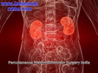
Percutaneous nephrolithotomy surgery india
- 2. The kidneys are a pair of vital organs that perform many functions to keep the blood clean and chemically balanced.
- 4. A kidney stone is a small stone, usually made up of calcium crystals, that forms inside the part of the kidney where urine collects. The stone usually causes little problem until it falls into the ureter, the tube that drains the kidney into the bladder, and causes an obstruction, preventing urine from draining out of the kidney and often causing severe pain.
- 5. Urolith: A stone anywhere within the urinary tract Nephrolith: A stone within the kidney Ureterolith: A stone within the ureter Calculus: A stone within the body
- 6. Changes in the acid-base balance (pH) of the urine, how concentrated it is, and the concentration of minerals and chemicals within the urine are all factors that can begin the formation of a stone.
- 7. In Percutaneous Nephrolithotomy or nephrolithotripsy, the surgeon makes a small incision in your back to remove kidney stones.
- 8. India offers very low Cost Percutaneous Nephrolithotomy Surgery compared to Western countries Like UK, USA
- 9. Write Us on at :Call: + 91 9029304141 (10 am. To 8 pm. IST) Email : info@indiasurgerytour.com (Preferred) Go to Site:- www.indiasurgerytour.com Like us on Facebook:- www.facebook.com/Indiasurgerytour (Only for international patients seeking treatment in India)