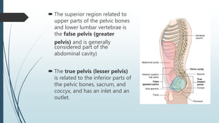
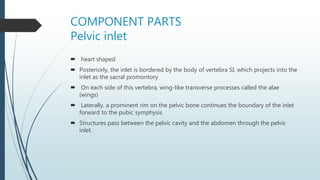
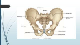
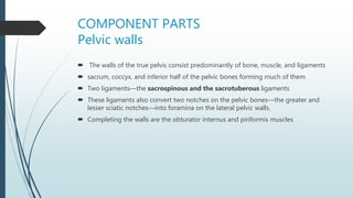
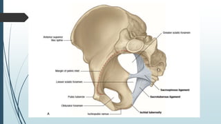
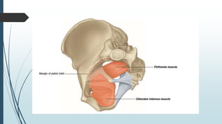
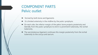
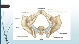
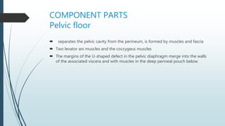
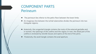
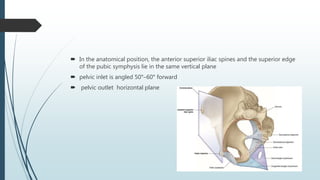
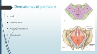
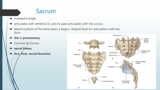
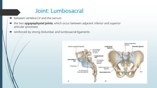
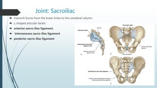
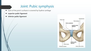
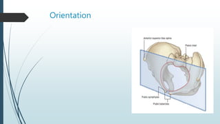
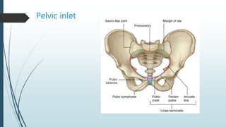
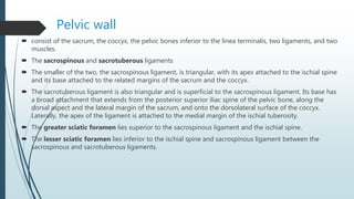
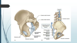
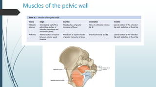
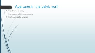
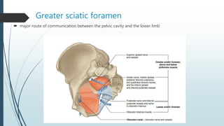
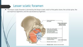
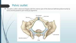
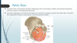
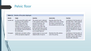
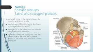
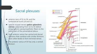
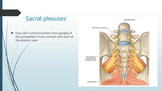
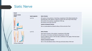
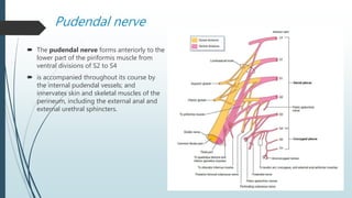
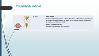
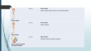
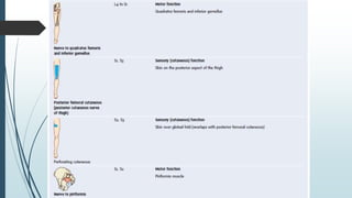
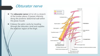
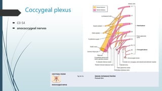
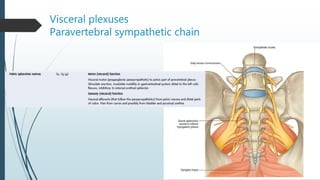
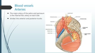
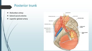
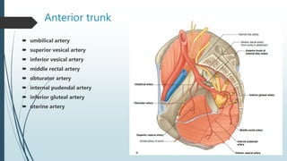
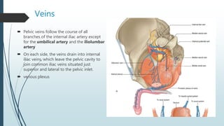
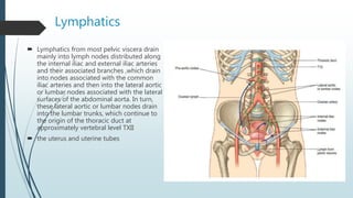
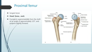
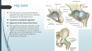
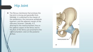
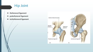
Pelvic & Hip Anatomy
Dr. Mohammad Mahdi Shater
 The superior region related to
upper parts of the pelvic bones
and lower lumbar vertebrae is
the false pelvis (greater
pelvis) and is generally
considered part of the
abdominal cavity)
 The true pelvis (lesser pelvis)
is related to the inferior parts of
the pelvic bones, sacrum, and
coccyx, and has an inlet and an
outlet.
COMPONENT PARTS
Pelvic inlet
 heart shaped
 Posteriorly, the inlet is bordered by the body of vertebra SI, which projects into the
inlet as the sacral promontory
 On each side of this vertebra, wing-like transverse processes called the alae
(wings)
 Laterally, a prominent rim on the pelvic bone continues the boundary of the inlet
forward to the pubic symphysis
 Structures pass between the pelvic cavity and the abdomen through the pelvic
inlet.
COMPONENT PARTS
Pelvic walls
 The walls of the true pelvis consist predominantly of bone, muscle, and ligaments
 sacrum, coccyx, and inferior half of the pelvic bones forming much of them
 Two ligaments—the sacrospinous and the sacrotuberous ligaments
 These ligaments also convert two notches on the pelvic bones—the greater and
lesser sciatic notches—into foramina on the lateral pelvic walls.
 Completing the walls are the obturator internus and piriformis muscles
COMPONENT PARTS
Pelvic outlet
 formed by both bone and ligaments
 It is limited anteriorly in the midline by the pubic symphysis.
 On each side, the inferior margin of the pelvic bone projects posteriorly and
laterally from the pubic symphysis to end in a prominent tuberosity, the ischial
tuberosity.
 The sacrotuberous ligament continues this margin posteriorly from the ischial
tuberosity to the coccyx and sacrum.
COMPONENT PARTS
Pelvic floor
 separates the pelvic cavity from the perineum, is formed by muscles and fascia
 Two levator ani muscles and the coccygeus muscles
 The margins of the U-shaped defect in the pelvic diaphragm merge into the walls
of the associated viscera and with muscles in the deep perineal pouch below.
COMPONENT PARTS
Perineum
 The perineum lies inferior to the pelvic floor between the lower limbs
 An imaginary line between the ischial tuberosities divides the perineum into two
triangular regions.
 Anteriorly, the urogenital triangle contains the roots of the external genitalia and,
in women, the openings of the urethra and the vagina. In men, the distal part of the
urethra is enclosed by erectile tissues and opens at the end of the penis.
 Posteriorly, the anal triangle contains the anal aperture.
 In the anatomical position, the anterior superior iliac spines and the superior edge
of the pubic symphysis lie in the same vertical plane
 pelvic inlet is angled 50°–60° forward
 pelvic outlet horizontal plane
Dermatomes of perineum
 Level
 Pudendal Nerve
 Parasympathetic Nerve
 Perineal body
Pelvic Bones
 The bones of the pelvis consist of the right and left pelvic (hip) bones, the sacrum,
and the coccyx.
 The sacrum articulates superiorly with vertebra LV at the lumbosacral joint.
 The pelvic bones articulate posteriorly with the sacrum at the sacro-iliac joints and
with each other anteriorly at the pubic symphysis.
Pelvic Bones
 irregular in shape and has two major parts separated by an oblique line on the
medial surface of the bone
 The linea terminalis is the lower two-thirds of this line
 The lateral surface of the pelvic bone has a large articular socket, the acetabulum,
which, together with the head of the femur, forms the hip joint
 Inferior to the acetabulum is the large obturator foramen, most of which is closed
by a flat connective tissue membrane, the obturator membrane
 obturator canal
 The posterior margin of the bone is marked by two notches separated by the
ischial spine: the greater sciatic notch, and the lesser sciatic notch.
 The posterior margin: ischial tuberosity
 The irregular anterior margin of the pelvic bone is marked by the anterior superior
iliac spine, the anterior inferior iliac spine, and the pubic tubercle.
Pelvic Bones
Ilium
 The bones of the pelvis consist of the right and left pelvic (hip)
bones, the sacrum, and the coccyx.
 The sacrum articulates superiorly with vertebra LV at the
lumbosacral joint.
 The pelvic bones articulate posteriorly with the sacrum at the
sacro-iliac joints and with each other anteriorly at the pubic
symphysis.
Ilium
 The ilium is separated into upper and lower parts by a ridge on the medial surface
 Anteriorly, arcuate line.
 Posteriorly, large L-shaped facet
Ilium
 The ilium is separated into upper and lower parts by a ridge on the medial surface
 Anteriorly, arcuate line.
 Posteriorly, large L-shaped facet
 Iliac fossa, iliac crest, tuberculum of the iliac crest
 anterior inferior iliac spine rectus femoris &
iliofemoral ligament
 Inf.Ant,Post Gluteal Line
Pubis
 The anterior and inferior part of the pelvic bone is the pubis. It has a body and two
arms (rami).
 pubic symphysis
 pubic tubercle
 Sup,inf rami
 pecten pubis (pectineal line)
Ischium
 The ischium is the posterior and inferior part of the pelvic bone
 Ramus of ischium
 Ischial spine
 Tuberosity
Ischium
Sacrum
 inverted triangle
 articulates with vertebra LV, and its apex articulates with the coccyx.
 lateral surfaces of the bone bears a large L-shaped facet for articulation with the
ilium
 Ala & promontory
 Concave & Convex
 sacral hiatus
 Ant, Post, sacral foramina
Coccyx
 consists of four fused coccygeal vertebrae
 inverted triangle
 two horns, or cornua
 small rudimentary transverse process
Joint: Lumbosacral
 between vertebra LV and the sacrum
 the two zygapophysial joints, which occur between adjacent inferior and superior
articular processes
 reinforced by strong iliolumbar and lumbosacral ligaments
Joint: Sacroiliac
 transmit forces from the lower limbs to the vertebral column.
 L-shaped articular facets
 anterior sacro-iliac ligament,
 interosseous sacro-iliac ligament
 posterior sacro-iliac ligament
Joint: Pubic symphysis
 Each of the joint’s surfaces is covered by hyaline cartilage
 superior pubic ligament
 inferior pubic ligament
Orientation
Differences between men and women
True pelvis
 The true pelvis is cylindrical and has an inlet, a wall, and an outlet. The inlet is open, whereas the pelvic
floor closes the outlet and separates the pelvic cavity, above, from the perineum, below.
Pelvic inlet
Pelvic wall
 consist of the sacrum, the coccyx, the pelvic bones inferior to the linea terminalis, two ligaments, and two
muscles.
 The sacrospinous and sacrotuberous ligaments
 The smaller of the two, the sacrospinous ligament, is triangular, with its apex attached to the ischial spine
and its base attached to the related margins of the sacrum and the coccyx.
 The sacrotuberous ligament is also triangular and is superficial to the sacrospinous ligament. Its base has
a broad attachment that extends from the posterior superior iliac spine of the pelvic bone, along the
dorsal aspect and the lateral margin of the sacrum, and onto the dorsolateral surface of the coccyx.
Laterally, the apex of the ligament is attached to the medial margin of the ischial tuberosity.
 The greater sciatic foramen lies superior to the sacrospinous ligament and the ischial spine.
 The lesser sciatic foramen lies inferior to the ischial spine and sacrospinous ligament between the
sacrospinous and sacrotuberous ligaments.
Muscles of the pelvic wall
Apertures in the pelvic wall
 the obturator canal,
 the greater sciatic foramen, and
 the lesser sciatic foramen.
Greater sciatic foramen
 major route of communication between the pelvic cavity and the lower limb
Lesser sciatic foramen
 The lesser sciatic foramen is formed by the lesser sciatic notch of the pelvic bone, the ischial spine, the
sacrospinous ligament, and the sacrotuberous ligament
Pelvic outlet
 The pelvic outlet is diamond shaped, with the anterior part of the diamond defined predominantly by
bone and the posterior part mainly by ligaments
Pelvic floor
 The pelvic floor is formed by the pelvic diaphragm and, in the anterior midline, the perineal membrane
and the muscles in the deep perineal pouch.
 The pelvic diaphragm is formed by the levator ani and the coccygeus muscles from both sides. The pelvic
floor separates the pelvic cavity, above, from the perineum, below
Pelvic floor
Nerves
Somatic plexuses
Sacral and coccygeal plexuses
 generally occur in the plane between the
muscles and blood vessels.
 ventral rami of S1 to Co, with a significant
contribution from L4 and L5,
 innervation of the lower limb and muscles
of the pelvis and perineum.
 Cutaneous branches supply skin over the
medial side of the foot, the posterior
aspect of the lower limb, and most of the
perineum
Sacral plexuses
 anterior rami of S1 to S4, and the
lumbosacral trunk (L4 and L5)
 special visceral nerves (pelvic splanchnic
nerves) originating from S2 to S4 deliver
preganglionic parasympathetic fibers to the
pelvic part of the prevertebral plexus
 Each anterior ramus has ventral and dorsal
divisions that combine with similar divisions
from other levels to form terminal nerves
 The anterior ramus of S4 has only a ventral
division
Sacral plexuses
 Gray rami communicantes from ganglia of
the sympathetic trunk connect with each of
the anterior rami
Siatic Nerve
 the largest nerve of the body and carries
contributions from L4 to S3
 forms on the anterior surface of the
piriformis muscle and leaves the pelvic
cavity through the greater sciatic foramen
inferior to the piriformis
 two major branches, the common fibular
nerve (common peroneal nerve) and the
tibial nerve
Siatic Nerve
Pudendal nerve
 The pudendal nerve forms anteriorly to the
lower part of the piriformis muscle from
ventral divisions of S2 to S4
 is accompanied throughout its course by
the internal pudendal vessels; and
innervates skin and skeletal muscles of the
perineum, including the external anal and
external urethral sphincters.
Pudendal nerve
Obturator nerve
 The obturator nerve (L2 to L4) is a branch
of the lumbar plexus. It passes inferiorly
along the posterior abdominal wall within
the psoas muscle
 It leaves the pelvic cavity by traveling
through the obturator canal and supplies
the adductor region of the thigh.
Coccygeal plexus
 C0-S4
 anococcygeal nerves
Visceral plexuses
Paravertebral sympathetic chain
Blood vessels
Arteries
 The major artery of the pelvis and perineum
is the internal iliac artery on each side
 divides into anterior and posterior trunks
Posterior trunk
 iliolumbar artery
 lateral sacral arteries
 superior gluteal artery
Anterior trunk
 umbilical artery
 superior vesical artery
 inferior vesical artery
 middle rectal artery
 obturator artery
 internal pudendal artery
 inferior gluteal artery
 uterine artery
Veins
 Pelvic veins follow the course of all
branches of the internal iliac artery except
for the umbilical artery and the iliolumbar
artery
 On each side, the veins drain into internal
iliac veins, which leave the pelvic cavity to
join common iliac veins situated just
superior and lateral to the pelvic inlet.
 venous plexus
Lymphatics
 Lymphatics from most pelvic viscera drain
mainly into lymph nodes distributed along
the internal iliac and external iliac arteries
and their associated branches ,which drain
into nodes associated with the common
iliac arteries and then into the lateral aortic
or lumbar nodes associated with the lateral
surfaces of the abdominal aorta. In turn,
these lateral aortic or lumbar nodes drain
into the lumbar trunks, which continue to
the origin of the thoracic duct at
approximately vertebral level TXII
 the uterus and uterine tubes
Dermatomes
 Flexion of the hip is controlled primarily
by L1 and L2
 over the inguinal ligament—L1
 skin over the gluteal fold—S3
Acetabulum
 The large cup-shaped acetabulum for
articulation with the head of the femur is on
the lateral surface of the pelvic bone in the
region where the ilium, pubis, and ischium
fuse
 The margin of the acetabulum is marked
inferiorly by a prominent notch (acetabular
notch).
 The wall of the acetabulum consists of
nonarticular and articular parts
 acetabular fossa
 lunate surface
 The acetabular fossa provides attachment
for the ligament of the head of the femur,
whereas blood vessels and nerves pass
through the acetabular notch.
Proximal femur
 longest bone
 Head, fovea , neck
 It projects superomedially from the shaft
at an angle of approximately 125°, and
projects slightly forward.
Hip Joint
 The hip joint is a synovial articulation
between the head of the femur and the
acetabulum of the pelvic bone
 transverse acetabular ligament
 ligament of the head of the femur
 The synovial membrane attaches to the
margins of the articular surfaces of the
femur and acetabulum, forms a tubular
covering around the ligament of the head
of the femur, and lines the fibrous
membrane of the joint
Hip Joint
 The fibrous membrane that encloses the
hip joint is strong and generally thick.
Medially, it is attached to the margin of
the acetabulum, the transverse acetabular
ligament, and the adjacent margin of the
obturator foramen .Laterally, it is
attached to the intertrochanteric line on
the anterior aspect of the femur and to
the neck of the femur just proximal to the
intertrochanteric crest on the posterior
surface.
Hip Joint
 iliofemoral ligament
 pubofemoral ligament
 ischiofemoral ligament
Hip Joint
 Vascular supply to the hip joint is
predominantly through branches of the
obturator artery, medial and lateral
circumflex femoral arteries, superior and
inferior gluteal arteries, and the first
perforating branch of the deep artery of
the thigh. The articular branches of these
vessels form a network around the joint
 The hip joint is innervated by articular
branches from the femoral, obturator,
and superior gluteal nerves, and the
nerve to the quadratus femoris
GLUTEAL REGION
 The gluteal region lies posterolateral to the bony pelvis and proximal end of the femur Muscles in
the region mainly abduct, extend, and laterally rotate the femur relative to the pelvic bone
 a deep group of small muscles, which are mainly lateral rotators of the femur at the hip joint and
include the piriformis, obturator internus, gemellus superior, gemellus inferior, and quadratus
femoris;
 a more superficial group of larger muscles, which mainly abduct and extend the hip and include
the gluteus minimus, gluteus medius, and gluteus maximus; an additional muscle in this group,
the tensor fasciae latae, stabilizes the knee in extension by acting on a specialized longitudinal
band of deep fascia (the iliotibial tract) that passes down the lateral side of the thigh to attach to
the proximal end of the tibia in the leg
GLUTEAL REGION
GLUTEAL REGION

Pelvic and hip anatomy

  • 2.
    Pelvic & HipAnatomy Dr. Mohammad Mahdi Shater
  • 3.
     The superiorregion related to upper parts of the pelvic bones and lower lumbar vertebrae is the false pelvis (greater pelvis) and is generally considered part of the abdominal cavity)  The true pelvis (lesser pelvis) is related to the inferior parts of the pelvic bones, sacrum, and coccyx, and has an inlet and an outlet.
  • 4.
    COMPONENT PARTS Pelvic inlet heart shaped  Posteriorly, the inlet is bordered by the body of vertebra SI, which projects into the inlet as the sacral promontory  On each side of this vertebra, wing-like transverse processes called the alae (wings)  Laterally, a prominent rim on the pelvic bone continues the boundary of the inlet forward to the pubic symphysis  Structures pass between the pelvic cavity and the abdomen through the pelvic inlet.
  • 6.
    COMPONENT PARTS Pelvic walls The walls of the true pelvis consist predominantly of bone, muscle, and ligaments  sacrum, coccyx, and inferior half of the pelvic bones forming much of them  Two ligaments—the sacrospinous and the sacrotuberous ligaments  These ligaments also convert two notches on the pelvic bones—the greater and lesser sciatic notches—into foramina on the lateral pelvic walls.  Completing the walls are the obturator internus and piriformis muscles
  • 9.
    COMPONENT PARTS Pelvic outlet formed by both bone and ligaments  It is limited anteriorly in the midline by the pubic symphysis.  On each side, the inferior margin of the pelvic bone projects posteriorly and laterally from the pubic symphysis to end in a prominent tuberosity, the ischial tuberosity.  The sacrotuberous ligament continues this margin posteriorly from the ischial tuberosity to the coccyx and sacrum.
  • 11.
    COMPONENT PARTS Pelvic floor separates the pelvic cavity from the perineum, is formed by muscles and fascia  Two levator ani muscles and the coccygeus muscles  The margins of the U-shaped defect in the pelvic diaphragm merge into the walls of the associated viscera and with muscles in the deep perineal pouch below.
  • 12.
    COMPONENT PARTS Perineum  Theperineum lies inferior to the pelvic floor between the lower limbs  An imaginary line between the ischial tuberosities divides the perineum into two triangular regions.  Anteriorly, the urogenital triangle contains the roots of the external genitalia and, in women, the openings of the urethra and the vagina. In men, the distal part of the urethra is enclosed by erectile tissues and opens at the end of the penis.  Posteriorly, the anal triangle contains the anal aperture.
  • 13.
     In theanatomical position, the anterior superior iliac spines and the superior edge of the pubic symphysis lie in the same vertical plane  pelvic inlet is angled 50°–60° forward  pelvic outlet horizontal plane
  • 14.
    Dermatomes of perineum Level  Pudendal Nerve  Parasympathetic Nerve  Perineal body
  • 15.
    Pelvic Bones  Thebones of the pelvis consist of the right and left pelvic (hip) bones, the sacrum, and the coccyx.  The sacrum articulates superiorly with vertebra LV at the lumbosacral joint.  The pelvic bones articulate posteriorly with the sacrum at the sacro-iliac joints and with each other anteriorly at the pubic symphysis.
  • 16.
    Pelvic Bones  irregularin shape and has two major parts separated by an oblique line on the medial surface of the bone  The linea terminalis is the lower two-thirds of this line  The lateral surface of the pelvic bone has a large articular socket, the acetabulum, which, together with the head of the femur, forms the hip joint  Inferior to the acetabulum is the large obturator foramen, most of which is closed by a flat connective tissue membrane, the obturator membrane  obturator canal  The posterior margin of the bone is marked by two notches separated by the ischial spine: the greater sciatic notch, and the lesser sciatic notch.  The posterior margin: ischial tuberosity  The irregular anterior margin of the pelvic bone is marked by the anterior superior iliac spine, the anterior inferior iliac spine, and the pubic tubercle.
  • 17.
  • 18.
    Ilium  The bonesof the pelvis consist of the right and left pelvic (hip) bones, the sacrum, and the coccyx.  The sacrum articulates superiorly with vertebra LV at the lumbosacral joint.  The pelvic bones articulate posteriorly with the sacrum at the sacro-iliac joints and with each other anteriorly at the pubic symphysis.
  • 19.
    Ilium  The iliumis separated into upper and lower parts by a ridge on the medial surface  Anteriorly, arcuate line.  Posteriorly, large L-shaped facet
  • 20.
    Ilium  The iliumis separated into upper and lower parts by a ridge on the medial surface  Anteriorly, arcuate line.  Posteriorly, large L-shaped facet  Iliac fossa, iliac crest, tuberculum of the iliac crest  anterior inferior iliac spine rectus femoris & iliofemoral ligament  Inf.Ant,Post Gluteal Line
  • 21.
    Ilium  The iliumis separated into upper and lower parts by a ridge on the medial surface  Anteriorly, arcuate line.  Posteriorly, large L-shaped facet  Iliac fossa, iliac crest, tuberculum of the iliac crest  anterior inferior iliac spine rectus femoris & iliofemoral ligament  Inf.Ant,Post Gluteal Line
  • 22.
    Pubis  The anteriorand inferior part of the pelvic bone is the pubis. It has a body and two arms (rami).  pubic symphysis  pubic tubercle  Sup,inf rami  pecten pubis (pectineal line)
  • 23.
    Ischium  The ischiumis the posterior and inferior part of the pelvic bone  Ramus of ischium  Ischial spine  Tuberosity
  • 24.
  • 25.
    Sacrum  inverted triangle articulates with vertebra LV, and its apex articulates with the coccyx.  lateral surfaces of the bone bears a large L-shaped facet for articulation with the ilium  Ala & promontory  Concave & Convex  sacral hiatus  Ant, Post, sacral foramina
  • 26.
    Coccyx  consists offour fused coccygeal vertebrae  inverted triangle  two horns, or cornua  small rudimentary transverse process
  • 27.
    Joint: Lumbosacral  betweenvertebra LV and the sacrum  the two zygapophysial joints, which occur between adjacent inferior and superior articular processes  reinforced by strong iliolumbar and lumbosacral ligaments
  • 28.
    Joint: Sacroiliac  transmitforces from the lower limbs to the vertebral column.  L-shaped articular facets  anterior sacro-iliac ligament,  interosseous sacro-iliac ligament  posterior sacro-iliac ligament
  • 29.
    Joint: Pubic symphysis Each of the joint’s surfaces is covered by hyaline cartilage  superior pubic ligament  inferior pubic ligament
  • 30.
  • 31.
  • 32.
    True pelvis  Thetrue pelvis is cylindrical and has an inlet, a wall, and an outlet. The inlet is open, whereas the pelvic floor closes the outlet and separates the pelvic cavity, above, from the perineum, below.
  • 33.
  • 34.
    Pelvic wall  consistof the sacrum, the coccyx, the pelvic bones inferior to the linea terminalis, two ligaments, and two muscles.  The sacrospinous and sacrotuberous ligaments  The smaller of the two, the sacrospinous ligament, is triangular, with its apex attached to the ischial spine and its base attached to the related margins of the sacrum and the coccyx.  The sacrotuberous ligament is also triangular and is superficial to the sacrospinous ligament. Its base has a broad attachment that extends from the posterior superior iliac spine of the pelvic bone, along the dorsal aspect and the lateral margin of the sacrum, and onto the dorsolateral surface of the coccyx. Laterally, the apex of the ligament is attached to the medial margin of the ischial tuberosity.  The greater sciatic foramen lies superior to the sacrospinous ligament and the ischial spine.  The lesser sciatic foramen lies inferior to the ischial spine and sacrospinous ligament between the sacrospinous and sacrotuberous ligaments.
  • 36.
    Muscles of thepelvic wall
  • 37.
    Apertures in thepelvic wall  the obturator canal,  the greater sciatic foramen, and  the lesser sciatic foramen.
  • 38.
    Greater sciatic foramen major route of communication between the pelvic cavity and the lower limb
  • 39.
    Lesser sciatic foramen The lesser sciatic foramen is formed by the lesser sciatic notch of the pelvic bone, the ischial spine, the sacrospinous ligament, and the sacrotuberous ligament
  • 40.
    Pelvic outlet  Thepelvic outlet is diamond shaped, with the anterior part of the diamond defined predominantly by bone and the posterior part mainly by ligaments
  • 41.
    Pelvic floor  Thepelvic floor is formed by the pelvic diaphragm and, in the anterior midline, the perineal membrane and the muscles in the deep perineal pouch.  The pelvic diaphragm is formed by the levator ani and the coccygeus muscles from both sides. The pelvic floor separates the pelvic cavity, above, from the perineum, below
  • 42.
  • 43.
    Nerves Somatic plexuses Sacral andcoccygeal plexuses  generally occur in the plane between the muscles and blood vessels.  ventral rami of S1 to Co, with a significant contribution from L4 and L5,  innervation of the lower limb and muscles of the pelvis and perineum.  Cutaneous branches supply skin over the medial side of the foot, the posterior aspect of the lower limb, and most of the perineum
  • 44.
    Sacral plexuses  anteriorrami of S1 to S4, and the lumbosacral trunk (L4 and L5)  special visceral nerves (pelvic splanchnic nerves) originating from S2 to S4 deliver preganglionic parasympathetic fibers to the pelvic part of the prevertebral plexus  Each anterior ramus has ventral and dorsal divisions that combine with similar divisions from other levels to form terminal nerves  The anterior ramus of S4 has only a ventral division
  • 45.
    Sacral plexuses  Grayrami communicantes from ganglia of the sympathetic trunk connect with each of the anterior rami
  • 46.
    Siatic Nerve  thelargest nerve of the body and carries contributions from L4 to S3  forms on the anterior surface of the piriformis muscle and leaves the pelvic cavity through the greater sciatic foramen inferior to the piriformis  two major branches, the common fibular nerve (common peroneal nerve) and the tibial nerve
  • 47.
  • 48.
    Pudendal nerve  Thepudendal nerve forms anteriorly to the lower part of the piriformis muscle from ventral divisions of S2 to S4  is accompanied throughout its course by the internal pudendal vessels; and innervates skin and skeletal muscles of the perineum, including the external anal and external urethral sphincters.
  • 49.
  • 52.
    Obturator nerve  Theobturator nerve (L2 to L4) is a branch of the lumbar plexus. It passes inferiorly along the posterior abdominal wall within the psoas muscle  It leaves the pelvic cavity by traveling through the obturator canal and supplies the adductor region of the thigh.
  • 53.
    Coccygeal plexus  C0-S4 anococcygeal nerves
  • 54.
  • 55.
    Blood vessels Arteries  Themajor artery of the pelvis and perineum is the internal iliac artery on each side  divides into anterior and posterior trunks
  • 56.
    Posterior trunk  iliolumbarartery  lateral sacral arteries  superior gluteal artery
  • 57.
    Anterior trunk  umbilicalartery  superior vesical artery  inferior vesical artery  middle rectal artery  obturator artery  internal pudendal artery  inferior gluteal artery  uterine artery
  • 58.
    Veins  Pelvic veinsfollow the course of all branches of the internal iliac artery except for the umbilical artery and the iliolumbar artery  On each side, the veins drain into internal iliac veins, which leave the pelvic cavity to join common iliac veins situated just superior and lateral to the pelvic inlet.  venous plexus
  • 59.
    Lymphatics  Lymphatics frommost pelvic viscera drain mainly into lymph nodes distributed along the internal iliac and external iliac arteries and their associated branches ,which drain into nodes associated with the common iliac arteries and then into the lateral aortic or lumbar nodes associated with the lateral surfaces of the abdominal aorta. In turn, these lateral aortic or lumbar nodes drain into the lumbar trunks, which continue to the origin of the thoracic duct at approximately vertebral level TXII  the uterus and uterine tubes
  • 60.
    Dermatomes  Flexion ofthe hip is controlled primarily by L1 and L2  over the inguinal ligament—L1  skin over the gluteal fold—S3
  • 61.
    Acetabulum  The largecup-shaped acetabulum for articulation with the head of the femur is on the lateral surface of the pelvic bone in the region where the ilium, pubis, and ischium fuse  The margin of the acetabulum is marked inferiorly by a prominent notch (acetabular notch).  The wall of the acetabulum consists of nonarticular and articular parts  acetabular fossa  lunate surface  The acetabular fossa provides attachment for the ligament of the head of the femur, whereas blood vessels and nerves pass through the acetabular notch.
  • 62.
    Proximal femur  longestbone  Head, fovea , neck  It projects superomedially from the shaft at an angle of approximately 125°, and projects slightly forward.
  • 63.
    Hip Joint  Thehip joint is a synovial articulation between the head of the femur and the acetabulum of the pelvic bone  transverse acetabular ligament  ligament of the head of the femur  The synovial membrane attaches to the margins of the articular surfaces of the femur and acetabulum, forms a tubular covering around the ligament of the head of the femur, and lines the fibrous membrane of the joint
  • 64.
    Hip Joint  Thefibrous membrane that encloses the hip joint is strong and generally thick. Medially, it is attached to the margin of the acetabulum, the transverse acetabular ligament, and the adjacent margin of the obturator foramen .Laterally, it is attached to the intertrochanteric line on the anterior aspect of the femur and to the neck of the femur just proximal to the intertrochanteric crest on the posterior surface.
  • 65.
    Hip Joint  Thefibrous membrane that encloses the hip joint is strong and generally thick. Medially, it is attached to the margin of the acetabulum, the transverse acetabular ligament, and the adjacent margin of the obturator foramen .Laterally, it is attached to the intertrochanteric line on the anterior aspect of the femur and to the neck of the femur just proximal to the intertrochanteric crest on the posterior surface.
  • 66.
    Hip Joint  iliofemoralligament  pubofemoral ligament  ischiofemoral ligament
  • 67.
    Hip Joint  Vascularsupply to the hip joint is predominantly through branches of the obturator artery, medial and lateral circumflex femoral arteries, superior and inferior gluteal arteries, and the first perforating branch of the deep artery of the thigh. The articular branches of these vessels form a network around the joint  The hip joint is innervated by articular branches from the femoral, obturator, and superior gluteal nerves, and the nerve to the quadratus femoris
  • 68.
    GLUTEAL REGION  Thegluteal region lies posterolateral to the bony pelvis and proximal end of the femur Muscles in the region mainly abduct, extend, and laterally rotate the femur relative to the pelvic bone  a deep group of small muscles, which are mainly lateral rotators of the femur at the hip joint and include the piriformis, obturator internus, gemellus superior, gemellus inferior, and quadratus femoris;  a more superficial group of larger muscles, which mainly abduct and extend the hip and include the gluteus minimus, gluteus medius, and gluteus maximus; an additional muscle in this group, the tensor fasciae latae, stabilizes the knee in extension by acting on a specialized longitudinal band of deep fascia (the iliotibial tract) that passes down the lateral side of the thigh to attach to the proximal end of the tibia in the leg
  • 69.
    GLUTEAL REGION  Thegluteal region lies posterolateral to the bony pelvis and proximal end of the femur Muscles in the region mainly abduct, extend, and laterally rotate the femur relative to the pelvic bone  a deep group of small muscles, which are mainly lateral rotators of the femur at the hip joint and include the piriformis, obturator internus, gemellus superior, gemellus inferior, and quadratus femoris;  a more superficial group of larger muscles, which mainly abduct and extend the hip and include the gluteus minimus, gluteus medius, and gluteus maximus; an additional muscle in this group, the tensor fasciae latae, stabilizes the knee in extension by acting on a specialized longitudinal band of deep fascia (the iliotibial tract) that passes down the lateral side of the thigh to attach to the proximal end of the tibia in the leg
  • 70.
  • 71.