Downloaded 286 times


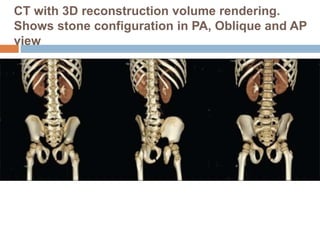


This document provides an overview of pediatric urolithiasis (urinary stones in children). Some key points: - Infection and metabolic abnormalities are major causes of stones in children. - Metabolic evaluation is essential for every child to identify underlying conditions. - Stones are most commonly calcium-containing, struvite, or uric acid. - Common metabolic abnormalities include hypercalciuria, hyperoxaluria, cystinuria, and hypocitraturia. - Treatment involves managing the underlying condition, increasing fluid intake, and sometimes surgery or lithotripsy as in adults. Thorough evaluation is needed to identify causes and guide management.


