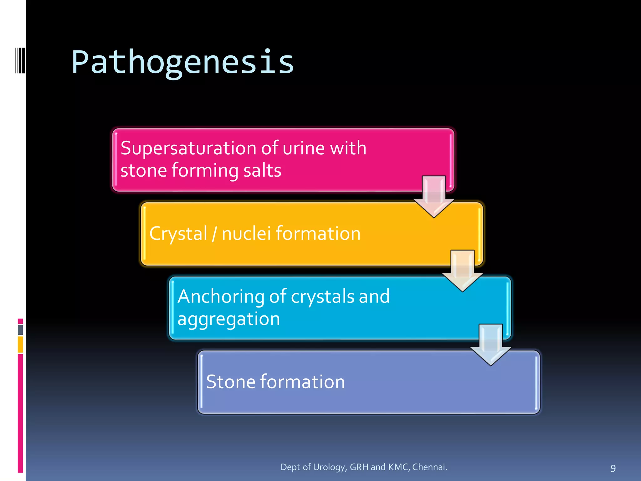
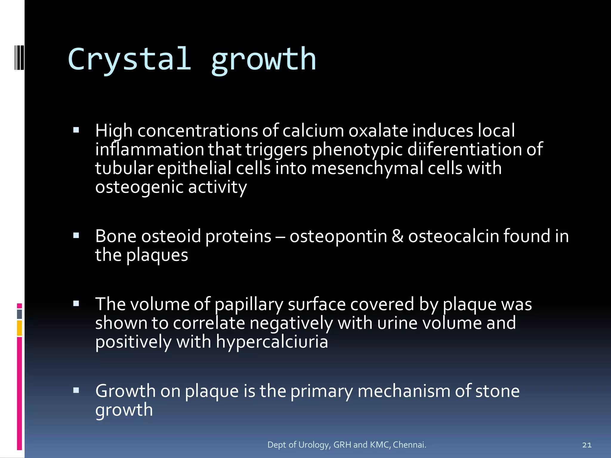
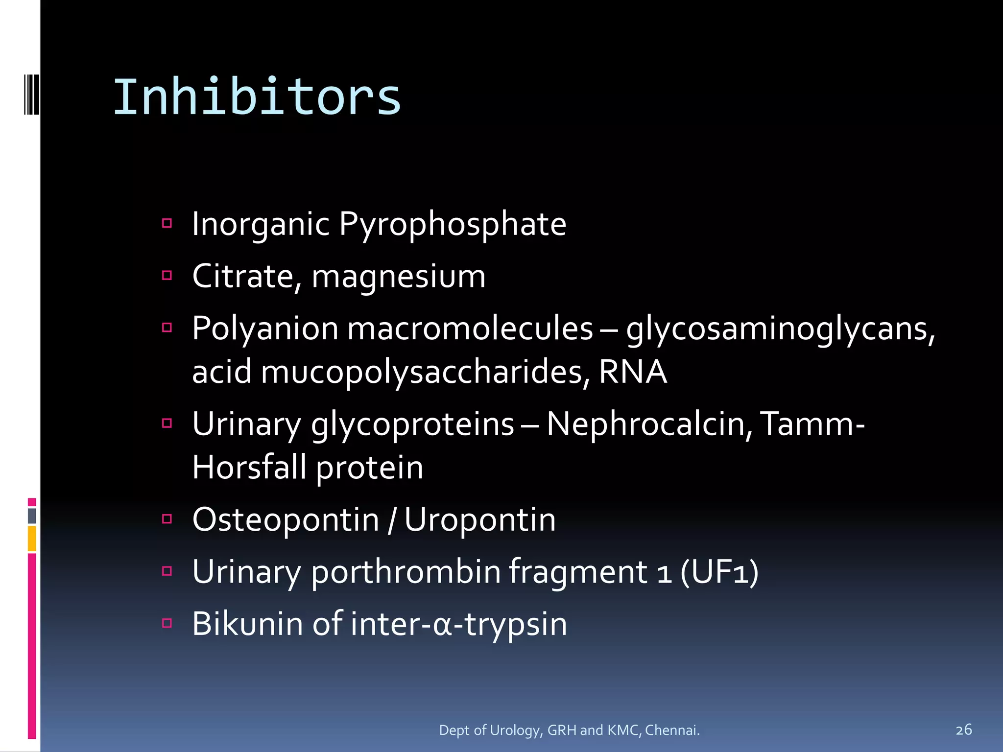
SlideShare a Scribd company logo
ETIOPATHOGENESIS OF
UROLITHIASIS
Dept of Urology
Govt RoyapettahHospital and KilpaukMedicalCollege
Chennai
Moderators:
Professors:
 Prof. Dr. G. Sivasankar, M.S., M.Ch.,
 Prof. Dr. A. Senthilvel, M.S., M.Ch.,
Asst Professors:
 Dr. J. Sivabalan, M.S., M.Ch.,
 Dr. R. Bhargavi, M.S., M.Ch.,
 Dr. S. Raju, M.S., M.Ch.,
 Dr. K. Muthurathinam, M.S., M.Ch.,
 Dr. D.Tamilselvan, M.S., M.Ch.,
 Dr. K. Senthilkumar, M.S., M.Ch.
Dept of Urology, GRH and KMC,Chennai. 2
Epidemiology
 Site – migrated from lower to upper urinary
tract
 Gender gap narrowing
 Rise in incidence – rise in the detection of
asymptomaticcalculithrough increased
radiographic imaging
 The incidence of symptomatic stones did not
increase significantly
3
Dept of Urology, GRH and KMC,Chennai.
Gender & Age
 Uncommon before 20 years
 Peak – 4th to 6th decade
 Women – bimodaldistribution
 30-39years & 60-69 years
 Second peak corresponding to the onset of
menopause
 Estrogen influence
 Protective against stone formation
 Enhanced renal calcium absorption & reduced
bone resorption
4
Dept of Urology, GRH and KMC,Chennai.
GEOGRAPHY
 Geography - hot, arid, dry climates
 Climate & Seasonal variation
 Summer (July to September)
 High temperature & Sunlight induced increase in
vitamin D
 Peak occuring within 1 to 2 months of maximal
mean temperature.
 Temperature dependence – primarily attributed to
effect on men likely due to differential sunlight
exposure, occupation, and hydration status.
5
Dept of Urology, GRH and KMC,Chennai.
Occupation
 Risk factors - Heat exposure & Dehydration
 Occupation – Cooks, Engineering room personnel,
Steel workers, Glass plant workers
 Exposure to higher temperature Higher incidence
of low urine volume, low pH, higher uric acid levels,
higher urine specific gravity and hypocitraturia 
higher urinary saturation of uric acid. (High incidence
of uric acid stones)
 Sedentary occupations – increased risk reflective of
more indulgent diet and lifestyle
6
Dept of Urology, GRH and KMC,Chennai.
Diet & Metabolic association
 Decreased risk
 High fluid intake
 Low protein intake
 Increased risk
 Obesity
 Metabolic syndrome (hyperlipidemia,
hypertriglyceridemia,hyperglycemia, and/or
hypertension)
 Type 2 Diabetes mellitus – higher urinary oxalate
and lower urine pH
7
Dept of Urology, GRH and KMC,Chennai.
 Exact pathophysiologicmechanism – not completely
elucidated.
 Most probably – Metabolic state of Insulin resistance
 Higher BMI
 Excreted more urinary oxalate, uric acid, sodiumand
phosphorous.
 Urinary supersaturationof uric acid inreased with BMI
 Association of obesity & calcium oxalate - primarily
due to increased excretion of promoters of stone
formation.
 Association of obesity & Uric acid – primarily
influenced by urinary pH
8
Dept of Urology, GRH and KMC,Chennai.
Pathogenesis
Supersaturation of urine with
stone forming salts
Crystal / nuclei formation
Anchoring of crystals and
aggregation
Stone formation
9
Dept of Urology, GRH and KMC,Chennai.
State of Saturation
 Saturated solution
 Pure aqueous solution when it reaches the point at
which no further added salt crystals will dissolve
 Solubility Product
 Concentration product at the point of saturation
 The point at which dissolved and crystalline
components are in equilibrium for a specific set of
conditions.
 Formation product
 As the concentration product increase, the point at
which the solution can be no longer be held in solution
and crystals form
10
Dept of Urology, GRH and KMC,Chennai.
States Of Saturation In
Urine
 Undersaturated
 Below the solubility product – crystal will not form
 Metastable
 Between the solubility product & formation product
 Spontaneous nucleation or precipitation does not
occur despite urine is supersaturated
 It is in this area modulation of factors controlling stone
formation can take place and therapeutic intervention
is directed
 Unstable
 At concentrations above formation product – crystals
will form
11
Dept of Urology, GRH and KMC,Chennai.
States of Saturation
12
Dept of Urology, GRH and KMC,Chennai.
Nucleation & Crystal Growth,
Aggregation & Retention
13
Dept of Urology, GRH and KMC,Chennai.
Crystal Nucleation
 Homogenous Nucleation
 Nuclei form in pure solution
 Heterogenous Nucleation
 Nuclei formation by adsorption onto existing
surfaces of epithelial cells, cell debris or other
crystals
14
Dept of Urology, GRH and KMC,Chennai.
 Within the timeframe of transit of urine
throughthe nephron, estimated at 5 to 7
minutes, crystal cannot grow to reach a size
sufficient to occludethe tubularlumen
 In the presence of urinary inhibitors , calcium
oxalate precipitation occursonly when
supersaturation exceed solubility by 7 to 11
times
15
Dept of Urology, GRH and KMC,Chennai.
Stone growth Theories
 Free particle growth theory
 Recent studies – free crystalline particles can be
formed that are large enough to be retained during
normal transit time through the kidney
 Findings based on recalculation using current nephron
dimensions, supersaturation and crystal growth rates
 Fixed particle growth theory
 Anchoring site to which crystals bind  prolongs the
time the crystals are exposed to supersaturated urine
 crystal aggregation & growth
16
Dept of Urology, GRH and KMC,Chennai.
Crystal Fixation
 Oxalate induced injury to renal tubular epithelial
cells mediated by reactive oxygen species–
adherence of calcium oxalate crystals
 Role of antioxidants
 Neutralises cytotoxicity & allow retention of renal cell
integrity (N-acetyl cysteine)
 Lower levels of serum antioxidants (α-carotene, β-
carotene, β-cryptoxanthin) – increased risk
 Oxidative stress implicated in – diabetes, metabolic
syndrome, coronary heart disease
 Gender disparity – differential antioxidant production
associated with testosterone and estradiol
 Increase testosterone  increased oxidative stress &
stones
 Increased estradiol  suppressed both
17
Dept of Urology, GRH and KMC,Chennai.
 Crystal bind preferentially to regenerating /
redifferentiatingrenal tubularcells
 Mediated by – luminal membrane molecules
 Hyaluronic acid
 Osteopontin
 Annexin II
 Nucleolin related protein
18
Dept of Urology, GRH and KMC,Chennai.
Randall Plaques
 Randall (1937) first observed areas of damage
associated with subepithelial plaques on the
renal papillae
 Initiating event – vascular injury to the vasa
recta near the renal papilla
 Repair of damaged vessel wall 
atheroscleroticlike reaction  calcification of
endothelial wall  erosion into papillary
interstitiumand then into collectingducts 
serves as nidus for stone formation
19
Dept of Urology, GRH and KMC,Chennai.
Structure
 Crystal component – calciumapatite
 Deposits consisted of individual laminated
particles with mineral and organic layers
 All crystals were coated with organic material
 Osteopontin was identified on the outer
surface of the crystal at the junction of
overlying organic molecularlayer.
20
Dept of Urology, GRH and KMC,Chennai.
Crystal growth
 High concentrations of calcium oxalate induces local
inflammation that triggers phenotypic diiferentiation of
tubular epithelial cells into mesenchymal cells with
osteogenic activity
 Bone osteoid proteins – osteopontin & osteocalcin found in
the plaques
 The volume of papillary surface covered by plaque was
shown to correlate negatively with urine volume and
positively with hypercalciuria
 Growth on plaque is the primary mechanism of stone
growth
21
Dept of Urology, GRH and KMC,Chennai.
Calcifying Nanoparticle
(CNPs)
 Self propagating particles that precipitate
calciumapatite on their exterior membrane
 Have been detected in blood, blood
products,renal stones as well as in pathologic
calcifications
 Promote rapid precipitationof calcium
phosphate from bloodunder physiologically
unfavorableconditions
22
Dept of Urology, GRH and KMC,Chennai.
 Enteric hyperoxaluria
 No plaque
 Apatite crystal deposits plugging inner medullary collecting
duct lumens, along with associated epithelialcell damage
with interstitial inflammationand fibrosis
 Brushite stone formers
 Intermediate pathology
 Interstitial apatite plaque and apatite crystal plugging of
inner medullary and terminal collecting ducts along with
associatedcollecting duct injury and interstitial fibrosis.
 Distal RTA
 Extensive renal calcifications
 Papillary changes – minimal to papillae pitted and
contained calcium phosphate plugs protruding from dilated
collecting ducts with extensive surrounding fibrosis
 Randall plaques were rarely encountered
23
Dept of Urology, GRH and KMC,Chennai.
 Cystinuria
 Plugging of terminal collecting ducts of Bellini
with masses of cystine crystals; apatite deposits
were also identified in the inner medullary
collecting ducts and in the thin ascending limbs of
the loops of Henle
 Primary hyperparathyroidism
 Similar to brushite stone formers with interstitial
deposits of plaque and associated stone
overgrowth traditionally seen with idiopathic
calcium oxalate stone formers.
24
Dept of Urology, GRH and KMC,Chennai.
Inhibitors
 Molecules that raise the level of
supersaturation needed to initiate crystal
nucleationor reduce the rate of crystal
growth or aggregation & prevent stone
formation from occurringon a routine basis.
 No specific inhibitors for uric acid
crystallisation
25
Dept of Urology, GRH and KMC,Chennai.
Inhibitors
 Inorganic Pyrophosphate
 Citrate, magnesium
 Polyanion macromolecules – glycosaminoglycans,
acid mucopolysaccharides, RNA
 Urinary glycoproteins – Nephrocalcin,Tamm-
Horsfall protein
 Osteopontin / Uropontin
 Urinary porthrombin fragment 1 (UF1)
 Bikunin of inter-α-trypsin
26
Dept of Urology, GRH and KMC,Chennai.
Citrate
 Reduces the availability of ionic calciumto
interact with oxalate or phosphate
 Inhibits the spontaneous precipitation of
calciumoxalate
 Prevents crystal aggregation
 Prevents heterogenous nucleationof calcium
oxalate by monosodiumurate
27
Dept of Urology, GRH and KMC,Chennai.
Magnesium
 It form complexationwith oxalate  reduces
ionic oxalate concentration and calcium
oxalate supersaturation
 Reduces the contact time between calcium
and oxalate moleculesin vitro
 Pyrophosphate,citrate, magnesium – inhibit
crystal growth
 High concentration of magnesium &
pyrophosphate– inhibit aggregation
28
Dept of Urology, GRH and KMC,Chennai.
Polyanion macromolecules
 Bonds with surface calciumions and inhibits
crystal nucleation and growth
 Most prominent glycosaminoglycanin
human urine – chondroitinsulfate
 Heparin sulfate interacts most strongly with
calciumoxalate monohydratecrystals
29
Dept of Urology, GRH and KMC,Chennai.
Nephrocalcin
 Acidic glycoprotein containing predominantly acidic
amino acids that is synthesized in the proximal renal
tubules and thick ascending limb
 Inhibits growth , nucleation and aggregation
 Four isoforms
 Non stone formers – excrete greater quantities of 2
isoforms with most inhibitory activity
 Isoforms with inhibitory activity were found to
contain γ-carboxyglutamic acid residues that were
lacking in the isoforms of stone formers
30
Dept of Urology, GRH and KMC,Chennai.
Tamm Horsfall protein
 Expressed by renal epithelial cells in the thick
ascending limb and the distal convoluted tubule
as a membrane anchored protein
 Released into the urine after cleavage of the
anchoring site by phopholipases or proteases.
 Potent inhibitor of calcium oxalate monohydrate
crystal aggregation, but not growth.
 Function varies
 Alkaline urine – inhibitor
 Acidic urine – polymerizes into a configuration that
promotes crystal aggregation.
31
Dept of Urology, GRH and KMC,Chennai.
Osteopontin or Uropontin
 Acidic phosphorylatedglycoprotein
 Expressed in bone matrix and renal epithelial
cells of the ascending limb of loop of Henle
and the distal tubule.
 Inhibit nucleation,growth, and aggregation
of calcium oxalate crystals, as well as to
reduce binding of crystals to renal epithelial
cells in vitro
 May work in conjunction with constitutively
expressedTamm-Horsfallprotein
32
Dept of Urology, GRH and KMC,Chennai.
Urinary Prothrombin Fragment 1
(F1)
 Crystal matrix protein
 Named for its resemblance to the F1
degradation productof prothrombin
 Inhibits crystal aggregation and deposition
33
Dept of Urology, GRH and KMC,Chennai.
Bikunin
 Comprises the light chain of inter-α-trypsin, a
glycoproteinsynthesized in liver
 Inhibitor of calcium oxalatecrystallisation,
aggregation and growth
34
Dept of Urology, GRH and KMC,Chennai.
MATRIX
 The noncrystalline component of renal calculi
 Accounts for about 2.5% of the weight of the
stone.
 In some, matrix comprises the majority – usually
in association with chronic UTIs
 Heterogenous mixture
 Protein – 65%
 Non amino sugars – 9%
 Glucosamine – 5%
 Bound water – 10%
 Organic ash – 12%
35
Dept of Urology, GRH and KMC,Chennai.
 Proteins –Tamm-Horsfall, neprhocalcin, renal
lithostathine, albumin, glycosaminoglycans,
mucoprotein matrix substance A
 Substance A is immunologically unique and
present in the matrix component of all stone
formers
 Inflammatory proteins – immunoglobulins,
defensin-3, clusterin, complement C3a,
kininogen and fibrinogen; comprises the
predominant protein in calcium oxalate and
phosphate stones
 Exact role of matrix yet to be elucidatied.
36
Dept of Urology, GRH and KMC,Chennai.
Summary
 Urine must be supersaturated for stones to
form
 Supersaturationalone is insufficientowing to
the presence of urinary inhibitors
 Nucleation in stone formation is
heterogenous
 Subepithelial plaques serve as an anchor on
which calcium oxalate aggregate and stone
growth occurs
 Noncrystalline component is matrix
37
Dept of Urology, GRH and KMC,Chennai.
38
Dept of Urology, GRH and KMC,Chennai.

More Related Content

What's hot

Urolithiasis lecture DR TARIK ELDARAT
Urolithiasis lecture DR TARIK ELDARATUrolithiasis lecture DR TARIK ELDARAT
Urolithiasis lecture DR TARIK ELDARAT
arabmed,BMC
 

What's hot (20)

Urolithiasis epidemology and pathogenesis
Urolithiasis epidemology and pathogenesisUrolithiasis epidemology and pathogenesis
Urolithiasis epidemology and pathogenesis
 
Bladder anatomy & embryology of bladder and urethra-converted
Bladder  anatomy & embryology of bladder and urethra-convertedBladder  anatomy & embryology of bladder and urethra-converted
Bladder anatomy & embryology of bladder and urethra-converted
 
Metabolic workup & medical management of urolithiasis
Metabolic workup & medical management of urolithiasisMetabolic workup & medical management of urolithiasis
Metabolic workup & medical management of urolithiasis
 
Urolithiasis (urinary stones disease) presentation
Urolithiasis (urinary stones disease) presentationUrolithiasis (urinary stones disease) presentation
Urolithiasis (urinary stones disease) presentation
 
Pediatric urology pujo- pyeloplasty
Pediatric urology  pujo- pyeloplastyPediatric urology  pujo- pyeloplasty
Pediatric urology pujo- pyeloplasty
 
Urinary obstruction pathophysiology
Urinary obstruction pathophysiologyUrinary obstruction pathophysiology
Urinary obstruction pathophysiology
 
Retrograde Intrarenal Ureteroscopic Surgery (RIRS)
Retrograde Intrarenal Ureteroscopic Surgery (RIRS)Retrograde Intrarenal Ureteroscopic Surgery (RIRS)
Retrograde Intrarenal Ureteroscopic Surgery (RIRS)
 
Acute urinary retention atila ppt
Acute urinary retention atila pptAcute urinary retention atila ppt
Acute urinary retention atila ppt
 
Tips and tricks semirigid urs final
Tips and tricks semirigid urs finalTips and tricks semirigid urs final
Tips and tricks semirigid urs final
 
Metabolic evaluation of stone
Metabolic evaluation  of stoneMetabolic evaluation  of stone
Metabolic evaluation of stone
 
Megaureter ppt. Types, pathophysiology, evaluation and management.
Megaureter ppt. Types, pathophysiology, evaluation and management.Megaureter ppt. Types, pathophysiology, evaluation and management.
Megaureter ppt. Types, pathophysiology, evaluation and management.
 
Pediatric urology : Vesicoureteric reflux(vur)- overview
Pediatric urology : Vesicoureteric reflux(vur)- overviewPediatric urology : Vesicoureteric reflux(vur)- overview
Pediatric urology : Vesicoureteric reflux(vur)- overview
 
Urinary stone disease
Urinary stone diseaseUrinary stone disease
Urinary stone disease
 
Urolithiasis lecture DR TARIK ELDARAT
Urolithiasis lecture DR TARIK ELDARATUrolithiasis lecture DR TARIK ELDARAT
Urolithiasis lecture DR TARIK ELDARAT
 
Prostate prostatitis,cpps,ic
Prostate  prostatitis,cpps,icProstate  prostatitis,cpps,ic
Prostate prostatitis,cpps,ic
 
PCNL - the Perfect Puncture
PCNL - the Perfect PuncturePCNL - the Perfect Puncture
PCNL - the Perfect Puncture
 
Ureter stricture- management
Ureter  stricture- managementUreter  stricture- management
Ureter stricture- management
 
Pfudd
PfuddPfudd
Pfudd
 
Urinary stone management
Urinary  stone managementUrinary  stone management
Urinary stone management
 
SMALL RENAL MASS
SMALL RENAL MASSSMALL RENAL MASS
SMALL RENAL MASS
 

Similar to Urolithiasis etiopathogenesis

Similar to Urolithiasis etiopathogenesis (20)

Urolithiasis pediatric
Urolithiasis  pediatricUrolithiasis  pediatric
Urolithiasis pediatric
 
Urolithiasis: Renal calculi rsd
Urolithiasis: Renal calculi rsdUrolithiasis: Renal calculi rsd
Urolithiasis: Renal calculi rsd
 
Urolithiasis biochemistry
Urolithiasis  biochemistryUrolithiasis  biochemistry
Urolithiasis biochemistry
 
Urolithiasis :anatomical predisposition
Urolithiasis :anatomical predispositionUrolithiasis :anatomical predisposition
Urolithiasis :anatomical predisposition
 
Urolithiasis evaluation
Urolithiasis  evaluationUrolithiasis  evaluation
Urolithiasis evaluation
 
ANOMALY OF COLLECTING DUCT
ANOMALY OF COLLECTING DUCTANOMALY OF COLLECTING DUCT
ANOMALY OF COLLECTING DUCT
 
Staghorn calculi
Staghorn calculiStaghorn calculi
Staghorn calculi
 
Nephrolithiasis
NephrolithiasisNephrolithiasis
Nephrolithiasis
 
Pediatric urolithiasis
Pediatric urolithiasisPediatric urolithiasis
Pediatric urolithiasis
 
Non Surgical or Medical Management of Uretric Stone: A Review
Non Surgical or Medical Management of Uretric Stone: A ReviewNon Surgical or Medical Management of Uretric Stone: A Review
Non Surgical or Medical Management of Uretric Stone: A Review
 
Urolithiasis by prof dr ahmed ragab
Urolithiasis by prof dr ahmed ragabUrolithiasis by prof dr ahmed ragab
Urolithiasis by prof dr ahmed ragab
 
Bladder carcinoma- augmentation cystoplasty complications
Bladder  carcinoma- augmentation cystoplasty complicationsBladder  carcinoma- augmentation cystoplasty complications
Bladder carcinoma- augmentation cystoplasty complications
 
Animal Models For Urolithiasis – A Short Review
Animal Models For Urolithiasis – A Short ReviewAnimal Models For Urolithiasis – A Short Review
Animal Models For Urolithiasis – A Short Review
 
Physicochemistry of renal stones
Physicochemistry of renal stonesPhysicochemistry of renal stones
Physicochemistry of renal stones
 
Pediatric urology:Pujo- etiopathogenesis and presentation
Pediatric urology:Pujo- etiopathogenesis and presentationPediatric urology:Pujo- etiopathogenesis and presentation
Pediatric urology:Pujo- etiopathogenesis and presentation
 
RENAL STONES & STONES IN PREGNANCY .pptx
RENAL STONES & STONES IN PREGNANCY .pptxRENAL STONES & STONES IN PREGNANCY .pptx
RENAL STONES & STONES IN PREGNANCY .pptx
 
09. RENAL CALCULI.ppt
09. RENAL CALCULI.ppt09. RENAL CALCULI.ppt
09. RENAL CALCULI.ppt
 
Renal calculi
Renal calculiRenal calculi
Renal calculi
 
UROLITHIASIS.pptx
UROLITHIASIS.pptxUROLITHIASIS.pptx
UROLITHIASIS.pptx
 
How do kidney stones form
How do kidney stones formHow do kidney stones form
How do kidney stones form
 

More from GovtRoyapettahHospit

More from GovtRoyapettahHospit (20)

RENOGRAM
RENOGRAMRENOGRAM
RENOGRAM
 
X RAY KUB 1
X RAY KUB 1X RAY KUB 1
X RAY KUB 1
 
X RAY KUB 2
X RAY KUB 2X RAY KUB 2
X RAY KUB 2
 
VOIDING CYSTO URETHROGRAM
VOIDING CYSTO URETHROGRAMVOIDING CYSTO URETHROGRAM
VOIDING CYSTO URETHROGRAM
 
ULTRASOUND IN UROLOGY
ULTRASOUND IN UROLOGYULTRASOUND IN UROLOGY
ULTRASOUND IN UROLOGY
 
URODYNAMICS
URODYNAMICSURODYNAMICS
URODYNAMICS
 
MRI IN UROLOGY
MRI IN UROLOGYMRI IN UROLOGY
MRI IN UROLOGY
 
INTRAVENOUS UROGRAPHY 1
INTRAVENOUS UROGRAPHY 1INTRAVENOUS UROGRAPHY 1
INTRAVENOUS UROGRAPHY 1
 
ANTEGRADE URETHROGRAM
ANTEGRADE URETHROGRAMANTEGRADE URETHROGRAM
ANTEGRADE URETHROGRAM
 
INTRAVENOUS UROGRAPHY
INTRAVENOUS UROGRAPHYINTRAVENOUS UROGRAPHY
INTRAVENOUS UROGRAPHY
 
Urinary extravasation
Urinary extravasationUrinary extravasation
Urinary extravasation
 
URODYNAMIC EVALUATION
URODYNAMIC EVALUATIONURODYNAMIC EVALUATION
URODYNAMIC EVALUATION
 
Tumour markers in urology
Tumour markers in urology Tumour markers in urology
Tumour markers in urology
 
Transitional urology 1
Transitional urology 1 Transitional urology 1
Transitional urology 1
 
Retroperitoneal fibrosis
Retroperitoneal fibrosis Retroperitoneal fibrosis
Retroperitoneal fibrosis
 
URODYNAMICS
URODYNAMICSURODYNAMICS
URODYNAMICS
 
Uroflowmetry
UroflowmetryUroflowmetry
Uroflowmetry
 
Pathophysiology of pneumoperitoneum and complications of laproscopic surgery
Pathophysiology of pneumoperitoneum and complications of laproscopic surgeryPathophysiology of pneumoperitoneum and complications of laproscopic surgery
Pathophysiology of pneumoperitoneum and complications of laproscopic surgery
 
Optics in urology
Optics in urologyOptics in urology
Optics in urology
 
Positioning in urological procedures
Positioning in urological procedures Positioning in urological procedures
Positioning in urological procedures
 

Recently uploaded

Circulation through Special Regions -characteristics and regulation
Circulation through Special Regions -characteristics and regulationCirculation through Special Regions -characteristics and regulation
Circulation through Special Regions -characteristics and regulation
MedicoseAcademics
 
Cardiac Impulse: Rhythmical Excitation and Conduction in the Heart
Cardiac Impulse: Rhythmical Excitation and Conduction in the HeartCardiac Impulse: Rhythmical Excitation and Conduction in the Heart
Cardiac Impulse: Rhythmical Excitation and Conduction in the Heart
MedicoseAcademics
 
Alcohol_Dr. Jeenal Mistry MD Pharmacology.pdf
Alcohol_Dr. Jeenal Mistry MD Pharmacology.pdfAlcohol_Dr. Jeenal Mistry MD Pharmacology.pdf
Alcohol_Dr. Jeenal Mistry MD Pharmacology.pdf
Dr Jeenal Mistry
 

Recently uploaded (20)

CURRENT HEALTH PROBLEMS AND ITS SOLUTION BY AYURVEDA.pptx
CURRENT HEALTH PROBLEMS AND ITS SOLUTION BY AYURVEDA.pptxCURRENT HEALTH PROBLEMS AND ITS SOLUTION BY AYURVEDA.pptx
CURRENT HEALTH PROBLEMS AND ITS SOLUTION BY AYURVEDA.pptx
 
Retinal consideration in cataract surgery
Retinal consideration in cataract surgeryRetinal consideration in cataract surgery
Retinal consideration in cataract surgery
 
Circulation through Special Regions -characteristics and regulation
Circulation through Special Regions -characteristics and regulationCirculation through Special Regions -characteristics and regulation
Circulation through Special Regions -characteristics and regulation
 
Arterial health throughout cancer treatment and exercise rehabilitation in wo...
Arterial health throughout cancer treatment and exercise rehabilitation in wo...Arterial health throughout cancer treatment and exercise rehabilitation in wo...
Arterial health throughout cancer treatment and exercise rehabilitation in wo...
 
DECIPHERING COMMON ECG FINDINGS IN ED.pptx
DECIPHERING COMMON ECG FINDINGS IN ED.pptxDECIPHERING COMMON ECG FINDINGS IN ED.pptx
DECIPHERING COMMON ECG FINDINGS IN ED.pptx
 
Cardiac Impulse: Rhythmical Excitation and Conduction in the Heart
Cardiac Impulse: Rhythmical Excitation and Conduction in the HeartCardiac Impulse: Rhythmical Excitation and Conduction in the Heart
Cardiac Impulse: Rhythmical Excitation and Conduction in the Heart
 
End Feel -joint end feel - Normal and Abnormal end feel
End Feel -joint end feel - Normal and Abnormal end feelEnd Feel -joint end feel - Normal and Abnormal end feel
End Feel -joint end feel - Normal and Abnormal end feel
 
Compare home pulse pressure components collected directly from home
Compare home pulse pressure components collected directly from homeCompare home pulse pressure components collected directly from home
Compare home pulse pressure components collected directly from home
 
Final CAPNOCYTOPHAGA INFECTION by Gauri Gawande.pptx
Final CAPNOCYTOPHAGA INFECTION by Gauri Gawande.pptxFinal CAPNOCYTOPHAGA INFECTION by Gauri Gawande.pptx
Final CAPNOCYTOPHAGA INFECTION by Gauri Gawande.pptx
 
Alcohol_Dr. Jeenal Mistry MD Pharmacology.pdf
Alcohol_Dr. Jeenal Mistry MD Pharmacology.pdfAlcohol_Dr. Jeenal Mistry MD Pharmacology.pdf
Alcohol_Dr. Jeenal Mistry MD Pharmacology.pdf
 
Multiple sclerosis diet.230524.ppt3.pptx
Multiple sclerosis diet.230524.ppt3.pptxMultiple sclerosis diet.230524.ppt3.pptx
Multiple sclerosis diet.230524.ppt3.pptx
 
DIGITAL RADIOGRAPHY-SABBU KHATOON .pptx
DIGITAL RADIOGRAPHY-SABBU KHATOON  .pptxDIGITAL RADIOGRAPHY-SABBU KHATOON  .pptx
DIGITAL RADIOGRAPHY-SABBU KHATOON .pptx
 
Effects of vaping e-cigarettes on arterial health
Effects of vaping e-cigarettes on arterial healthEffects of vaping e-cigarettes on arterial health
Effects of vaping e-cigarettes on arterial health
 
Is preeclampsia and spontaneous preterm delivery associate with vascular and ...
Is preeclampsia and spontaneous preterm delivery associate with vascular and ...Is preeclampsia and spontaneous preterm delivery associate with vascular and ...
Is preeclampsia and spontaneous preterm delivery associate with vascular and ...
 
1130525--家醫計畫2.0糖尿病照護研討會-社團法人高雄市醫師公會.pdf
1130525--家醫計畫2.0糖尿病照護研討會-社團法人高雄市醫師公會.pdf1130525--家醫計畫2.0糖尿病照護研討會-社團法人高雄市醫師公會.pdf
1130525--家醫計畫2.0糖尿病照護研討會-社團法人高雄市醫師公會.pdf
 
PT MANAGEMENT OF URINARY INCONTINENCE.pptx
PT MANAGEMENT OF URINARY INCONTINENCE.pptxPT MANAGEMENT OF URINARY INCONTINENCE.pptx
PT MANAGEMENT OF URINARY INCONTINENCE.pptx
 
Hemodialysis: Chapter 2, Extracorporeal Blood Circuit - Dr.Gawad
Hemodialysis: Chapter 2, Extracorporeal Blood Circuit - Dr.GawadHemodialysis: Chapter 2, Extracorporeal Blood Circuit - Dr.Gawad
Hemodialysis: Chapter 2, Extracorporeal Blood Circuit - Dr.Gawad
 
Gauri Gawande(9) Constipation Final.pptx
Gauri Gawande(9) Constipation Final.pptxGauri Gawande(9) Constipation Final.pptx
Gauri Gawande(9) Constipation Final.pptx
 
Antiplatelets in IHD, Dose Duration, DAPT vs SAPT
Antiplatelets in IHD, Dose Duration, DAPT vs SAPTAntiplatelets in IHD, Dose Duration, DAPT vs SAPT
Antiplatelets in IHD, Dose Duration, DAPT vs SAPT
 
Cardiovascular Physiology - Regulation of Cardiac Pumping
Cardiovascular Physiology - Regulation of Cardiac PumpingCardiovascular Physiology - Regulation of Cardiac Pumping
Cardiovascular Physiology - Regulation of Cardiac Pumping
 

Urolithiasis etiopathogenesis

  • 1. ETIOPATHOGENESIS OF UROLITHIASIS Dept of Urology Govt RoyapettahHospital and KilpaukMedicalCollege Chennai
  • 2. Moderators: Professors:  Prof. Dr. G. Sivasankar, M.S., M.Ch.,  Prof. Dr. A. Senthilvel, M.S., M.Ch., Asst Professors:  Dr. J. Sivabalan, M.S., M.Ch.,  Dr. R. Bhargavi, M.S., M.Ch.,  Dr. S. Raju, M.S., M.Ch.,  Dr. K. Muthurathinam, M.S., M.Ch.,  Dr. D.Tamilselvan, M.S., M.Ch.,  Dr. K. Senthilkumar, M.S., M.Ch. Dept of Urology, GRH and KMC,Chennai. 2
  • 3. Epidemiology  Site – migrated from lower to upper urinary tract  Gender gap narrowing  Rise in incidence – rise in the detection of asymptomaticcalculithrough increased radiographic imaging  The incidence of symptomatic stones did not increase significantly 3 Dept of Urology, GRH and KMC,Chennai.
  • 4. Gender & Age  Uncommon before 20 years  Peak – 4th to 6th decade  Women – bimodaldistribution  30-39years & 60-69 years  Second peak corresponding to the onset of menopause  Estrogen influence  Protective against stone formation  Enhanced renal calcium absorption & reduced bone resorption 4 Dept of Urology, GRH and KMC,Chennai.
  • 5. GEOGRAPHY  Geography - hot, arid, dry climates  Climate & Seasonal variation  Summer (July to September)  High temperature & Sunlight induced increase in vitamin D  Peak occuring within 1 to 2 months of maximal mean temperature.  Temperature dependence – primarily attributed to effect on men likely due to differential sunlight exposure, occupation, and hydration status. 5 Dept of Urology, GRH and KMC,Chennai.
  • 6. Occupation  Risk factors - Heat exposure & Dehydration  Occupation – Cooks, Engineering room personnel, Steel workers, Glass plant workers  Exposure to higher temperature Higher incidence of low urine volume, low pH, higher uric acid levels, higher urine specific gravity and hypocitraturia  higher urinary saturation of uric acid. (High incidence of uric acid stones)  Sedentary occupations – increased risk reflective of more indulgent diet and lifestyle 6 Dept of Urology, GRH and KMC,Chennai.
  • 7. Diet & Metabolic association  Decreased risk  High fluid intake  Low protein intake  Increased risk  Obesity  Metabolic syndrome (hyperlipidemia, hypertriglyceridemia,hyperglycemia, and/or hypertension)  Type 2 Diabetes mellitus – higher urinary oxalate and lower urine pH 7 Dept of Urology, GRH and KMC,Chennai.
  • 8.  Exact pathophysiologicmechanism – not completely elucidated.  Most probably – Metabolic state of Insulin resistance  Higher BMI  Excreted more urinary oxalate, uric acid, sodiumand phosphorous.  Urinary supersaturationof uric acid inreased with BMI  Association of obesity & calcium oxalate - primarily due to increased excretion of promoters of stone formation.  Association of obesity & Uric acid – primarily influenced by urinary pH 8 Dept of Urology, GRH and KMC,Chennai.
  • 9. Pathogenesis Supersaturation of urine with stone forming salts Crystal / nuclei formation Anchoring of crystals and aggregation Stone formation 9 Dept of Urology, GRH and KMC,Chennai.
  • 10. State of Saturation  Saturated solution  Pure aqueous solution when it reaches the point at which no further added salt crystals will dissolve  Solubility Product  Concentration product at the point of saturation  The point at which dissolved and crystalline components are in equilibrium for a specific set of conditions.  Formation product  As the concentration product increase, the point at which the solution can be no longer be held in solution and crystals form 10 Dept of Urology, GRH and KMC,Chennai.
  • 11. States Of Saturation In Urine  Undersaturated  Below the solubility product – crystal will not form  Metastable  Between the solubility product & formation product  Spontaneous nucleation or precipitation does not occur despite urine is supersaturated  It is in this area modulation of factors controlling stone formation can take place and therapeutic intervention is directed  Unstable  At concentrations above formation product – crystals will form 11 Dept of Urology, GRH and KMC,Chennai.
  • 12. States of Saturation 12 Dept of Urology, GRH and KMC,Chennai.
  • 13. Nucleation & Crystal Growth, Aggregation & Retention 13 Dept of Urology, GRH and KMC,Chennai.
  • 14. Crystal Nucleation  Homogenous Nucleation  Nuclei form in pure solution  Heterogenous Nucleation  Nuclei formation by adsorption onto existing surfaces of epithelial cells, cell debris or other crystals 14 Dept of Urology, GRH and KMC,Chennai.
  • 15.  Within the timeframe of transit of urine throughthe nephron, estimated at 5 to 7 minutes, crystal cannot grow to reach a size sufficient to occludethe tubularlumen  In the presence of urinary inhibitors , calcium oxalate precipitation occursonly when supersaturation exceed solubility by 7 to 11 times 15 Dept of Urology, GRH and KMC,Chennai.
  • 16. Stone growth Theories  Free particle growth theory  Recent studies – free crystalline particles can be formed that are large enough to be retained during normal transit time through the kidney  Findings based on recalculation using current nephron dimensions, supersaturation and crystal growth rates  Fixed particle growth theory  Anchoring site to which crystals bind  prolongs the time the crystals are exposed to supersaturated urine  crystal aggregation & growth 16 Dept of Urology, GRH and KMC,Chennai.
  • 17. Crystal Fixation  Oxalate induced injury to renal tubular epithelial cells mediated by reactive oxygen species– adherence of calcium oxalate crystals  Role of antioxidants  Neutralises cytotoxicity & allow retention of renal cell integrity (N-acetyl cysteine)  Lower levels of serum antioxidants (α-carotene, β- carotene, β-cryptoxanthin) – increased risk  Oxidative stress implicated in – diabetes, metabolic syndrome, coronary heart disease  Gender disparity – differential antioxidant production associated with testosterone and estradiol  Increase testosterone  increased oxidative stress & stones  Increased estradiol  suppressed both 17 Dept of Urology, GRH and KMC,Chennai.
  • 18.  Crystal bind preferentially to regenerating / redifferentiatingrenal tubularcells  Mediated by – luminal membrane molecules  Hyaluronic acid  Osteopontin  Annexin II  Nucleolin related protein 18 Dept of Urology, GRH and KMC,Chennai.
  • 19. Randall Plaques  Randall (1937) first observed areas of damage associated with subepithelial plaques on the renal papillae  Initiating event – vascular injury to the vasa recta near the renal papilla  Repair of damaged vessel wall  atheroscleroticlike reaction  calcification of endothelial wall  erosion into papillary interstitiumand then into collectingducts  serves as nidus for stone formation 19 Dept of Urology, GRH and KMC,Chennai.
  • 20. Structure  Crystal component – calciumapatite  Deposits consisted of individual laminated particles with mineral and organic layers  All crystals were coated with organic material  Osteopontin was identified on the outer surface of the crystal at the junction of overlying organic molecularlayer. 20 Dept of Urology, GRH and KMC,Chennai.
  • 21. Crystal growth  High concentrations of calcium oxalate induces local inflammation that triggers phenotypic diiferentiation of tubular epithelial cells into mesenchymal cells with osteogenic activity  Bone osteoid proteins – osteopontin & osteocalcin found in the plaques  The volume of papillary surface covered by plaque was shown to correlate negatively with urine volume and positively with hypercalciuria  Growth on plaque is the primary mechanism of stone growth 21 Dept of Urology, GRH and KMC,Chennai.
  • 22. Calcifying Nanoparticle (CNPs)  Self propagating particles that precipitate calciumapatite on their exterior membrane  Have been detected in blood, blood products,renal stones as well as in pathologic calcifications  Promote rapid precipitationof calcium phosphate from bloodunder physiologically unfavorableconditions 22 Dept of Urology, GRH and KMC,Chennai.
  • 23.  Enteric hyperoxaluria  No plaque  Apatite crystal deposits plugging inner medullary collecting duct lumens, along with associated epithelialcell damage with interstitial inflammationand fibrosis  Brushite stone formers  Intermediate pathology  Interstitial apatite plaque and apatite crystal plugging of inner medullary and terminal collecting ducts along with associatedcollecting duct injury and interstitial fibrosis.  Distal RTA  Extensive renal calcifications  Papillary changes – minimal to papillae pitted and contained calcium phosphate plugs protruding from dilated collecting ducts with extensive surrounding fibrosis  Randall plaques were rarely encountered 23 Dept of Urology, GRH and KMC,Chennai.
  • 24.  Cystinuria  Plugging of terminal collecting ducts of Bellini with masses of cystine crystals; apatite deposits were also identified in the inner medullary collecting ducts and in the thin ascending limbs of the loops of Henle  Primary hyperparathyroidism  Similar to brushite stone formers with interstitial deposits of plaque and associated stone overgrowth traditionally seen with idiopathic calcium oxalate stone formers. 24 Dept of Urology, GRH and KMC,Chennai.
  • 25. Inhibitors  Molecules that raise the level of supersaturation needed to initiate crystal nucleationor reduce the rate of crystal growth or aggregation & prevent stone formation from occurringon a routine basis.  No specific inhibitors for uric acid crystallisation 25 Dept of Urology, GRH and KMC,Chennai.
  • 26. Inhibitors  Inorganic Pyrophosphate  Citrate, magnesium  Polyanion macromolecules – glycosaminoglycans, acid mucopolysaccharides, RNA  Urinary glycoproteins – Nephrocalcin,Tamm- Horsfall protein  Osteopontin / Uropontin  Urinary porthrombin fragment 1 (UF1)  Bikunin of inter-α-trypsin 26 Dept of Urology, GRH and KMC,Chennai.
  • 27. Citrate  Reduces the availability of ionic calciumto interact with oxalate or phosphate  Inhibits the spontaneous precipitation of calciumoxalate  Prevents crystal aggregation  Prevents heterogenous nucleationof calcium oxalate by monosodiumurate 27 Dept of Urology, GRH and KMC,Chennai.
  • 28. Magnesium  It form complexationwith oxalate  reduces ionic oxalate concentration and calcium oxalate supersaturation  Reduces the contact time between calcium and oxalate moleculesin vitro  Pyrophosphate,citrate, magnesium – inhibit crystal growth  High concentration of magnesium & pyrophosphate– inhibit aggregation 28 Dept of Urology, GRH and KMC,Chennai.
  • 29. Polyanion macromolecules  Bonds with surface calciumions and inhibits crystal nucleation and growth  Most prominent glycosaminoglycanin human urine – chondroitinsulfate  Heparin sulfate interacts most strongly with calciumoxalate monohydratecrystals 29 Dept of Urology, GRH and KMC,Chennai.
  • 30. Nephrocalcin  Acidic glycoprotein containing predominantly acidic amino acids that is synthesized in the proximal renal tubules and thick ascending limb  Inhibits growth , nucleation and aggregation  Four isoforms  Non stone formers – excrete greater quantities of 2 isoforms with most inhibitory activity  Isoforms with inhibitory activity were found to contain γ-carboxyglutamic acid residues that were lacking in the isoforms of stone formers 30 Dept of Urology, GRH and KMC,Chennai.
  • 31. Tamm Horsfall protein  Expressed by renal epithelial cells in the thick ascending limb and the distal convoluted tubule as a membrane anchored protein  Released into the urine after cleavage of the anchoring site by phopholipases or proteases.  Potent inhibitor of calcium oxalate monohydrate crystal aggregation, but not growth.  Function varies  Alkaline urine – inhibitor  Acidic urine – polymerizes into a configuration that promotes crystal aggregation. 31 Dept of Urology, GRH and KMC,Chennai.
  • 32. Osteopontin or Uropontin  Acidic phosphorylatedglycoprotein  Expressed in bone matrix and renal epithelial cells of the ascending limb of loop of Henle and the distal tubule.  Inhibit nucleation,growth, and aggregation of calcium oxalate crystals, as well as to reduce binding of crystals to renal epithelial cells in vitro  May work in conjunction with constitutively expressedTamm-Horsfallprotein 32 Dept of Urology, GRH and KMC,Chennai.
  • 33. Urinary Prothrombin Fragment 1 (F1)  Crystal matrix protein  Named for its resemblance to the F1 degradation productof prothrombin  Inhibits crystal aggregation and deposition 33 Dept of Urology, GRH and KMC,Chennai.
  • 34. Bikunin  Comprises the light chain of inter-α-trypsin, a glycoproteinsynthesized in liver  Inhibitor of calcium oxalatecrystallisation, aggregation and growth 34 Dept of Urology, GRH and KMC,Chennai.
  • 35. MATRIX  The noncrystalline component of renal calculi  Accounts for about 2.5% of the weight of the stone.  In some, matrix comprises the majority – usually in association with chronic UTIs  Heterogenous mixture  Protein – 65%  Non amino sugars – 9%  Glucosamine – 5%  Bound water – 10%  Organic ash – 12% 35 Dept of Urology, GRH and KMC,Chennai.
  • 36.  Proteins –Tamm-Horsfall, neprhocalcin, renal lithostathine, albumin, glycosaminoglycans, mucoprotein matrix substance A  Substance A is immunologically unique and present in the matrix component of all stone formers  Inflammatory proteins – immunoglobulins, defensin-3, clusterin, complement C3a, kininogen and fibrinogen; comprises the predominant protein in calcium oxalate and phosphate stones  Exact role of matrix yet to be elucidatied. 36 Dept of Urology, GRH and KMC,Chennai.
  • 37. Summary  Urine must be supersaturated for stones to form  Supersaturationalone is insufficientowing to the presence of urinary inhibitors  Nucleation in stone formation is heterogenous  Subepithelial plaques serve as an anchor on which calcium oxalate aggregate and stone growth occurs  Noncrystalline component is matrix 37 Dept of Urology, GRH and KMC,Chennai.
  • 38. 38 Dept of Urology, GRH and KMC,Chennai.