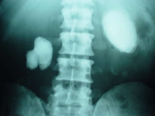
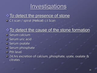
1
UROLITHIASIS
2
Urinary stones have afflicted humankind since
antiquity with the earliest recorded example
being bladder and kidney stones detected in
Egyptian mummies dated to 4800 BC.
The specialty of urologic surgery was
recognized even by Hippocrates, who wrote, in
his famous oath for the physician, “I will not cut,
even for the stone, but leave such procedure to
the practitioner of the craft”
The recurrence rate without treatment for
calcium oxalate renal stones is about 10% at 1
year, 35% at 5 years, and 50% at 10 years.
3
Age & Sex
Incidence
 20 – 40 years
Male Female
3 1
Testosterone UTI
↑ Endogenous production
of oxalate by liver
Hyperparathyroidism
Cystinuria
4
PHYSICAL CHEMISTRY
Phenomena
Nucleation will occur
Inhibitors not generally effective
Crystal growth will occur
Crystal aggregation will occur
Inhibitors will impede or prevent crystallization
De novo nucleation is very slow
Hetrogeneous nucleation may occur
Matrix may be involved
Crystals will for form
Existing stones may dissolve
Formation
Product
Solubility
Product
Concentration
Product
State of Saturation
5
RENAL STONES
Second commonest site of stone formation in
the body.
Prevalence 2 to 3 percent.
5 % lifetime risk of developing renal stones.
Accounts for 10 to 15% of CRF patients. (1 t0 2
% in western countries)
6
AETIOLOGY
(Multifactorial)
Genetic Factors
Environmental Factors
Anatomical Factors in the Urinary Tract
7
AETIOLOGY
(Genetic Factors)
25% have family history of renal stones
RTA
Hyperoxaluria
Cysteinuria
Xanthine Stones
8
AETIOLOGY
(Environmental Factors)
Hot Climate
 Warm desert region
 Mountains & Tropical Areas
Water Intake
 Areas having Chronic / Endemic Diarrhea leading to dehydration
 ?? Hard Water
 Zinc in consumed Water
Diet
 Excessive Intake of Calcium
 Excessive Intake of Oxalates
 Excessive Intake of meat/meat products (purines)
9
AETIOLOGY
(Factors in the Urinary Tract)
Obstruction
 Horse-shoe Kidney
 PUJ Obstruction
 Medullary Sponge Kidney
 Subtle anatomic Abnormality in the kidney not visible to the
naked eye
Infection
 Urease Splitting Organisms
Nidus for Stone Formation
 Randal Plaque
10
Supersaturated Urine
HyperOxaluria
 Primary (genetic disorder)
 Enteric
Short Bowl Syndrome
Malabosrption
Crohn’s Disease
Hyperuricosuria
 Gouty Diathesis
 Myeloproliferative Disorders
 Anti Cancer Chemotherapy
 ↑ meat intake
Hypercysteinuria
 Autosomal recessive disorder
11
Supersaturated Urine
Hypercalciuria
 Absorptive
 Resorptive
 Renal
Absorptive Hypercalciuria
 Hypervitaminosis D
 Sarcoidosis
 Hyperparathyrodism
 Excessive intake of calcium in diet e.g. milk & milk products
Resorptive Hypercalciuria
 Hyperparathyrodism
 Prolonged Immobilization
 Metastatic Malignancy
 ↑ Glucocorticoids
Renal Hypercalciuria
 Renal Calcium leak
12
Inhibiters of Stone Formation
Citrates
Mg
Tamm - Horsefall Proteins
Nephrocalcin
Glycoprotein
Orthophosphates / Cellulose Phosphate
13
Types of Renal Stones
Calcium Oxalate
 75 % to 80 % of renal stones
 Irregular / Spiky Surface
 Can be single or multiple
 Hyper Oxaluria & Acidic urine predisposing factors
 Exists as monohydrate / dihydrate
 Radiopaque on Radiography
Calcium Phosphate & Triple Phosphate
 15 % o 20 % of renal stones
 Struvite stones
 Require alkaline urine, so formed in infection with urease splitting
organisms & RTA
 Can attain very large size & account for the majority of staghorn calculi
 Usually single but may be multiple
 Relatively fragile stone and amenable to lithotripsy
 Radiopaque on Radiography
14
Types of Renal Stones
Uric Acid Stones
 Account for 5 % to 10 % of renal stones
 Typically radiolucent
 Usually have smooth surface
 Can be single or multiple
 Formed in acidic urine
Cysteine Stones
 Account for 1 % to 2 % of renal stones
 Formed in hypercystineuria which is an autosomal recessive disorder
 Faintly radiopaque
 Usually single
 Amenable to medical treatment
 Formed in acidic urine
Rare Stones
 Xanthine
 Di-hydroxy adenine
 Triamterine
15
Clinical Features
Asymptomatic
Flank pain (Mild, Moderate, Severe)
Haematuria
pyuria
16
Complications
Renal / ureteric colic
Haematuria
Hydronephrosis
Infection
Progressive loss of kidney function (ESRD)
17
Investigations
To detect the presence of stone
Urine analysis
Ultrasound scan
i.v.u
18
19
20
21
22
23
24
25
Investigations
To detect the presence of stone
C.t scan / spiral (Helical) c.t Scan
To detect the cause of the stone formation
Serum calcium
Serum uric acid
Serum oxalate
Serum phosphate
Pth level
24 hrs excretion of calcium, phosphate, urate, oxalate &
citrates
26
Medical Treatment
General measure of prevention
 Hydration
 Diet
Dietary protein
Dietary calcium
Dietary sodium
Dietary oxalate
Dietary phosphate
Dietary fiber
 Special Measures
Thiazides
Orthophosphates
Sodium cellulose phosphate
Allopurinol
Citrates
Magnesium
27
Surgical Treatment
Minimally invasive techniques
 ESWL (lithotripsy)
 Intracarporeal lithotripsy
PCNL
URS / LC
Open surgical techniques
 Pyelolithotomy
 Nephrolithotomy
 Pyelo-nephrolithotomy
 Partial Nephrectomy
 Correction of associated anatomic
abnormality e.g. pyeloplasty of PUJ
obstruction

UROLITHIASIS.ppt

  • 1.
  • 2.
    2 Urinary stones haveafflicted humankind since antiquity with the earliest recorded example being bladder and kidney stones detected in Egyptian mummies dated to 4800 BC. The specialty of urologic surgery was recognized even by Hippocrates, who wrote, in his famous oath for the physician, “I will not cut, even for the stone, but leave such procedure to the practitioner of the craft” The recurrence rate without treatment for calcium oxalate renal stones is about 10% at 1 year, 35% at 5 years, and 50% at 10 years.
  • 3.
    3 Age & Sex Incidence 20 – 40 years Male Female 3 1 Testosterone UTI ↑ Endogenous production of oxalate by liver Hyperparathyroidism Cystinuria
  • 4.
    4 PHYSICAL CHEMISTRY Phenomena Nucleation willoccur Inhibitors not generally effective Crystal growth will occur Crystal aggregation will occur Inhibitors will impede or prevent crystallization De novo nucleation is very slow Hetrogeneous nucleation may occur Matrix may be involved Crystals will for form Existing stones may dissolve Formation Product Solubility Product Concentration Product State of Saturation
  • 5.
    5 RENAL STONES Second commonestsite of stone formation in the body. Prevalence 2 to 3 percent. 5 % lifetime risk of developing renal stones. Accounts for 10 to 15% of CRF patients. (1 t0 2 % in western countries)
  • 6.
  • 7.
    7 AETIOLOGY (Genetic Factors) 25% havefamily history of renal stones RTA Hyperoxaluria Cysteinuria Xanthine Stones
  • 8.
    8 AETIOLOGY (Environmental Factors) Hot Climate Warm desert region  Mountains & Tropical Areas Water Intake  Areas having Chronic / Endemic Diarrhea leading to dehydration  ?? Hard Water  Zinc in consumed Water Diet  Excessive Intake of Calcium  Excessive Intake of Oxalates  Excessive Intake of meat/meat products (purines)
  • 9.
    9 AETIOLOGY (Factors in theUrinary Tract) Obstruction  Horse-shoe Kidney  PUJ Obstruction  Medullary Sponge Kidney  Subtle anatomic Abnormality in the kidney not visible to the naked eye Infection  Urease Splitting Organisms Nidus for Stone Formation  Randal Plaque
  • 10.
    10 Supersaturated Urine HyperOxaluria  Primary(genetic disorder)  Enteric Short Bowl Syndrome Malabosrption Crohn’s Disease Hyperuricosuria  Gouty Diathesis  Myeloproliferative Disorders  Anti Cancer Chemotherapy  ↑ meat intake Hypercysteinuria  Autosomal recessive disorder
  • 11.
    11 Supersaturated Urine Hypercalciuria  Absorptive Resorptive  Renal Absorptive Hypercalciuria  Hypervitaminosis D  Sarcoidosis  Hyperparathyrodism  Excessive intake of calcium in diet e.g. milk & milk products Resorptive Hypercalciuria  Hyperparathyrodism  Prolonged Immobilization  Metastatic Malignancy  ↑ Glucocorticoids Renal Hypercalciuria  Renal Calcium leak
  • 12.
    12 Inhibiters of StoneFormation Citrates Mg Tamm - Horsefall Proteins Nephrocalcin Glycoprotein Orthophosphates / Cellulose Phosphate
  • 13.
    13 Types of RenalStones Calcium Oxalate  75 % to 80 % of renal stones  Irregular / Spiky Surface  Can be single or multiple  Hyper Oxaluria & Acidic urine predisposing factors  Exists as monohydrate / dihydrate  Radiopaque on Radiography Calcium Phosphate & Triple Phosphate  15 % o 20 % of renal stones  Struvite stones  Require alkaline urine, so formed in infection with urease splitting organisms & RTA  Can attain very large size & account for the majority of staghorn calculi  Usually single but may be multiple  Relatively fragile stone and amenable to lithotripsy  Radiopaque on Radiography
  • 14.
    14 Types of RenalStones Uric Acid Stones  Account for 5 % to 10 % of renal stones  Typically radiolucent  Usually have smooth surface  Can be single or multiple  Formed in acidic urine Cysteine Stones  Account for 1 % to 2 % of renal stones  Formed in hypercystineuria which is an autosomal recessive disorder  Faintly radiopaque  Usually single  Amenable to medical treatment  Formed in acidic urine Rare Stones  Xanthine  Di-hydroxy adenine  Triamterine
  • 15.
    15 Clinical Features Asymptomatic Flank pain(Mild, Moderate, Severe) Haematuria pyuria
  • 16.
    16 Complications Renal / uretericcolic Haematuria Hydronephrosis Infection Progressive loss of kidney function (ESRD)
  • 17.
    17 Investigations To detect thepresence of stone Urine analysis Ultrasound scan i.v.u
  • 18.
  • 19.
  • 20.
  • 21.
  • 22.
  • 23.
  • 24.
  • 25.
    25 Investigations To detect thepresence of stone C.t scan / spiral (Helical) c.t Scan To detect the cause of the stone formation Serum calcium Serum uric acid Serum oxalate Serum phosphate Pth level 24 hrs excretion of calcium, phosphate, urate, oxalate & citrates
  • 26.
    26 Medical Treatment General measureof prevention  Hydration  Diet Dietary protein Dietary calcium Dietary sodium Dietary oxalate Dietary phosphate Dietary fiber  Special Measures Thiazides Orthophosphates Sodium cellulose phosphate Allopurinol Citrates Magnesium
  • 27.
    27 Surgical Treatment Minimally invasivetechniques  ESWL (lithotripsy)  Intracarporeal lithotripsy PCNL URS / LC Open surgical techniques  Pyelolithotomy  Nephrolithotomy  Pyelo-nephrolithotomy  Partial Nephrectomy  Correction of associated anatomic abnormality e.g. pyeloplasty of PUJ obstruction