Downloaded 257 times
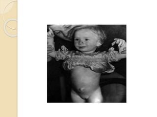
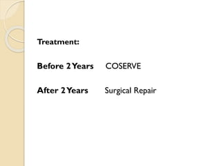
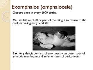
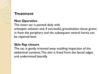
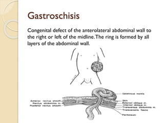
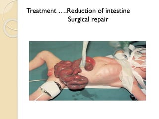
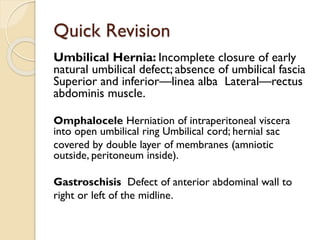


This document discusses several congenital abdominal wall anomalies: - Umbilical hernias are caused by incomplete closure of the umbilical defect and absence of fascia. They are covered by skin and bounded superiorly/inferiorly by the linea alba and laterally by the rectus muscles. Treatment before 2 years is observation, after 2 years is surgical repair. - Omphaloceles involve herniation of intestines into an open umbilical ring through a very thin sac. Non-operative treatment involves cleaning the sac daily until it closes. Surgical options include skin flap closure or sac removal to inspect organs. - Gastroschisis is a defect to the right or