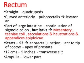
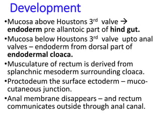
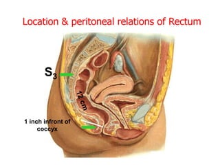
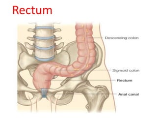
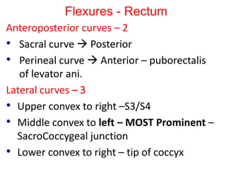
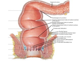
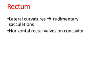
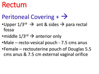
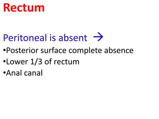
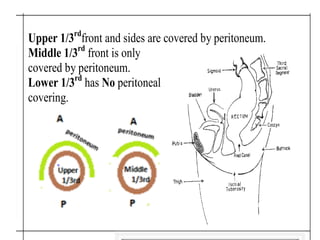
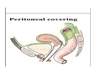
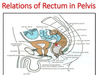
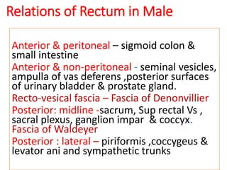
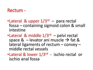
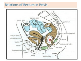
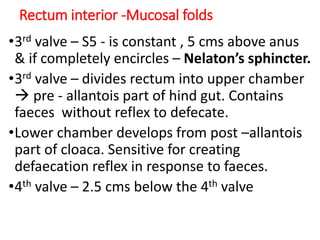
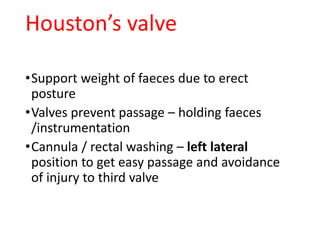
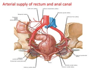
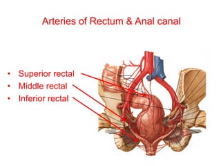
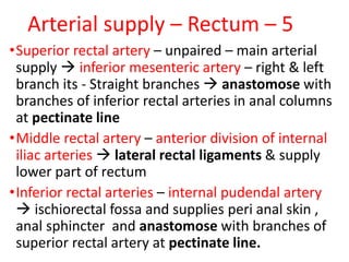
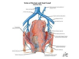
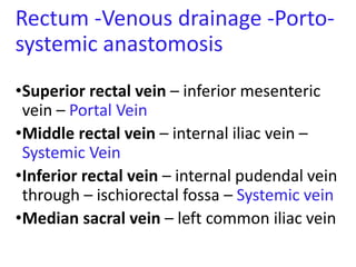
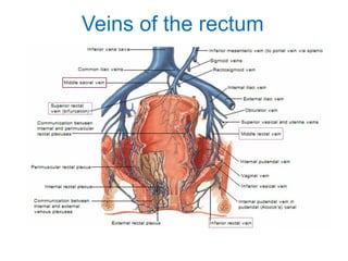
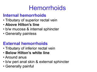
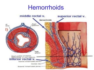
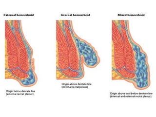
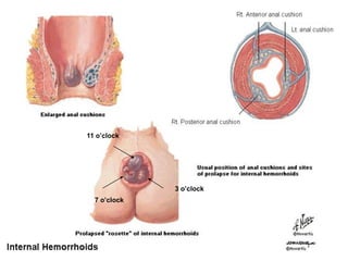
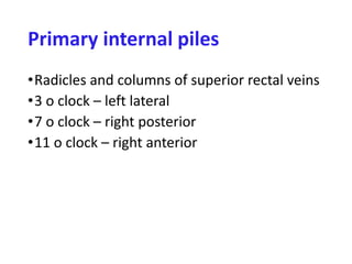
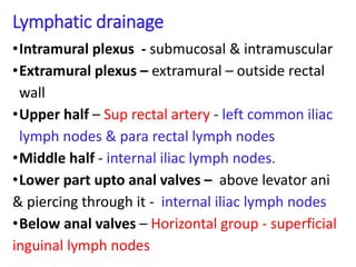
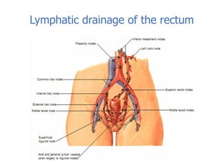
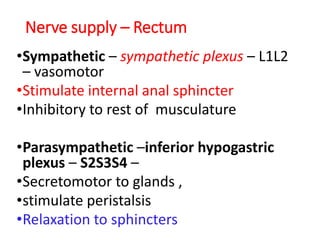
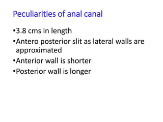
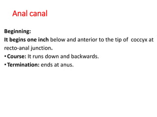
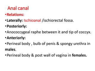
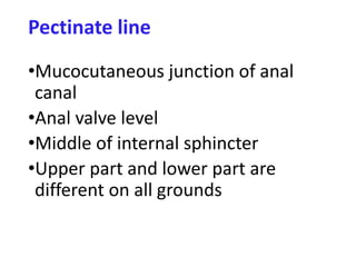
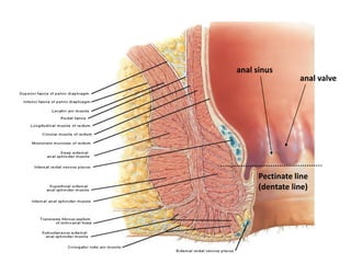
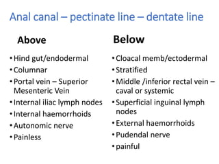
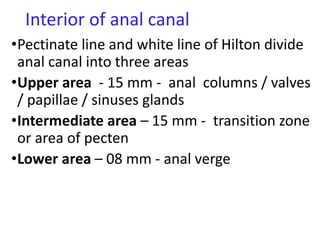
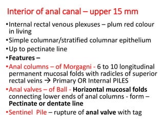
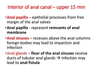
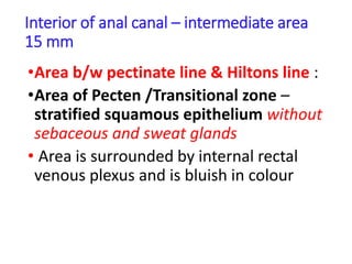
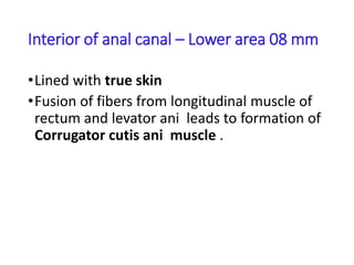
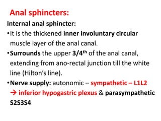
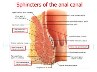
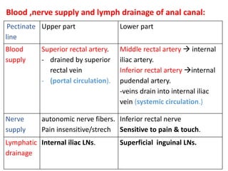
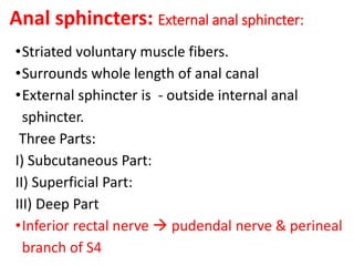
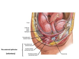
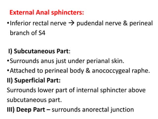
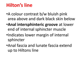
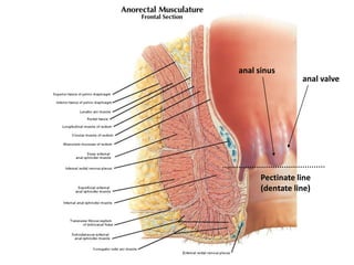
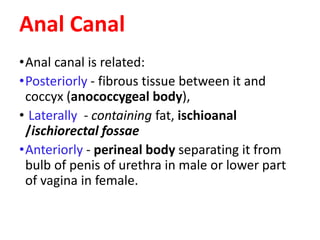
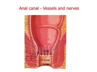
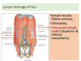
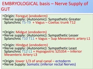
SlideShare a Scribd company logo
Rectum & Anal
canal
Dr Brijendra Singh
Prof & Head Anatomy
AIIMS Rishikesh
27/04/2019
EMBRYOLOGICAL basis – Nerve Supply of
GUT
•Origin: Foregut (endoderm)
•Nerve supply: (Autonomic): Sympathetic Greater
Splanchnic T5-T9 + Vagus – Coeliac trunk T12
•Origin: Midgut (endoderm)
•Nerve supply: (Autonomic): Sympathetic Lesser
Splanchnic T10 T11 + Vagus – Sup Mesenteric artery L1
•Origin: Hindgut (endoderm)
•Nerve supply: (Autonomic): Sympathetic Least
Splanchnic T12 L1 + Hypogastric S2S3S4 – Inferior
Mesenteric Artery L3
•Origin :lower 1/3 of anal canal – ectoderm
•Nerve Supply: Somatic (inferior rectal Nerves)
Rectum
•Straight – quadrupeds
•Curved anteriorly – puborectalis  levator
ani
•Part of large intestine – continuation of
sigmoid colon , but lacks  Mesentery ,
taeniae coli , sacculations & haustrations &
appendices epiploicae.
•Starts – S3  anorectal junction – ant to tip
of coccyx – apex of prostate
•12 cms – 5 inches - transverse slit
•Ampulla – lower part
Development
•Mucosa above Houstons 3rd valve 
endoderm pre allantoic part of hind gut.
•Mucosa below Houstons 3rd valve upto anal
valves – endoderm from dorsal part of
endodermal cloaca.
•Musculature of rectum is derived from
splanchnic mesoderm surrounding cloaca.
•Proctodeum the surface ectoderm – muco-
cutaneous junction.
•Anal membrane disappears – and rectum
communicates outside through anal canal.
1 inch infront of
coccyx
Location & peritoneal relations of Rectum
S3
• Beginning: continuation of sigmoid
colon at S3.
• Termination: continues as anal canal,
• one inch below & front of tip of
coccyx.
• End is dilated to form rectal ampulla.
• Length: 13 cm(5 inches)
Rectum
Rectum
Flexures - Rectum
Anteroposterior curves – 2
• Sacral curve  Posterior
• Perineal curve  Anterior – puborectalis
of levator ani.
Lateral curves – 3
• Upper convex to right –S3/S4
• Middle convex to left – MOST Prominent –
SacroCoccygeal junction
• Lower convex to right – tip of coccyx
Rectum
•Lateral curvatures  rudimentary
sacculations
•Horizontal rectal valves on concavity
Rectum
Peritoneal Covering + 
•Upper 1/3rd  ant & sides  para rectal
fossa
•middle 1/3rd  anterior only
•Male – recto-vesical pouch - 7.5 cms anus
•Female – rectouterine pouch of Douglas 5.5
cms anus & 7.5 cm external vaginal orifice
Rectum
Peritoneal is absent 
•Posterior surface complete absence
•Lower 1/3 of rectum
•Anal canal
ng Upper 1/3rd
front and sides are covered by peritoneum.
Middle 1/3rd
front is only
covered by peritoneum.
Lower 1/3rd
has No peritoneal
covering.
Peritoneal covering
Relations of Rectum in Pelvis
R
Relations of Rectum in Male
Anterior & peritoneal – sigmoid colon &
small intestine
Anterior & non-peritoneal - seminal vesicles,
ampulla of vas deferens ,posterior surfaces
of urinary bladder & prostate gland.
Recto-vesical fascia – Fascia of Denonvillier
Posterior: midline -sacrum, Sup rectal Vs ,
sacral plexus, ganglion impar & coccyx.
Fascia of Waldeyer
Posterior : lateral – piriformis ,coccygeus &
levator ani and sympathetic trunks
Rectum -
•Lateral & upper 1/3rd – para rectal
fossa – containing sigmoid colon & small
intestine
•Lateral & middle 1/3rd – pelvi rectal
space & – levator ani muscle  fat &
lateral ligaments of rectum – convey –
middle rectal vessels
•Lateral & lower 1/3rd - ischio rectal or
ischio anal fossa
Relations of Rectum in Pelvis
R
Relations of Rectum in Pelvis
FEMALE PELVIS
Anterior: posterior wall of vagina & recto
vaginal fascia
Posterior: sacrum , sacral plexus & coccyx
Support of rectum
•Puborectal sling of levator ani –
anorectal flexure
•Reflection of pelvic fascia &
peritoneum
•Waldeyer fascia & lateral ligaments
of rectum
•Pelvi rectal and ischiorectal fat
Wall of rectum
•Serous , muscular ,submucosa & mucosa
•Serosa – peritoneum incomplete
•Muscular – outer longitudinal & inner circular
•Longitudinal muscle – anterior - blends at anorectal
junction with puborectalis part of levator ani 
corrugator ani cutis muscle & attached to white line of
Hilton
•Some fibres also form – urethra – recto urethral muscle
of Roux
•Coccyx – recto coccygeus muscle
•Circular muscle – entire rectum, in lower part 
internal anal sphincter – covers upper 3/4th of anal
canal
Rectum Interior -Mucosal folds
•Temporary – longitudinal & in lower part –
disappear in distension
•Permanent mucosal folds – Horizontal -
Houston’s valve - 04 ,
•1st & 3rd are constant.
•1st - S3 - recto sigmoid junction
•3rd - S5 - 5 cms above anus & if completely
encircles – Nelaton’s sphincter.
•2nd - about 2.5cms superior to 3rd valve
Rectum interior -Mucosal folds
•3rd valve – S5 - is constant , 5 cms above anus
& if completely encircles – Nelaton’s sphincter.
•3rd valve – divides rectum into upper chamber
 pre - allantois part of hind gut. Contains
faeces without reflex to defecate.
•Lower chamber develops from post –allantois
part of cloaca. Sensitive for creating
defaecation reflex in response to faeces.
•4th valve – 2.5 cms below the 4th valve
Houston’s valve
•Support weight of faeces due to erect
posture
•Valves prevent passage – holding faeces
/instrumentation
•Cannula / rectal washing – left lateral
position to get easy passage and avoidance
of injury to third valve
Arterial supply of rectum and anal canal
Arterial supply - Rectum
Median sacral artery – abdominal aorta
Superior rectal artery – continuation of inferior
mesenteric artery
Middle rectal artery – ant div of internal iliac
artery
Inferior rectal artery - internal pudendal artery
Inferior gluteal artery - internal iliac artery
Arteries of Rectum & Anal canal
• Superior rectal
• Middle rectal
• Inferior rectal
Arterial supply – Rectum – 5
•Superior rectal artery – unpaired – main arterial
supply  inferior mesenteric artery – right & left
branch its - Straight branches  anastomose with
branches of inferior rectal arteries in anal columns
at pectinate line
•Middle rectal artery – anterior division of internal
iliac arteries  lateral rectal ligaments & supply
lower part of rectum
•Inferior rectal arteries – internal pudendal artery
 ischiorectal fossa and supplies peri anal skin ,
anal sphincter and anastomose with branches of
superior rectal artery at pectinate line.
Rectum -Venous drainage -Porto-
systemic anastomosis
•Superior rectal vein – inferior mesenteric
vein – Portal Vein
•Middle rectal vein – internal iliac vein –
Systemic Vein
•Inferior rectal vein – internal pudendal vein
through – ischiorectal fossa – Systemic vein
•Median sacral vein – left common iliac vein
Veins of the rectum
Internal hemorrhoids
• Tributary of superior rectal vein
• Above Hilton’s line
• b/w mucosa & internal sphincter
• Generally painless
External hemorrhoids
• Tributary of inferior rectal vein
• Below Hilton’s white line
• Around anus
• b/w peri anal skin & external sphincter
• Generally painful
Hemorrhoids
Hemorrhoids
11 o’clock
7 o’clock
3 o’clock
Primary internal piles
•Radicles and columns of superior rectal veins
•3 o clock – left lateral
•7 o clock – right posterior
•11 o clock – right anterior
Lymphatic drainage
•Intramural plexus - submucosal & intramuscular
•Extramural plexus – extramural – outside rectal
wall
•Upper half – Sup rectal artery - left common iliac
lymph nodes & para rectal lymph nodes
•Middle half - internal iliac lymph nodes.
•Lower part upto anal valves – above levator ani
& piercing through it - internal iliac lymph nodes
•Below anal valves – Horizontal group - superficial
inguinal lymph nodes
Lymphatic drainage of the rectum
Nerve supply – Rectum
•Sympathetic – sympathetic plexus – L1L2
– vasomotor
•Stimulate internal anal sphincter
•Inhibitory to rest of musculature
•Parasympathetic –inferior hypogastric
plexus – S2S3S4 –
•Secretomotor to glands ,
•stimulate peristalsis
•Relaxation to sphincters
Peculiarities of anal canal
•3.8 cms in length
•Antero posterior slit as lateral walls are
approximated
•Anterior wall is shorter
•Posterior wall is longer
Anal canal
Beginning:
It begins one inch below and anterior to the tip of coccyx at
recto-anal junction.
•Course: It runs down and backwards.
•Termination: ends at anus.
Anal canal
•Relations:
•Laterally: Ischioanal /ischiorectal fossa.
•Posteriorly:
•Anococcygeal raphe between it and tip of coccyx.
•Anteriorly:
•Perineal body , bulb of penis & spongy urethra in
males.
•Perineal body & post wall of vagina in females.
Pectinate line
•Mucocutaneous junction of anal
canal
•Anal valve level
•Middle of internal sphincter
•Upper part and lower part are
different on all grounds
Pectinate line
(dentate line)
anal sinus
anal valve
Anal canal – pectinate line – dentate line
Above
•Hind gut/endodermal
•Columnar
•Portal vein – Superior
Mesenteric Vein
•Internal iliac lymph nodes
•Internal haemorrhoids
•Autonomic nerve
•Painless
Below
•Cloacal memb/ectodermal
•Stratified
•Middle /inferior rectal vein –
caval or systemic
•Superficial inguinal lymph
nodes
•External haemorrhoids
•Pudendal nerve
•painful
Interior of anal canal
•Pectinate line and white line of Hilton divide
anal canal into three areas
•Upper area - 15 mm - anal columns / valves
/ papillae / sinuses glands
•Intermediate area – 15 mm - transition zone
or area of pecten
•Lower area – 08 mm - anal verge
Interior of anal canal – upper 15 mm
•Internal rectal venous plexuses – plum red colour
in living
•Simple columnar/stratified columnar epithelium
•Up to pectinate line
•Features –
•Anal columns – of Morgagni - 6 to 10 longitudinal
permanent mucosal folds with radicles of superior
rectal veins  Primary OR Internal PILES
•Anal valves – of Ball - Horizontal mucosal folds
connecting lower ends of anal columns - form –
Pectinate or dentate line
•Sentinel Pile – rupture of anal valve with tag
Pectinate line
(dentate line)
anal sinus
anal valve
Interior of anal canal – upper 15 mm
•Anal papilla – epithelial processes from free
margin of the anal valves
•Anal papilla - represent remnants of anal
membrane
•Anal sinuses – recesses above the anal columns
foreign bodies may lead to impaction and
infection
•Anal glands – floor of the anal sinuses receive
ducts of tubular anal glands  infection may
lead to anal fistula
Interior of anal canal – intermediate area
15 mm
•Area b/w pectinate line & Hiltons line :
•Area of Pecten /Transitional zone –
stratified squamous epithelium without
sebaceous and sweat glands
• Area is surrounded by internal rectal
venous plexus and is bluish in colour
Interior of anal canal – Lower area 08 mm
•Lined with true skin
•Fusion of fibers from longitudinal muscle of
rectum and levator ani leads to formation of
Corrugator cutis ani muscle .
Anal sphincters:
Internal anal sphincter:
•It is the thickened inner involuntary circular
muscle layer of the anal canal.
•Surrounds the upper 3/4th of the anal canal,
extending from ano-rectal junction till the white
line (Hilton’s line).
•Nerve supply: autonomic – sympathetic – L1L2
 inferior hypogastric plexus & parasympathetic
S2S3S4
Sphincters of the anal canal
1
2
Pectinate
line
Upper part Lower part
Blood
supply
Superior rectal artery.
- drained by superior
rectal vein
- (portal circulation).
Middle rectal artery  internal
iliac artery.
Inferior rectal artery internal
pudendal artery.
-veins drain into internal iliac
vein (systemic circulation.)
Nerve
supply
autonomic nerve fibers.
Pain insensitive/strech
Inferior rectal nerve
Sensitive to pain & touch.
Lymphatic
drainage
Internal iliac LNs. Superficial inguinal LNs.
Blood ,nerve supply and lymph drainage of anal canal:
Anal sphincters: External anal sphincter:
•Striated voluntary muscle fibers.
•Surrounds whole length of anal canal
•External sphincter is - outside internal anal
sphincter.
Three Parts:
I) Subcutaneous Part:
II) Superficial Part:
III) Deep Part
•Inferior rectal nerve  pudendal nerve & perineal
branch of S4
Sphincters of the anal canal
1
2
The external sphincter
(voluntary)
External Anal sphincters:
•Inferior rectal nerve  pudendal nerve & perineal
branch of S4
I) Subcutaneous Part:
•Surrounds anus just under perianal skin.
•Attached to perineal body & anococcygeal raphe.
II) Superficial Part:
Surrounds lower part of internal sphincter above
subcutaneous part.
III) Deep Part – surrounds anorectal junction
Hilton’s line
•A colour contrast b/w bluish pink
area above and dark black skin below
•Anal intersphinteric groove at lower
end of internal sphincter muscle
•Indicates lower margin of internal
sphincter
•Anal fascia and lunate fascia extend
up to Hiltons line
Pectinate line
(dentate line)
anal sinus
anal valve
Anal Canal
•Anal canal is related:
•Posteriorly - fibrous tissue between it and
coccyx (anococcygeal body),
• Laterally - containing fat, ischioanal
/ischiorectal fossae
•Anteriorly - perineal body separating it from
bulb of penis of urethra in male or lower part
of vagina in female.
Anal canal - Vessels and nerves
2 cm
1 ½ cm
½ cm
Lymph drainage of Gut
•lymph vessels
follow arteries.
•Ultimately,
• Pre-aortic lymph
nodes (Superior &
Inferior
mesenteric).
EMBRYOLOGICAL basis – Nerve Supply of
GUT
•Origin: Foregut (endoderm)
•Nerve supply: (Autonomic): Sympathetic Greater
Splanchnic T5-T9 + Vagus – Coeliac trunk T12
•Origin: Midgut (endoderm)
•Nerve supply: (Autonomic): Sympathetic Lesser
Splanchnic T10 T11 + Vagus – Sup Mesenteric artery L1
•Origin: Hindgut (endoderm)
•Nerve supply: (Autonomic): Sympathetic Least
Splanchnic T12 L1 + Hypogastric S2S3S4 – Inferior
Mesenteric Artery L3
•Origin :lower 1/3 of anal canal – ectoderm
•Nerve Supply: Somatic (inferior rectal Nerves)

More Related Content

What's hot

ANATOMY OF DUODENUM
ANATOMY OF DUODENUMANATOMY OF DUODENUM
ANATOMY OF DUODENUM
Dr. sana yaseen
 
urinary Bladder.ppt
urinary Bladder.ppturinary Bladder.ppt
urinary Bladder.ppt
Dr. sana yaseen
 
Vessels of upper limb
Vessels of upper limbVessels of upper limb
Anatomy of inguinal canal - Dr nesar Ahmad
Anatomy of inguinal canal -  Dr nesar AhmadAnatomy of inguinal canal -  Dr nesar Ahmad
Anatomy of inguinal canal - Dr nesar Ahmad
Student
 
Anterior abdominal wall
Anterior abdominal wallAnterior abdominal wall
Anterior abdominal wall
Seemi Shah
 
Upper Limb
Upper LimbUpper Limb
Upper Limb
MBBS IMS MSU
 
Posterior abdominal wall
Posterior abdominal wallPosterior abdominal wall
Posterior abdominal wall
Suraj Dhara
 
Anatomy of duodenum and pancreas
Anatomy of duodenum and pancreas Anatomy of duodenum and pancreas
Anatomy of duodenum and pancreas
Dr. Mohammad Mahmoud
 
Urogenital triangle
Urogenital triangleUrogenital triangle
Urogenital triangle
GovtRoyapettahHospit
 
Venous drainage of the lower limb
Venous drainage of the lower limbVenous drainage of the lower limb
Venous drainage of the lower limb
mattygamer
 
Front of the thigh
Front of the thighFront of the thigh
Front of the thigh
Dr. Mushfiqul Hoque
 
Diencephalon 1
Diencephalon 1Diencephalon 1
Diencephalon 1
Dr. sana yaseen
 
Thoracic part of Sympathetic Chain (Anatomy of the Thorax)
Thoracic part of Sympathetic Chain (Anatomy of the Thorax)Thoracic part of Sympathetic Chain (Anatomy of the Thorax)
Thoracic part of Sympathetic Chain (Anatomy of the Thorax)
Dr. Sherif Fahmy
 
Peritoneum Dr. Mehul Tandel
Peritoneum Dr. Mehul TandelPeritoneum Dr. Mehul Tandel
Peritoneum Dr. Mehul Tandel
Mehul Tandel
 
Anatomy of perineum
Anatomy of perineumAnatomy of perineum
Anatomy of perineum
Ammedicine Medicine
 
Caecum and vermiform appendix
Caecum and vermiform appendixCaecum and vermiform appendix
Caecum and vermiform appendix
Dr Mohammad Amaan
 
Thoracic duct
Thoracic ductThoracic duct
Thoracic duct
Idris Siddiqui
 
Deep cervical fascia (fascia colli)
Deep cervical fascia (fascia colli)Deep cervical fascia (fascia colli)
Deep cervical fascia (fascia colli)
surajitkundu
 
Adductor canal
Adductor canalAdductor canal
Adductor canal
Dr. sana yaseen
 
ABDOMINAL WALL ANATOMY
ABDOMINAL WALL ANATOMY ABDOMINAL WALL ANATOMY

What's hot (20)

ANATOMY OF DUODENUM
ANATOMY OF DUODENUMANATOMY OF DUODENUM
ANATOMY OF DUODENUM
 
urinary Bladder.ppt
urinary Bladder.ppturinary Bladder.ppt
urinary Bladder.ppt
 
Vessels of upper limb
Vessels of upper limbVessels of upper limb
Vessels of upper limb
 
Anatomy of inguinal canal - Dr nesar Ahmad
Anatomy of inguinal canal -  Dr nesar AhmadAnatomy of inguinal canal -  Dr nesar Ahmad
Anatomy of inguinal canal - Dr nesar Ahmad
 
Anterior abdominal wall
Anterior abdominal wallAnterior abdominal wall
Anterior abdominal wall
 
Upper Limb
Upper LimbUpper Limb
Upper Limb
 
Posterior abdominal wall
Posterior abdominal wallPosterior abdominal wall
Posterior abdominal wall
 
Anatomy of duodenum and pancreas
Anatomy of duodenum and pancreas Anatomy of duodenum and pancreas
Anatomy of duodenum and pancreas
 
Urogenital triangle
Urogenital triangleUrogenital triangle
Urogenital triangle
 
Venous drainage of the lower limb
Venous drainage of the lower limbVenous drainage of the lower limb
Venous drainage of the lower limb
 
Front of the thigh
Front of the thighFront of the thigh
Front of the thigh
 
Diencephalon 1
Diencephalon 1Diencephalon 1
Diencephalon 1
 
Thoracic part of Sympathetic Chain (Anatomy of the Thorax)
Thoracic part of Sympathetic Chain (Anatomy of the Thorax)Thoracic part of Sympathetic Chain (Anatomy of the Thorax)
Thoracic part of Sympathetic Chain (Anatomy of the Thorax)
 
Peritoneum Dr. Mehul Tandel
Peritoneum Dr. Mehul TandelPeritoneum Dr. Mehul Tandel
Peritoneum Dr. Mehul Tandel
 
Anatomy of perineum
Anatomy of perineumAnatomy of perineum
Anatomy of perineum
 
Caecum and vermiform appendix
Caecum and vermiform appendixCaecum and vermiform appendix
Caecum and vermiform appendix
 
Thoracic duct
Thoracic ductThoracic duct
Thoracic duct
 
Deep cervical fascia (fascia colli)
Deep cervical fascia (fascia colli)Deep cervical fascia (fascia colli)
Deep cervical fascia (fascia colli)
 
Adductor canal
Adductor canalAdductor canal
Adductor canal
 
ABDOMINAL WALL ANATOMY
ABDOMINAL WALL ANATOMY ABDOMINAL WALL ANATOMY
ABDOMINAL WALL ANATOMY
 

Similar to 10.-Rectum-anal-canal-BS-Copy.pdf

Rectum and anal canal
Rectum and anal canalRectum and anal canal
Rectum and anal canal
Dr. Mushfiqul Hoque
 
Rectum & anal canal
Rectum & anal canalRectum & anal canal
Rectum & anal canal
Mehul Tandel
 
Applied anatomy in inguinoscrotal swelling
Applied anatomy in inguinoscrotal swellingApplied anatomy in inguinoscrotal swelling
Applied anatomy in inguinoscrotal swelling
sunil kumar daha
 
Abdomen & Pelvis II
Abdomen & Pelvis IIAbdomen & Pelvis II
Abdomen & Pelvis II
SaruGosain
 
Esophagus anatomy presentation full .pptx
Esophagus anatomy presentation full .pptxEsophagus anatomy presentation full .pptx
Esophagus anatomy presentation full .pptx
BhishmaKandoriya
 
ANATOMY OF PANCREAS
ANATOMY OF PANCREASANATOMY OF PANCREAS
ANATOMY OF PANCREAS
Aamir Hela
 
Small intestine lecture slides for medical students
Small intestine lecture slides for medical studentsSmall intestine lecture slides for medical students
Small intestine lecture slides for medical students
ymusa1334
 
Lect 2 anterior abdominal wall
Lect 2  anterior abdominal wallLect 2  anterior abdominal wall
Lect 2 anterior abdominal wall
Mohaned Lehya
 
Anatomy the small and large intestine.pptx
Anatomy the small and large intestine.pptxAnatomy the small and large intestine.pptx
Anatomy the small and large intestine.pptx
Pradeep Pande
 
2. ureter
2. ureter2. ureter
2. ureter
kanakmauritius
 
Urinary bladder, rectum and anal canal.pptx
Urinary bladder, rectum and anal canal.pptxUrinary bladder, rectum and anal canal.pptx
Urinary bladder, rectum and anal canal.pptx
IbeClement
 
LARGE INTESTINE in the human body of a person
LARGE INTESTINE in the human  body of a personLARGE INTESTINE in the human  body of a person
LARGE INTESTINE in the human body of a person
AtemJoshua
 
Abdomen Anatomy - Made Easy
Abdomen Anatomy - Made EasyAbdomen Anatomy - Made Easy
Abdomen Anatomy - Made Easy
RER MedApps
 
Anatomy of genitourinary system
Anatomy of genitourinary systemAnatomy of genitourinary system
Anatomy of genitourinary system
RamanGhimire3
 
analcanal anatomy (aspects of surgery) well described
analcanal anatomy (aspects of surgery) well describedanalcanal anatomy (aspects of surgery) well described
analcanal anatomy (aspects of surgery) well described
vishalvaishnavi2
 
GROSS ANATOMY OF THE INGUINAL CANAL.ppt
GROSS ANATOMY OF THE INGUINAL CANAL.pptGROSS ANATOMY OF THE INGUINAL CANAL.ppt
GROSS ANATOMY OF THE INGUINAL CANAL.ppt
IniibeheOKOKO1
 
Ventral abdominal hernia1
Ventral abdominal hernia1Ventral abdominal hernia1
Ventral abdominal hernia1
Rekha Pathak
 
Anatomy of Esophagus.pptx
Anatomy of Esophagus.pptxAnatomy of Esophagus.pptx
Anatomy of Esophagus.pptx
kesavanKesavan45
 
Anatomy of The Urinary System
Anatomy of The Urinary SystemAnatomy of The Urinary System
Anatomy of The Urinary System
Aswan University | جامعة أسوان
 
Applied anatomy and physiology of pancreas and spleen.pptx
Applied  anatomy and physiology of pancreas and spleen.pptxApplied  anatomy and physiology of pancreas and spleen.pptx
Applied anatomy and physiology of pancreas and spleen.pptx
Shishir Shrestha
 

Similar to 10.-Rectum-anal-canal-BS-Copy.pdf (20)

Rectum and anal canal
Rectum and anal canalRectum and anal canal
Rectum and anal canal
 
Rectum & anal canal
Rectum & anal canalRectum & anal canal
Rectum & anal canal
 
Applied anatomy in inguinoscrotal swelling
Applied anatomy in inguinoscrotal swellingApplied anatomy in inguinoscrotal swelling
Applied anatomy in inguinoscrotal swelling
 
Abdomen & Pelvis II
Abdomen & Pelvis IIAbdomen & Pelvis II
Abdomen & Pelvis II
 
Esophagus anatomy presentation full .pptx
Esophagus anatomy presentation full .pptxEsophagus anatomy presentation full .pptx
Esophagus anatomy presentation full .pptx
 
ANATOMY OF PANCREAS
ANATOMY OF PANCREASANATOMY OF PANCREAS
ANATOMY OF PANCREAS
 
Small intestine lecture slides for medical students
Small intestine lecture slides for medical studentsSmall intestine lecture slides for medical students
Small intestine lecture slides for medical students
 
Lect 2 anterior abdominal wall
Lect 2  anterior abdominal wallLect 2  anterior abdominal wall
Lect 2 anterior abdominal wall
 
Anatomy the small and large intestine.pptx
Anatomy the small and large intestine.pptxAnatomy the small and large intestine.pptx
Anatomy the small and large intestine.pptx
 
2. ureter
2. ureter2. ureter
2. ureter
 
Urinary bladder, rectum and anal canal.pptx
Urinary bladder, rectum and anal canal.pptxUrinary bladder, rectum and anal canal.pptx
Urinary bladder, rectum and anal canal.pptx
 
LARGE INTESTINE in the human body of a person
LARGE INTESTINE in the human  body of a personLARGE INTESTINE in the human  body of a person
LARGE INTESTINE in the human body of a person
 
Abdomen Anatomy - Made Easy
Abdomen Anatomy - Made EasyAbdomen Anatomy - Made Easy
Abdomen Anatomy - Made Easy
 
Anatomy of genitourinary system
Anatomy of genitourinary systemAnatomy of genitourinary system
Anatomy of genitourinary system
 
analcanal anatomy (aspects of surgery) well described
analcanal anatomy (aspects of surgery) well describedanalcanal anatomy (aspects of surgery) well described
analcanal anatomy (aspects of surgery) well described
 
GROSS ANATOMY OF THE INGUINAL CANAL.ppt
GROSS ANATOMY OF THE INGUINAL CANAL.pptGROSS ANATOMY OF THE INGUINAL CANAL.ppt
GROSS ANATOMY OF THE INGUINAL CANAL.ppt
 
Ventral abdominal hernia1
Ventral abdominal hernia1Ventral abdominal hernia1
Ventral abdominal hernia1
 
Anatomy of Esophagus.pptx
Anatomy of Esophagus.pptxAnatomy of Esophagus.pptx
Anatomy of Esophagus.pptx
 
Anatomy of The Urinary System
Anatomy of The Urinary SystemAnatomy of The Urinary System
Anatomy of The Urinary System
 
Applied anatomy and physiology of pancreas and spleen.pptx
Applied  anatomy and physiology of pancreas and spleen.pptxApplied  anatomy and physiology of pancreas and spleen.pptx
Applied anatomy and physiology of pancreas and spleen.pptx
 

More from elphaswalela

1_Introduction to Biostatistics-2 (2).pdf
1_Introduction to Biostatistics-2 (2).pdf1_Introduction to Biostatistics-2 (2).pdf
1_Introduction to Biostatistics-2 (2).pdf
elphaswalela
 
TOBIAS BIOSYNTHESIS OF AMINO ACIDS PRESENTATION.pptx
TOBIAS BIOSYNTHESIS OF AMINO ACIDS PRESENTATION.pptxTOBIAS BIOSYNTHESIS OF AMINO ACIDS PRESENTATION.pptx
TOBIAS BIOSYNTHESIS OF AMINO ACIDS PRESENTATION.pptx
elphaswalela
 
BIOSYNTHESIS NUCLEOTIDE.pdf
BIOSYNTHESIS  NUCLEOTIDE.pdfBIOSYNTHESIS  NUCLEOTIDE.pdf
BIOSYNTHESIS NUCLEOTIDE.pdf
elphaswalela
 
NEUROPHYSIOLOGY_GM_1_2018.pdf
NEUROPHYSIOLOGY_GM_1_2018.pdfNEUROPHYSIOLOGY_GM_1_2018.pdf
NEUROPHYSIOLOGY_GM_1_2018.pdf
elphaswalela
 
Topic 6 learning theory.pptx
Topic 6 learning theory.pptxTopic 6 learning theory.pptx
Topic 6 learning theory.pptx
elphaswalela
 
TOPIC 4 Psychosexualdevelopment.pptx
TOPIC 4 Psychosexualdevelopment.pptxTOPIC 4 Psychosexualdevelopment.pptx
TOPIC 4 Psychosexualdevelopment.pptx
elphaswalela
 
TOPIC 2 THE PSYCHOLOGICAL BASIS OF BEHAVIOR.pptx
TOPIC 2 THE PSYCHOLOGICAL BASIS OF BEHAVIOR.pptxTOPIC 2 THE PSYCHOLOGICAL BASIS OF BEHAVIOR.pptx
TOPIC 2 THE PSYCHOLOGICAL BASIS OF BEHAVIOR.pptx
elphaswalela
 
Topic 1 Introduction behavioural sciences-1.pptx
Topic 1 Introduction behavioural sciences-1.pptxTopic 1 Introduction behavioural sciences-1.pptx
Topic 1 Introduction behavioural sciences-1.pptx
elphaswalela
 
Topic 1 Introduction behavioural sciences-1.pptx
Topic 1 Introduction behavioural sciences-1.pptxTopic 1 Introduction behavioural sciences-1.pptx
Topic 1 Introduction behavioural sciences-1.pptx
elphaswalela
 
Lesson 9 Personality.pptx
Lesson 9 Personality.pptxLesson 9 Personality.pptx
Lesson 9 Personality.pptx
elphaswalela
 
TOPIC 4 DEFENSE MECHANISMS.pptx
TOPIC 4 DEFENSE MECHANISMS.pptxTOPIC 4 DEFENSE MECHANISMS.pptx
TOPIC 4 DEFENSE MECHANISMS.pptx
elphaswalela
 
HCM 124 lecture 5 .ppt
HCM 124 lecture 5 .pptHCM 124 lecture 5 .ppt
HCM 124 lecture 5 .ppt
elphaswalela
 
HCM 124 lect 4.pptx
HCM 124 lect 4.pptxHCM 124 lect 4.pptx
HCM 124 lect 4.pptx
elphaswalela
 
lact8protoza-130927064752-phpapp01.pdf
lact8protoza-130927064752-phpapp01.pdflact8protoza-130927064752-phpapp01.pdf
lact8protoza-130927064752-phpapp01.pdf
elphaswalela
 
Topic 8 mental disorders.pptx
Topic 8 mental disorders.pptxTopic 8 mental disorders.pptx
Topic 8 mental disorders.pptx
elphaswalela
 
GIT SECRETIONS, DIGESTION & ABSORPTION.pptx
GIT SECRETIONS, DIGESTION & ABSORPTION.pptxGIT SECRETIONS, DIGESTION & ABSORPTION.pptx
GIT SECRETIONS, DIGESTION & ABSORPTION.pptx
elphaswalela
 
Medical sociology.pptx
Medical sociology.pptxMedical sociology.pptx
Medical sociology.pptx
elphaswalela
 
HCM 124 lecture 8 Malaria.pptx
HCM 124 lecture 8 Malaria.pptxHCM 124 lecture 8 Malaria.pptx
HCM 124 lecture 8 Malaria.pptx
elphaswalela
 
HCM 124 lecture 6.pptx
HCM 124 lecture 6.pptxHCM 124 lecture 6.pptx
HCM 124 lecture 6.pptx
elphaswalela
 
Medical Mycology.pptx
Medical Mycology.pptxMedical Mycology.pptx
Medical Mycology.pptx
elphaswalela
 

More from elphaswalela (20)

1_Introduction to Biostatistics-2 (2).pdf
1_Introduction to Biostatistics-2 (2).pdf1_Introduction to Biostatistics-2 (2).pdf
1_Introduction to Biostatistics-2 (2).pdf
 
TOBIAS BIOSYNTHESIS OF AMINO ACIDS PRESENTATION.pptx
TOBIAS BIOSYNTHESIS OF AMINO ACIDS PRESENTATION.pptxTOBIAS BIOSYNTHESIS OF AMINO ACIDS PRESENTATION.pptx
TOBIAS BIOSYNTHESIS OF AMINO ACIDS PRESENTATION.pptx
 
BIOSYNTHESIS NUCLEOTIDE.pdf
BIOSYNTHESIS  NUCLEOTIDE.pdfBIOSYNTHESIS  NUCLEOTIDE.pdf
BIOSYNTHESIS NUCLEOTIDE.pdf
 
NEUROPHYSIOLOGY_GM_1_2018.pdf
NEUROPHYSIOLOGY_GM_1_2018.pdfNEUROPHYSIOLOGY_GM_1_2018.pdf
NEUROPHYSIOLOGY_GM_1_2018.pdf
 
Topic 6 learning theory.pptx
Topic 6 learning theory.pptxTopic 6 learning theory.pptx
Topic 6 learning theory.pptx
 
TOPIC 4 Psychosexualdevelopment.pptx
TOPIC 4 Psychosexualdevelopment.pptxTOPIC 4 Psychosexualdevelopment.pptx
TOPIC 4 Psychosexualdevelopment.pptx
 
TOPIC 2 THE PSYCHOLOGICAL BASIS OF BEHAVIOR.pptx
TOPIC 2 THE PSYCHOLOGICAL BASIS OF BEHAVIOR.pptxTOPIC 2 THE PSYCHOLOGICAL BASIS OF BEHAVIOR.pptx
TOPIC 2 THE PSYCHOLOGICAL BASIS OF BEHAVIOR.pptx
 
Topic 1 Introduction behavioural sciences-1.pptx
Topic 1 Introduction behavioural sciences-1.pptxTopic 1 Introduction behavioural sciences-1.pptx
Topic 1 Introduction behavioural sciences-1.pptx
 
Topic 1 Introduction behavioural sciences-1.pptx
Topic 1 Introduction behavioural sciences-1.pptxTopic 1 Introduction behavioural sciences-1.pptx
Topic 1 Introduction behavioural sciences-1.pptx
 
Lesson 9 Personality.pptx
Lesson 9 Personality.pptxLesson 9 Personality.pptx
Lesson 9 Personality.pptx
 
TOPIC 4 DEFENSE MECHANISMS.pptx
TOPIC 4 DEFENSE MECHANISMS.pptxTOPIC 4 DEFENSE MECHANISMS.pptx
TOPIC 4 DEFENSE MECHANISMS.pptx
 
HCM 124 lecture 5 .ppt
HCM 124 lecture 5 .pptHCM 124 lecture 5 .ppt
HCM 124 lecture 5 .ppt
 
HCM 124 lect 4.pptx
HCM 124 lect 4.pptxHCM 124 lect 4.pptx
HCM 124 lect 4.pptx
 
lact8protoza-130927064752-phpapp01.pdf
lact8protoza-130927064752-phpapp01.pdflact8protoza-130927064752-phpapp01.pdf
lact8protoza-130927064752-phpapp01.pdf
 
Topic 8 mental disorders.pptx
Topic 8 mental disorders.pptxTopic 8 mental disorders.pptx
Topic 8 mental disorders.pptx
 
GIT SECRETIONS, DIGESTION & ABSORPTION.pptx
GIT SECRETIONS, DIGESTION & ABSORPTION.pptxGIT SECRETIONS, DIGESTION & ABSORPTION.pptx
GIT SECRETIONS, DIGESTION & ABSORPTION.pptx
 
Medical sociology.pptx
Medical sociology.pptxMedical sociology.pptx
Medical sociology.pptx
 
HCM 124 lecture 8 Malaria.pptx
HCM 124 lecture 8 Malaria.pptxHCM 124 lecture 8 Malaria.pptx
HCM 124 lecture 8 Malaria.pptx
 
HCM 124 lecture 6.pptx
HCM 124 lecture 6.pptxHCM 124 lecture 6.pptx
HCM 124 lecture 6.pptx
 
Medical Mycology.pptx
Medical Mycology.pptxMedical Mycology.pptx
Medical Mycology.pptx
 

Recently uploaded

一比一原版(Chester毕业证书)切斯特大学毕业证如何办理
一比一原版(Chester毕业证书)切斯特大学毕业证如何办理一比一原版(Chester毕业证书)切斯特大学毕业证如何办理
一比一原版(Chester毕业证书)切斯特大学毕业证如何办理
74nqk8xf
 
原版制作(Deakin毕业证书)迪肯大学毕业证学位证一模一样
原版制作(Deakin毕业证书)迪肯大学毕业证学位证一模一样原版制作(Deakin毕业证书)迪肯大学毕业证学位证一模一样
原版制作(Deakin毕业证书)迪肯大学毕业证学位证一模一样
u86oixdj
 
Predictably Improve Your B2B Tech Company's Performance by Leveraging Data
Predictably Improve Your B2B Tech Company's Performance by Leveraging DataPredictably Improve Your B2B Tech Company's Performance by Leveraging Data
Predictably Improve Your B2B Tech Company's Performance by Leveraging Data
Kiwi Creative
 
STATATHON: Unleashing the Power of Statistics in a 48-Hour Knowledge Extravag...
STATATHON: Unleashing the Power of Statistics in a 48-Hour Knowledge Extravag...STATATHON: Unleashing the Power of Statistics in a 48-Hour Knowledge Extravag...
STATATHON: Unleashing the Power of Statistics in a 48-Hour Knowledge Extravag...
sameer shah
 
一比一原版(CBU毕业证)卡普顿大学毕业证如何办理
一比一原版(CBU毕业证)卡普顿大学毕业证如何办理一比一原版(CBU毕业证)卡普顿大学毕业证如何办理
一比一原版(CBU毕业证)卡普顿大学毕业证如何办理
ahzuo
 
一比一原版(Adelaide毕业证书)阿德莱德大学毕业证如何办理
一比一原版(Adelaide毕业证书)阿德莱德大学毕业证如何办理一比一原版(Adelaide毕业证书)阿德莱德大学毕业证如何办理
一比一原版(Adelaide毕业证书)阿德莱德大学毕业证如何办理
slg6lamcq
 
一比一原版(GWU,GW文凭证书)乔治·华盛顿大学毕业证如何办理
一比一原版(GWU,GW文凭证书)乔治·华盛顿大学毕业证如何办理一比一原版(GWU,GW文凭证书)乔治·华盛顿大学毕业证如何办理
一比一原版(GWU,GW文凭证书)乔治·华盛顿大学毕业证如何办理
bopyb
 
办(uts毕业证书)悉尼科技大学毕业证学历证书原版一模一样
办(uts毕业证书)悉尼科技大学毕业证学历证书原版一模一样办(uts毕业证书)悉尼科技大学毕业证学历证书原版一模一样
办(uts毕业证书)悉尼科技大学毕业证学历证书原版一模一样
apvysm8
 
一比一原版(UniSA毕业证书)南澳大学毕业证如何办理
一比一原版(UniSA毕业证书)南澳大学毕业证如何办理一比一原版(UniSA毕业证书)南澳大学毕业证如何办理
一比一原版(UniSA毕业证书)南澳大学毕业证如何办理
slg6lamcq
 
一比一原版(Bradford毕业证书)布拉德福德大学毕业证如何办理
一比一原版(Bradford毕业证书)布拉德福德大学毕业证如何办理一比一原版(Bradford毕业证书)布拉德福德大学毕业证如何办理
一比一原版(Bradford毕业证书)布拉德福德大学毕业证如何办理
mbawufebxi
 
一比一原版(Glasgow毕业证书)格拉斯哥大学毕业证如何办理
一比一原版(Glasgow毕业证书)格拉斯哥大学毕业证如何办理一比一原版(Glasgow毕业证书)格拉斯哥大学毕业证如何办理
一比一原版(Glasgow毕业证书)格拉斯哥大学毕业证如何办理
g4dpvqap0
 
一比一原版(UCSB文凭证书)圣芭芭拉分校毕业证如何办理
一比一原版(UCSB文凭证书)圣芭芭拉分校毕业证如何办理一比一原版(UCSB文凭证书)圣芭芭拉分校毕业证如何办理
一比一原版(UCSB文凭证书)圣芭芭拉分校毕业证如何办理
nuttdpt
 
一比一原版(UCSF文凭证书)旧金山分校毕业证如何办理
一比一原版(UCSF文凭证书)旧金山分校毕业证如何办理一比一原版(UCSF文凭证书)旧金山分校毕业证如何办理
一比一原版(UCSF文凭证书)旧金山分校毕业证如何办理
nuttdpt
 
一比一原版(牛布毕业证书)牛津布鲁克斯大学毕业证如何办理
一比一原版(牛布毕业证书)牛津布鲁克斯大学毕业证如何办理一比一原版(牛布毕业证书)牛津布鲁克斯大学毕业证如何办理
一比一原版(牛布毕业证书)牛津布鲁克斯大学毕业证如何办理
74nqk8xf
 
一比一原版(Harvard毕业证书)哈佛大学毕业证如何办理
一比一原版(Harvard毕业证书)哈佛大学毕业证如何办理一比一原版(Harvard毕业证书)哈佛大学毕业证如何办理
一比一原版(Harvard毕业证书)哈佛大学毕业证如何办理
zsjl4mimo
 
Enhanced Enterprise Intelligence with your personal AI Data Copilot.pdf
Enhanced Enterprise Intelligence with your personal AI Data Copilot.pdfEnhanced Enterprise Intelligence with your personal AI Data Copilot.pdf
Enhanced Enterprise Intelligence with your personal AI Data Copilot.pdf
GetInData
 
Intelligence supported media monitoring in veterinary medicine
Intelligence supported media monitoring in veterinary medicineIntelligence supported media monitoring in veterinary medicine
Intelligence supported media monitoring in veterinary medicine
AndrzejJarynowski
 
Influence of Marketing Strategy and Market Competition on Business Plan
Influence of Marketing Strategy and Market Competition on Business PlanInfluence of Marketing Strategy and Market Competition on Business Plan
Influence of Marketing Strategy and Market Competition on Business Plan
jerlynmaetalle
 
一比一原版(UO毕业证)渥太华大学毕业证如何办理
一比一原版(UO毕业证)渥太华大学毕业证如何办理一比一原版(UO毕业证)渥太华大学毕业证如何办理
一比一原版(UO毕业证)渥太华大学毕业证如何办理
aqzctr7x
 
State of Artificial intelligence Report 2023
State of Artificial intelligence Report 2023State of Artificial intelligence Report 2023
State of Artificial intelligence Report 2023
kuntobimo2016
 

Recently uploaded (20)

一比一原版(Chester毕业证书)切斯特大学毕业证如何办理
一比一原版(Chester毕业证书)切斯特大学毕业证如何办理一比一原版(Chester毕业证书)切斯特大学毕业证如何办理
一比一原版(Chester毕业证书)切斯特大学毕业证如何办理
 
原版制作(Deakin毕业证书)迪肯大学毕业证学位证一模一样
原版制作(Deakin毕业证书)迪肯大学毕业证学位证一模一样原版制作(Deakin毕业证书)迪肯大学毕业证学位证一模一样
原版制作(Deakin毕业证书)迪肯大学毕业证学位证一模一样
 
Predictably Improve Your B2B Tech Company's Performance by Leveraging Data
Predictably Improve Your B2B Tech Company's Performance by Leveraging DataPredictably Improve Your B2B Tech Company's Performance by Leveraging Data
Predictably Improve Your B2B Tech Company's Performance by Leveraging Data
 
STATATHON: Unleashing the Power of Statistics in a 48-Hour Knowledge Extravag...
STATATHON: Unleashing the Power of Statistics in a 48-Hour Knowledge Extravag...STATATHON: Unleashing the Power of Statistics in a 48-Hour Knowledge Extravag...
STATATHON: Unleashing the Power of Statistics in a 48-Hour Knowledge Extravag...
 
一比一原版(CBU毕业证)卡普顿大学毕业证如何办理
一比一原版(CBU毕业证)卡普顿大学毕业证如何办理一比一原版(CBU毕业证)卡普顿大学毕业证如何办理
一比一原版(CBU毕业证)卡普顿大学毕业证如何办理
 
一比一原版(Adelaide毕业证书)阿德莱德大学毕业证如何办理
一比一原版(Adelaide毕业证书)阿德莱德大学毕业证如何办理一比一原版(Adelaide毕业证书)阿德莱德大学毕业证如何办理
一比一原版(Adelaide毕业证书)阿德莱德大学毕业证如何办理
 
一比一原版(GWU,GW文凭证书)乔治·华盛顿大学毕业证如何办理
一比一原版(GWU,GW文凭证书)乔治·华盛顿大学毕业证如何办理一比一原版(GWU,GW文凭证书)乔治·华盛顿大学毕业证如何办理
一比一原版(GWU,GW文凭证书)乔治·华盛顿大学毕业证如何办理
 
办(uts毕业证书)悉尼科技大学毕业证学历证书原版一模一样
办(uts毕业证书)悉尼科技大学毕业证学历证书原版一模一样办(uts毕业证书)悉尼科技大学毕业证学历证书原版一模一样
办(uts毕业证书)悉尼科技大学毕业证学历证书原版一模一样
 
一比一原版(UniSA毕业证书)南澳大学毕业证如何办理
一比一原版(UniSA毕业证书)南澳大学毕业证如何办理一比一原版(UniSA毕业证书)南澳大学毕业证如何办理
一比一原版(UniSA毕业证书)南澳大学毕业证如何办理
 
一比一原版(Bradford毕业证书)布拉德福德大学毕业证如何办理
一比一原版(Bradford毕业证书)布拉德福德大学毕业证如何办理一比一原版(Bradford毕业证书)布拉德福德大学毕业证如何办理
一比一原版(Bradford毕业证书)布拉德福德大学毕业证如何办理
 
一比一原版(Glasgow毕业证书)格拉斯哥大学毕业证如何办理
一比一原版(Glasgow毕业证书)格拉斯哥大学毕业证如何办理一比一原版(Glasgow毕业证书)格拉斯哥大学毕业证如何办理
一比一原版(Glasgow毕业证书)格拉斯哥大学毕业证如何办理
 
一比一原版(UCSB文凭证书)圣芭芭拉分校毕业证如何办理
一比一原版(UCSB文凭证书)圣芭芭拉分校毕业证如何办理一比一原版(UCSB文凭证书)圣芭芭拉分校毕业证如何办理
一比一原版(UCSB文凭证书)圣芭芭拉分校毕业证如何办理
 
一比一原版(UCSF文凭证书)旧金山分校毕业证如何办理
一比一原版(UCSF文凭证书)旧金山分校毕业证如何办理一比一原版(UCSF文凭证书)旧金山分校毕业证如何办理
一比一原版(UCSF文凭证书)旧金山分校毕业证如何办理
 
一比一原版(牛布毕业证书)牛津布鲁克斯大学毕业证如何办理
一比一原版(牛布毕业证书)牛津布鲁克斯大学毕业证如何办理一比一原版(牛布毕业证书)牛津布鲁克斯大学毕业证如何办理
一比一原版(牛布毕业证书)牛津布鲁克斯大学毕业证如何办理
 
一比一原版(Harvard毕业证书)哈佛大学毕业证如何办理
一比一原版(Harvard毕业证书)哈佛大学毕业证如何办理一比一原版(Harvard毕业证书)哈佛大学毕业证如何办理
一比一原版(Harvard毕业证书)哈佛大学毕业证如何办理
 
Enhanced Enterprise Intelligence with your personal AI Data Copilot.pdf
Enhanced Enterprise Intelligence with your personal AI Data Copilot.pdfEnhanced Enterprise Intelligence with your personal AI Data Copilot.pdf
Enhanced Enterprise Intelligence with your personal AI Data Copilot.pdf
 
Intelligence supported media monitoring in veterinary medicine
Intelligence supported media monitoring in veterinary medicineIntelligence supported media monitoring in veterinary medicine
Intelligence supported media monitoring in veterinary medicine
 
Influence of Marketing Strategy and Market Competition on Business Plan
Influence of Marketing Strategy and Market Competition on Business PlanInfluence of Marketing Strategy and Market Competition on Business Plan
Influence of Marketing Strategy and Market Competition on Business Plan
 
一比一原版(UO毕业证)渥太华大学毕业证如何办理
一比一原版(UO毕业证)渥太华大学毕业证如何办理一比一原版(UO毕业证)渥太华大学毕业证如何办理
一比一原版(UO毕业证)渥太华大学毕业证如何办理
 
State of Artificial intelligence Report 2023
State of Artificial intelligence Report 2023State of Artificial intelligence Report 2023
State of Artificial intelligence Report 2023
 

10.-Rectum-anal-canal-BS-Copy.pdf

  • 1. Rectum & Anal canal Dr Brijendra Singh Prof & Head Anatomy AIIMS Rishikesh 27/04/2019
  • 2. EMBRYOLOGICAL basis – Nerve Supply of GUT •Origin: Foregut (endoderm) •Nerve supply: (Autonomic): Sympathetic Greater Splanchnic T5-T9 + Vagus – Coeliac trunk T12 •Origin: Midgut (endoderm) •Nerve supply: (Autonomic): Sympathetic Lesser Splanchnic T10 T11 + Vagus – Sup Mesenteric artery L1 •Origin: Hindgut (endoderm) •Nerve supply: (Autonomic): Sympathetic Least Splanchnic T12 L1 + Hypogastric S2S3S4 – Inferior Mesenteric Artery L3 •Origin :lower 1/3 of anal canal – ectoderm •Nerve Supply: Somatic (inferior rectal Nerves)
  • 3. Rectum •Straight – quadrupeds •Curved anteriorly – puborectalis  levator ani •Part of large intestine – continuation of sigmoid colon , but lacks  Mesentery , taeniae coli , sacculations & haustrations & appendices epiploicae. •Starts – S3  anorectal junction – ant to tip of coccyx – apex of prostate •12 cms – 5 inches - transverse slit •Ampulla – lower part
  • 4. Development •Mucosa above Houstons 3rd valve  endoderm pre allantoic part of hind gut. •Mucosa below Houstons 3rd valve upto anal valves – endoderm from dorsal part of endodermal cloaca. •Musculature of rectum is derived from splanchnic mesoderm surrounding cloaca. •Proctodeum the surface ectoderm – muco- cutaneous junction. •Anal membrane disappears – and rectum communicates outside through anal canal.
  • 5. 1 inch infront of coccyx Location & peritoneal relations of Rectum S3
  • 6. • Beginning: continuation of sigmoid colon at S3. • Termination: continues as anal canal, • one inch below & front of tip of coccyx. • End is dilated to form rectal ampulla. • Length: 13 cm(5 inches) Rectum
  • 8. Flexures - Rectum Anteroposterior curves – 2 • Sacral curve  Posterior • Perineal curve  Anterior – puborectalis of levator ani. Lateral curves – 3 • Upper convex to right –S3/S4 • Middle convex to left – MOST Prominent – SacroCoccygeal junction • Lower convex to right – tip of coccyx
  • 9.
  • 10. Rectum •Lateral curvatures  rudimentary sacculations •Horizontal rectal valves on concavity
  • 11. Rectum Peritoneal Covering +  •Upper 1/3rd  ant & sides  para rectal fossa •middle 1/3rd  anterior only •Male – recto-vesical pouch - 7.5 cms anus •Female – rectouterine pouch of Douglas 5.5 cms anus & 7.5 cm external vaginal orifice
  • 12. Rectum Peritoneal is absent  •Posterior surface complete absence •Lower 1/3 of rectum •Anal canal
  • 13. ng Upper 1/3rd front and sides are covered by peritoneum. Middle 1/3rd front is only covered by peritoneum. Lower 1/3rd has No peritoneal covering.
  • 15. Relations of Rectum in Pelvis R
  • 16. Relations of Rectum in Male Anterior & peritoneal – sigmoid colon & small intestine Anterior & non-peritoneal - seminal vesicles, ampulla of vas deferens ,posterior surfaces of urinary bladder & prostate gland. Recto-vesical fascia – Fascia of Denonvillier Posterior: midline -sacrum, Sup rectal Vs , sacral plexus, ganglion impar & coccyx. Fascia of Waldeyer Posterior : lateral – piriformis ,coccygeus & levator ani and sympathetic trunks
  • 17. Rectum - •Lateral & upper 1/3rd – para rectal fossa – containing sigmoid colon & small intestine •Lateral & middle 1/3rd – pelvi rectal space & – levator ani muscle  fat & lateral ligaments of rectum – convey – middle rectal vessels •Lateral & lower 1/3rd - ischio rectal or ischio anal fossa
  • 18. Relations of Rectum in Pelvis R
  • 19. Relations of Rectum in Pelvis FEMALE PELVIS Anterior: posterior wall of vagina & recto vaginal fascia Posterior: sacrum , sacral plexus & coccyx
  • 20. Support of rectum •Puborectal sling of levator ani – anorectal flexure •Reflection of pelvic fascia & peritoneum •Waldeyer fascia & lateral ligaments of rectum •Pelvi rectal and ischiorectal fat
  • 21. Wall of rectum •Serous , muscular ,submucosa & mucosa •Serosa – peritoneum incomplete •Muscular – outer longitudinal & inner circular •Longitudinal muscle – anterior - blends at anorectal junction with puborectalis part of levator ani  corrugator ani cutis muscle & attached to white line of Hilton •Some fibres also form – urethra – recto urethral muscle of Roux •Coccyx – recto coccygeus muscle •Circular muscle – entire rectum, in lower part  internal anal sphincter – covers upper 3/4th of anal canal
  • 22. Rectum Interior -Mucosal folds •Temporary – longitudinal & in lower part – disappear in distension •Permanent mucosal folds – Horizontal - Houston’s valve - 04 , •1st & 3rd are constant. •1st - S3 - recto sigmoid junction •3rd - S5 - 5 cms above anus & if completely encircles – Nelaton’s sphincter. •2nd - about 2.5cms superior to 3rd valve
  • 23. Rectum interior -Mucosal folds •3rd valve – S5 - is constant , 5 cms above anus & if completely encircles – Nelaton’s sphincter. •3rd valve – divides rectum into upper chamber  pre - allantois part of hind gut. Contains faeces without reflex to defecate. •Lower chamber develops from post –allantois part of cloaca. Sensitive for creating defaecation reflex in response to faeces. •4th valve – 2.5 cms below the 4th valve
  • 24. Houston’s valve •Support weight of faeces due to erect posture •Valves prevent passage – holding faeces /instrumentation •Cannula / rectal washing – left lateral position to get easy passage and avoidance of injury to third valve
  • 25. Arterial supply of rectum and anal canal
  • 26. Arterial supply - Rectum Median sacral artery – abdominal aorta Superior rectal artery – continuation of inferior mesenteric artery Middle rectal artery – ant div of internal iliac artery Inferior rectal artery - internal pudendal artery Inferior gluteal artery - internal iliac artery
  • 27. Arteries of Rectum & Anal canal • Superior rectal • Middle rectal • Inferior rectal
  • 28. Arterial supply – Rectum – 5 •Superior rectal artery – unpaired – main arterial supply  inferior mesenteric artery – right & left branch its - Straight branches  anastomose with branches of inferior rectal arteries in anal columns at pectinate line •Middle rectal artery – anterior division of internal iliac arteries  lateral rectal ligaments & supply lower part of rectum •Inferior rectal arteries – internal pudendal artery  ischiorectal fossa and supplies peri anal skin , anal sphincter and anastomose with branches of superior rectal artery at pectinate line.
  • 29.
  • 30. Rectum -Venous drainage -Porto- systemic anastomosis •Superior rectal vein – inferior mesenteric vein – Portal Vein •Middle rectal vein – internal iliac vein – Systemic Vein •Inferior rectal vein – internal pudendal vein through – ischiorectal fossa – Systemic vein •Median sacral vein – left common iliac vein
  • 31. Veins of the rectum
  • 32. Internal hemorrhoids • Tributary of superior rectal vein • Above Hilton’s line • b/w mucosa & internal sphincter • Generally painless External hemorrhoids • Tributary of inferior rectal vein • Below Hilton’s white line • Around anus • b/w peri anal skin & external sphincter • Generally painful Hemorrhoids
  • 34.
  • 36. Primary internal piles •Radicles and columns of superior rectal veins •3 o clock – left lateral •7 o clock – right posterior •11 o clock – right anterior
  • 37. Lymphatic drainage •Intramural plexus - submucosal & intramuscular •Extramural plexus – extramural – outside rectal wall •Upper half – Sup rectal artery - left common iliac lymph nodes & para rectal lymph nodes •Middle half - internal iliac lymph nodes. •Lower part upto anal valves – above levator ani & piercing through it - internal iliac lymph nodes •Below anal valves – Horizontal group - superficial inguinal lymph nodes
  • 38. Lymphatic drainage of the rectum
  • 39. Nerve supply – Rectum •Sympathetic – sympathetic plexus – L1L2 – vasomotor •Stimulate internal anal sphincter •Inhibitory to rest of musculature •Parasympathetic –inferior hypogastric plexus – S2S3S4 – •Secretomotor to glands , •stimulate peristalsis •Relaxation to sphincters
  • 40. Peculiarities of anal canal •3.8 cms in length •Antero posterior slit as lateral walls are approximated •Anterior wall is shorter •Posterior wall is longer
  • 41. Anal canal Beginning: It begins one inch below and anterior to the tip of coccyx at recto-anal junction. •Course: It runs down and backwards. •Termination: ends at anus.
  • 42. Anal canal •Relations: •Laterally: Ischioanal /ischiorectal fossa. •Posteriorly: •Anococcygeal raphe between it and tip of coccyx. •Anteriorly: •Perineal body , bulb of penis & spongy urethra in males. •Perineal body & post wall of vagina in females.
  • 43. Pectinate line •Mucocutaneous junction of anal canal •Anal valve level •Middle of internal sphincter •Upper part and lower part are different on all grounds
  • 45. Anal canal – pectinate line – dentate line Above •Hind gut/endodermal •Columnar •Portal vein – Superior Mesenteric Vein •Internal iliac lymph nodes •Internal haemorrhoids •Autonomic nerve •Painless Below •Cloacal memb/ectodermal •Stratified •Middle /inferior rectal vein – caval or systemic •Superficial inguinal lymph nodes •External haemorrhoids •Pudendal nerve •painful
  • 46. Interior of anal canal •Pectinate line and white line of Hilton divide anal canal into three areas •Upper area - 15 mm - anal columns / valves / papillae / sinuses glands •Intermediate area – 15 mm - transition zone or area of pecten •Lower area – 08 mm - anal verge
  • 47. Interior of anal canal – upper 15 mm •Internal rectal venous plexuses – plum red colour in living •Simple columnar/stratified columnar epithelium •Up to pectinate line •Features – •Anal columns – of Morgagni - 6 to 10 longitudinal permanent mucosal folds with radicles of superior rectal veins  Primary OR Internal PILES •Anal valves – of Ball - Horizontal mucosal folds connecting lower ends of anal columns - form – Pectinate or dentate line •Sentinel Pile – rupture of anal valve with tag
  • 49. Interior of anal canal – upper 15 mm •Anal papilla – epithelial processes from free margin of the anal valves •Anal papilla - represent remnants of anal membrane •Anal sinuses – recesses above the anal columns foreign bodies may lead to impaction and infection •Anal glands – floor of the anal sinuses receive ducts of tubular anal glands  infection may lead to anal fistula
  • 50. Interior of anal canal – intermediate area 15 mm •Area b/w pectinate line & Hiltons line : •Area of Pecten /Transitional zone – stratified squamous epithelium without sebaceous and sweat glands • Area is surrounded by internal rectal venous plexus and is bluish in colour
  • 51. Interior of anal canal – Lower area 08 mm •Lined with true skin •Fusion of fibers from longitudinal muscle of rectum and levator ani leads to formation of Corrugator cutis ani muscle .
  • 52. Anal sphincters: Internal anal sphincter: •It is the thickened inner involuntary circular muscle layer of the anal canal. •Surrounds the upper 3/4th of the anal canal, extending from ano-rectal junction till the white line (Hilton’s line). •Nerve supply: autonomic – sympathetic – L1L2  inferior hypogastric plexus & parasympathetic S2S3S4
  • 53. Sphincters of the anal canal 1 2
  • 54. Pectinate line Upper part Lower part Blood supply Superior rectal artery. - drained by superior rectal vein - (portal circulation). Middle rectal artery  internal iliac artery. Inferior rectal artery internal pudendal artery. -veins drain into internal iliac vein (systemic circulation.) Nerve supply autonomic nerve fibers. Pain insensitive/strech Inferior rectal nerve Sensitive to pain & touch. Lymphatic drainage Internal iliac LNs. Superficial inguinal LNs. Blood ,nerve supply and lymph drainage of anal canal:
  • 55. Anal sphincters: External anal sphincter: •Striated voluntary muscle fibers. •Surrounds whole length of anal canal •External sphincter is - outside internal anal sphincter. Three Parts: I) Subcutaneous Part: II) Superficial Part: III) Deep Part •Inferior rectal nerve  pudendal nerve & perineal branch of S4
  • 56. Sphincters of the anal canal 1 2
  • 58. External Anal sphincters: •Inferior rectal nerve  pudendal nerve & perineal branch of S4 I) Subcutaneous Part: •Surrounds anus just under perianal skin. •Attached to perineal body & anococcygeal raphe. II) Superficial Part: Surrounds lower part of internal sphincter above subcutaneous part. III) Deep Part – surrounds anorectal junction
  • 59. Hilton’s line •A colour contrast b/w bluish pink area above and dark black skin below •Anal intersphinteric groove at lower end of internal sphincter muscle •Indicates lower margin of internal sphincter •Anal fascia and lunate fascia extend up to Hiltons line
  • 61. Anal Canal •Anal canal is related: •Posteriorly - fibrous tissue between it and coccyx (anococcygeal body), • Laterally - containing fat, ischioanal /ischiorectal fossae •Anteriorly - perineal body separating it from bulb of penis of urethra in male or lower part of vagina in female.
  • 62. Anal canal - Vessels and nerves 2 cm 1 ½ cm ½ cm
  • 63. Lymph drainage of Gut •lymph vessels follow arteries. •Ultimately, • Pre-aortic lymph nodes (Superior & Inferior mesenteric).
  • 64. EMBRYOLOGICAL basis – Nerve Supply of GUT •Origin: Foregut (endoderm) •Nerve supply: (Autonomic): Sympathetic Greater Splanchnic T5-T9 + Vagus – Coeliac trunk T12 •Origin: Midgut (endoderm) •Nerve supply: (Autonomic): Sympathetic Lesser Splanchnic T10 T11 + Vagus – Sup Mesenteric artery L1 •Origin: Hindgut (endoderm) •Nerve supply: (Autonomic): Sympathetic Least Splanchnic T12 L1 + Hypogastric S2S3S4 – Inferior Mesenteric Artery L3 •Origin :lower 1/3 of anal canal – ectoderm •Nerve Supply: Somatic (inferior rectal Nerves)