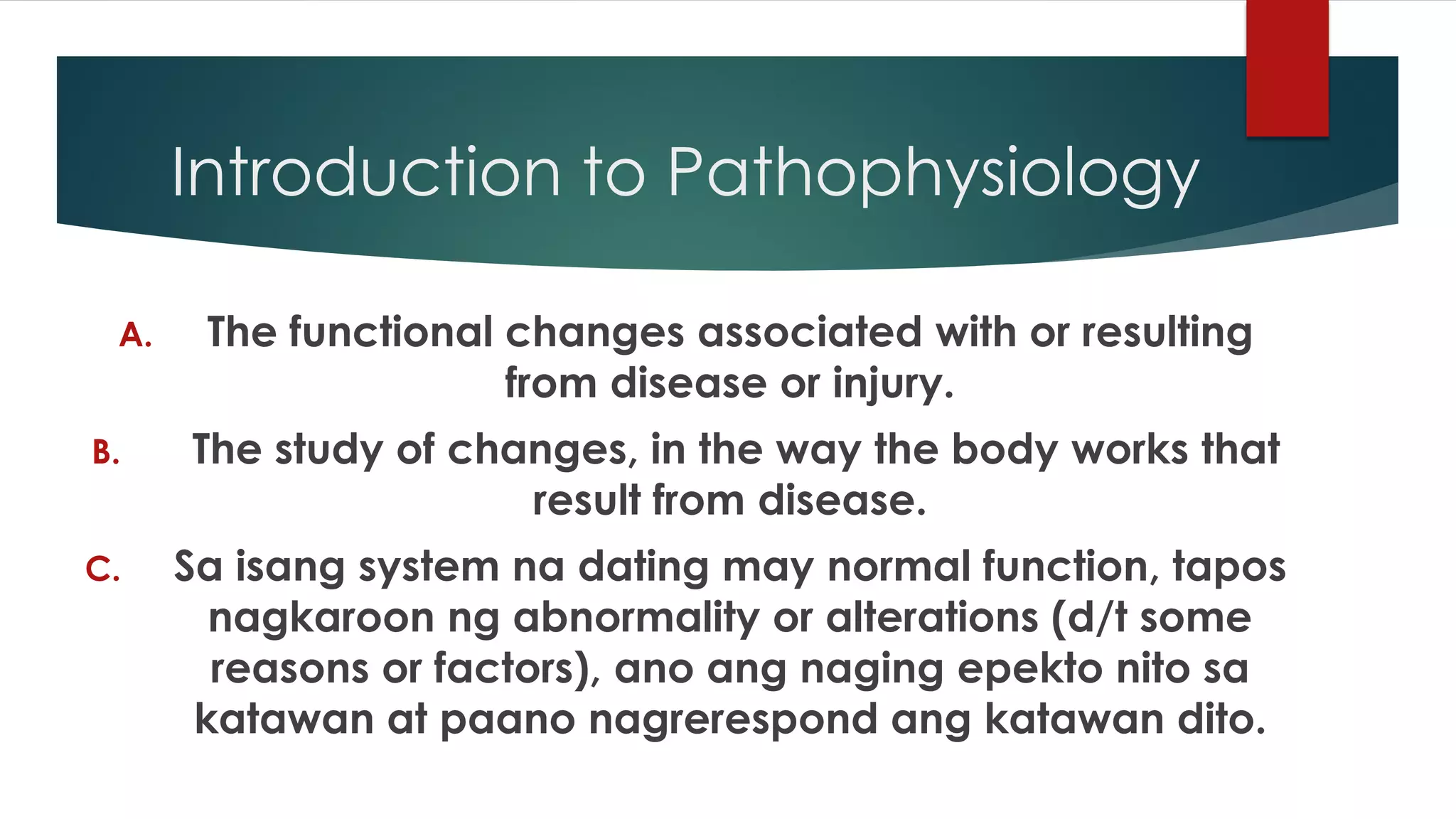
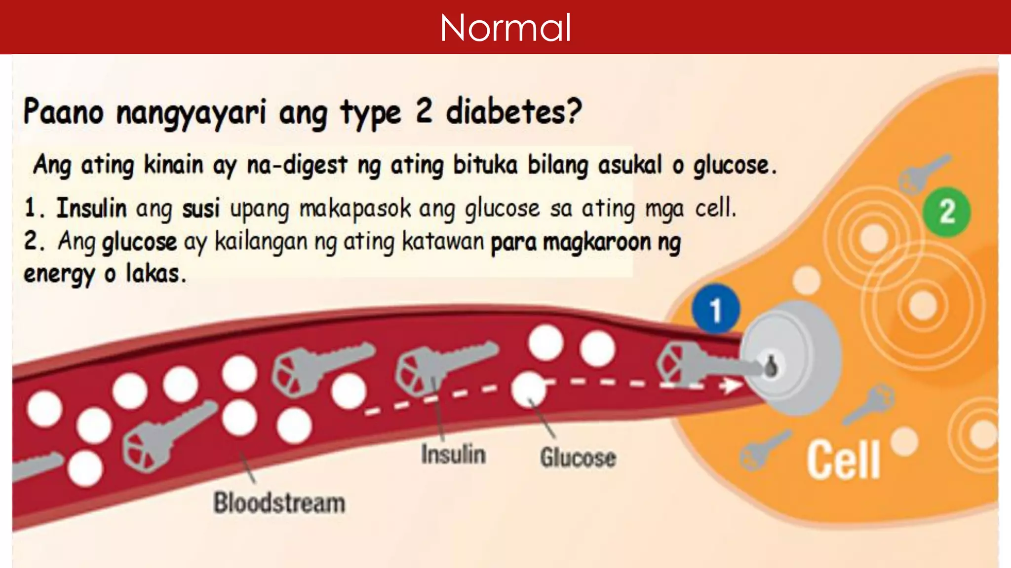
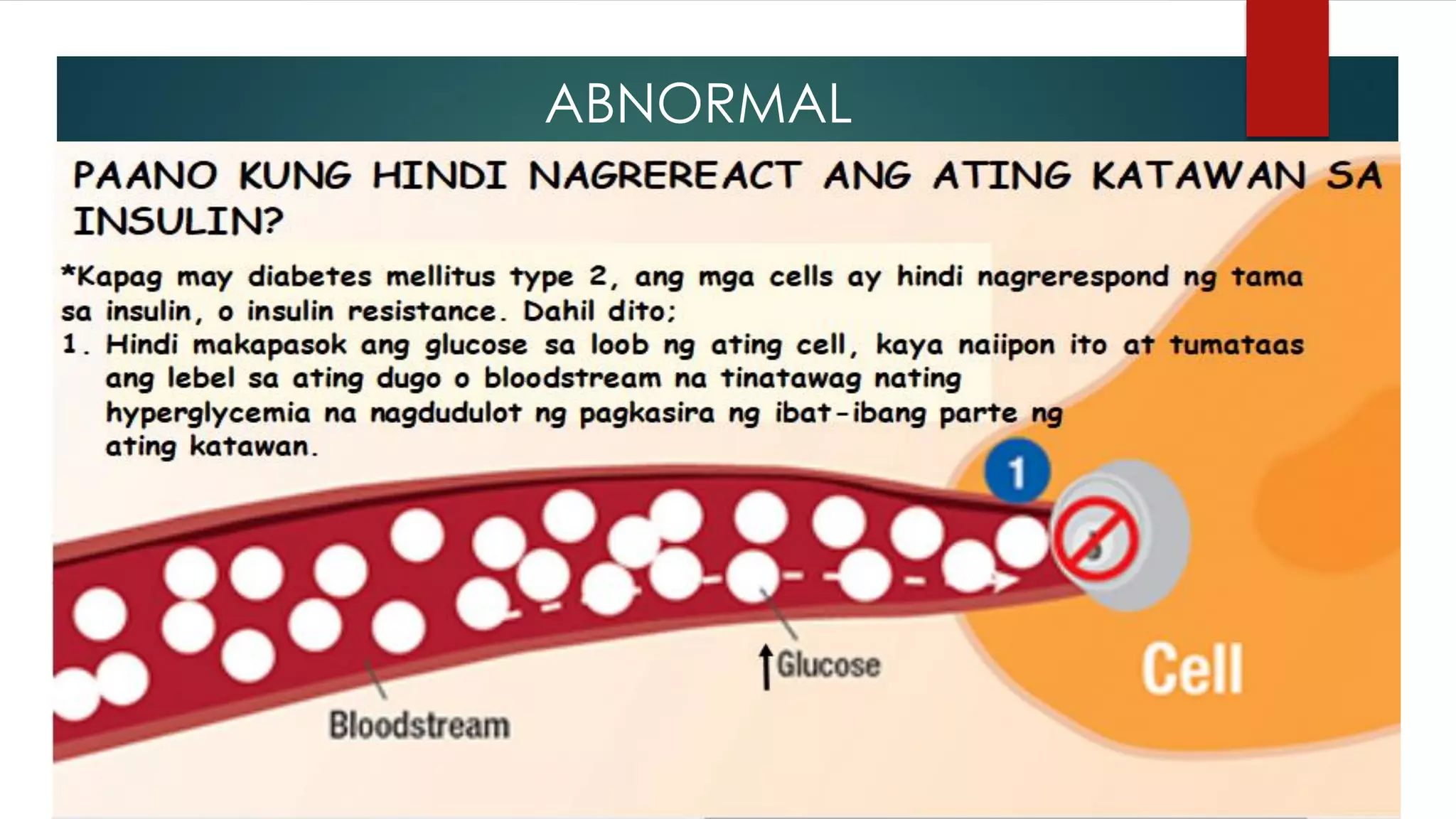
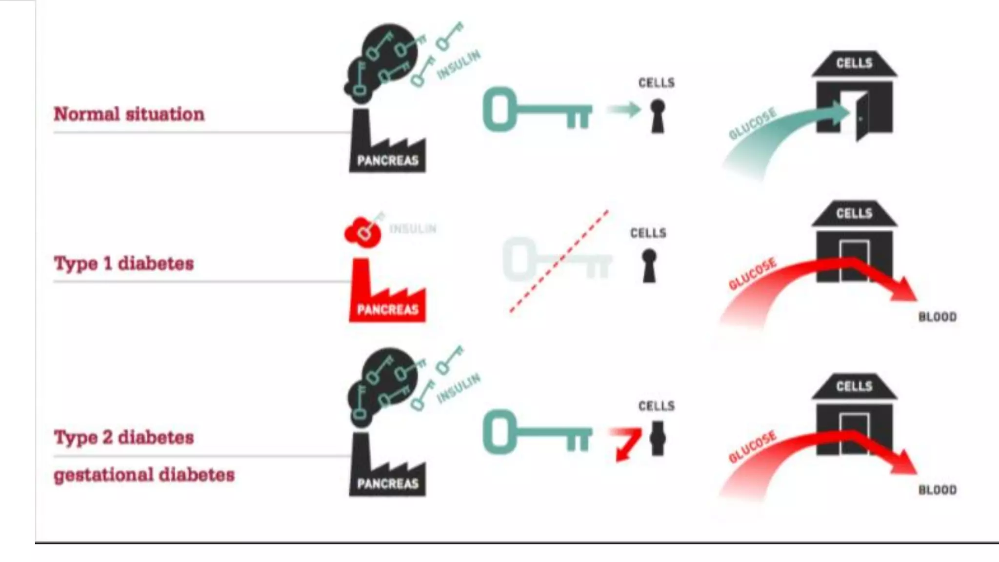
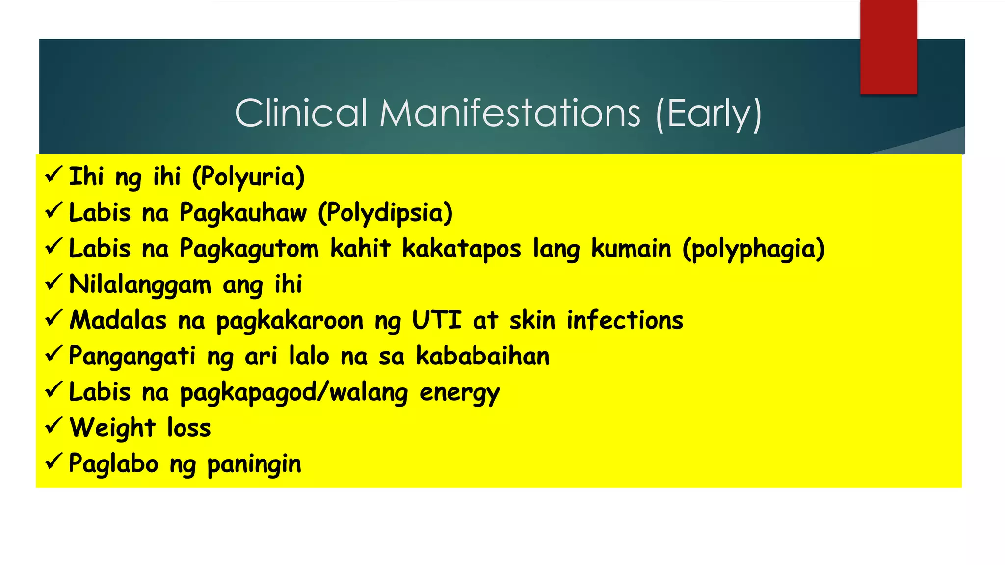
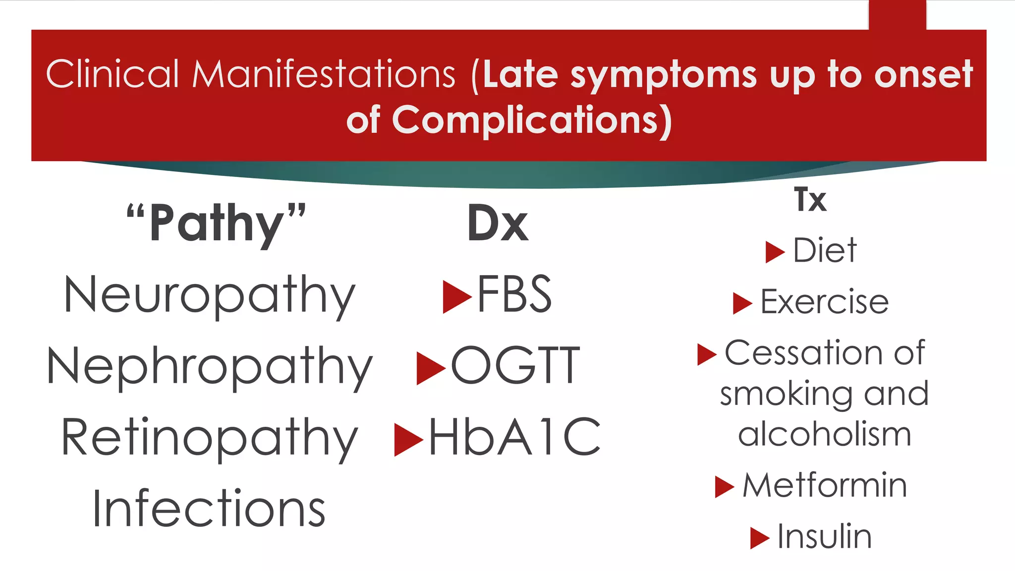
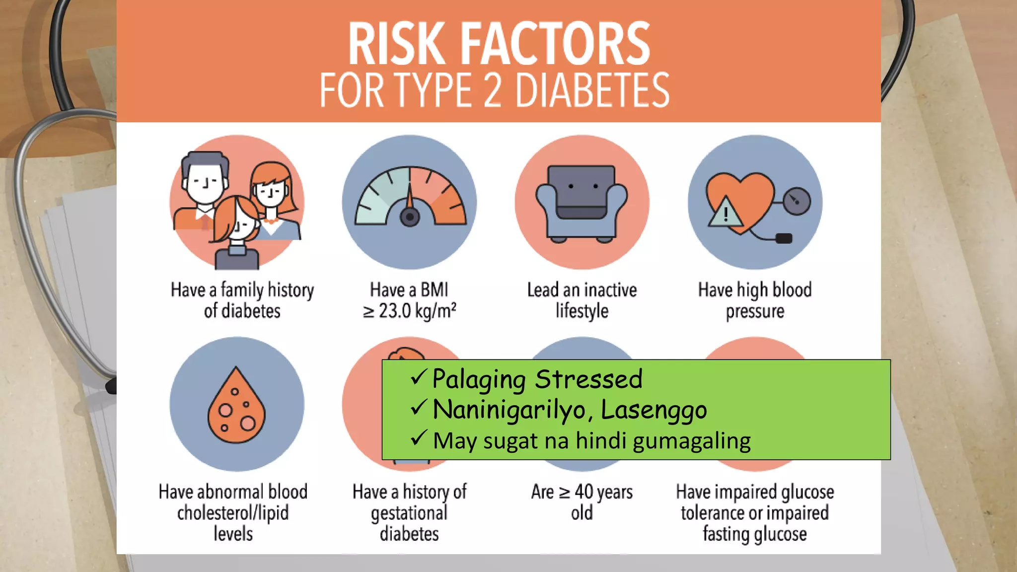
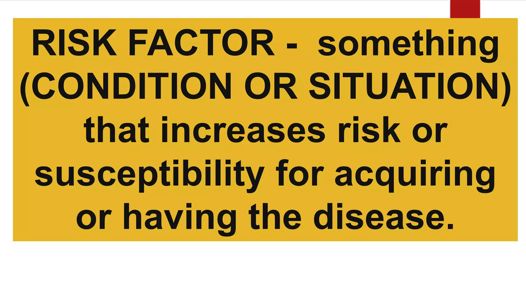
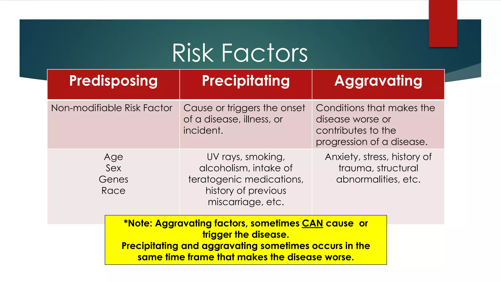
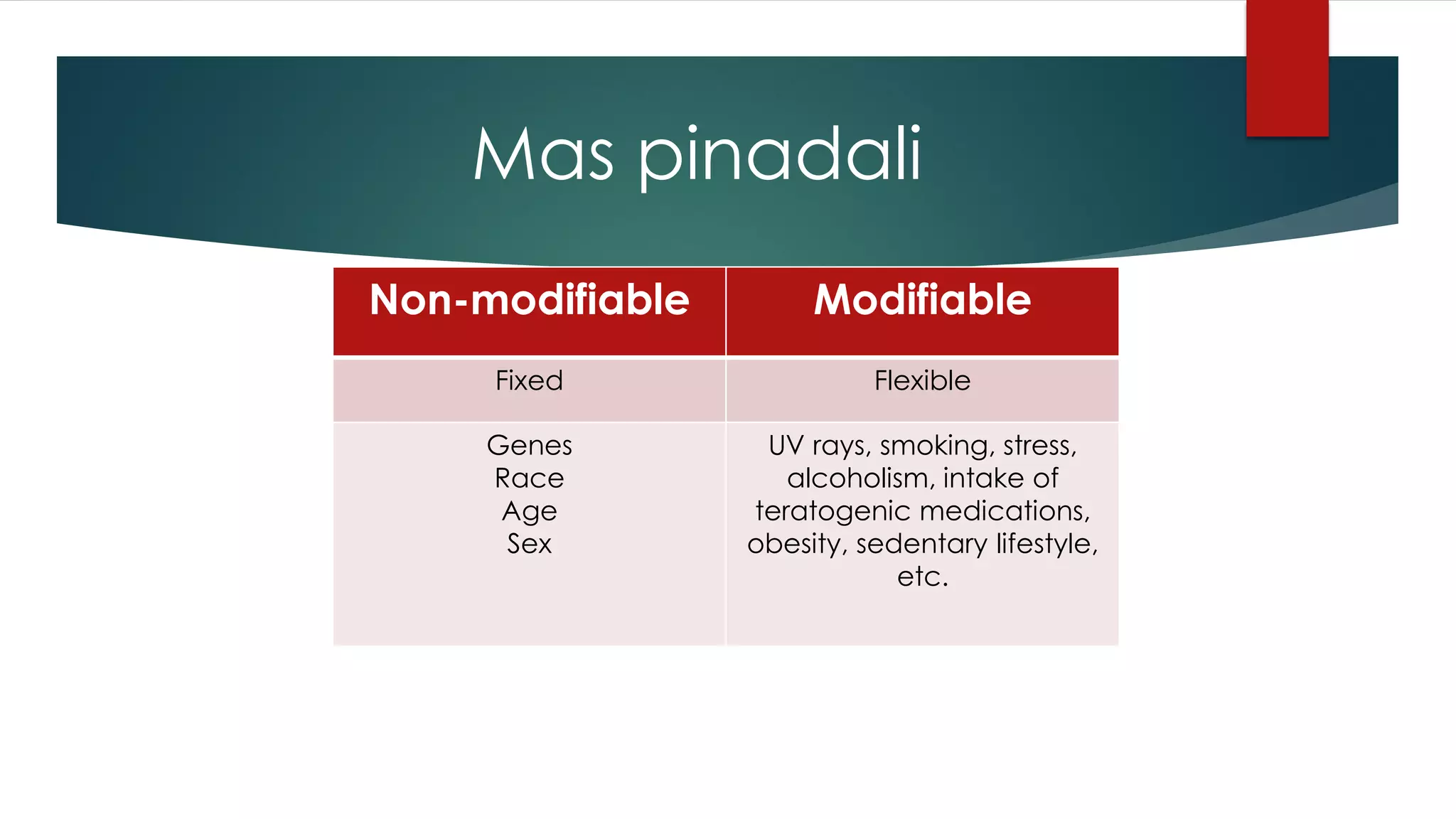
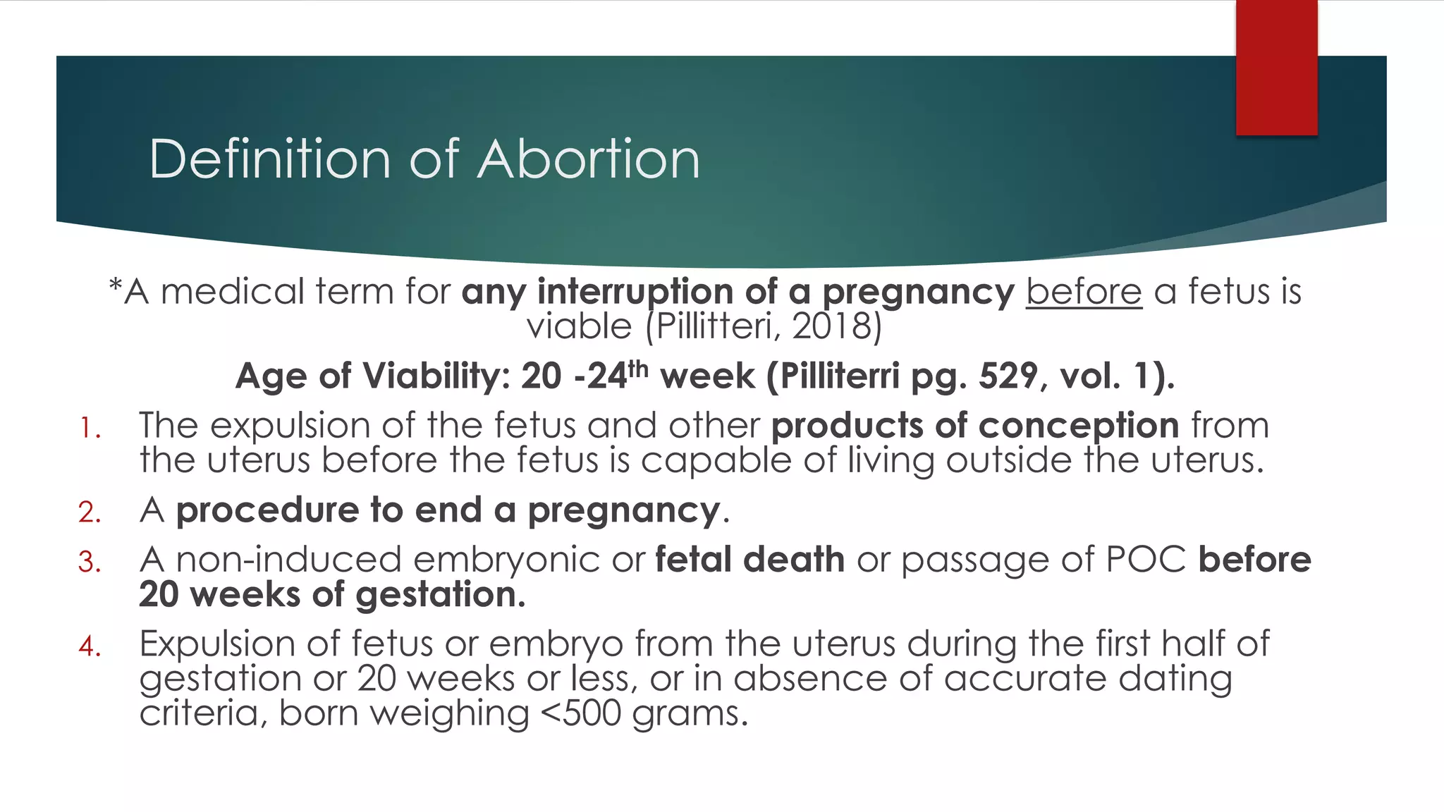
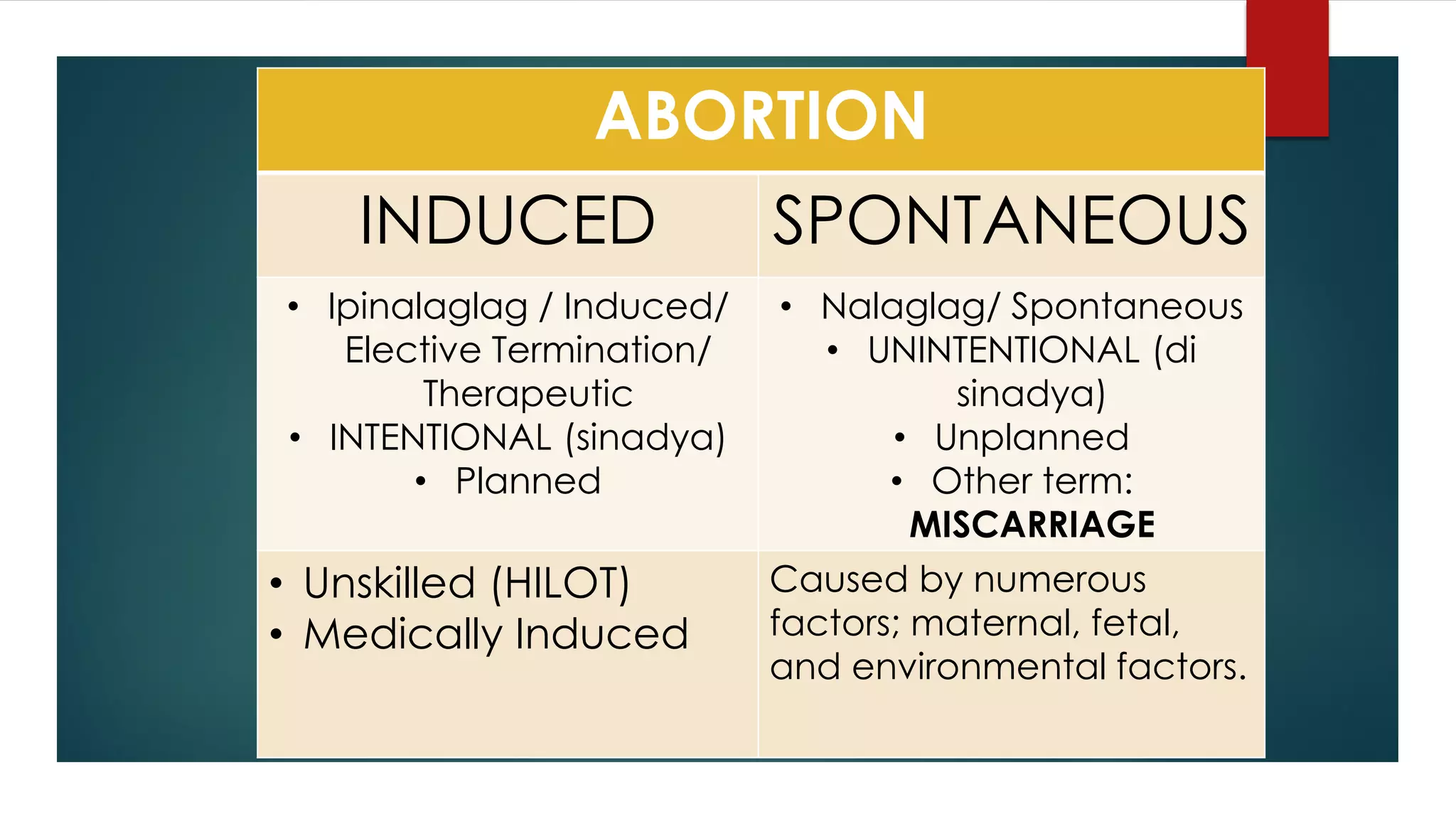
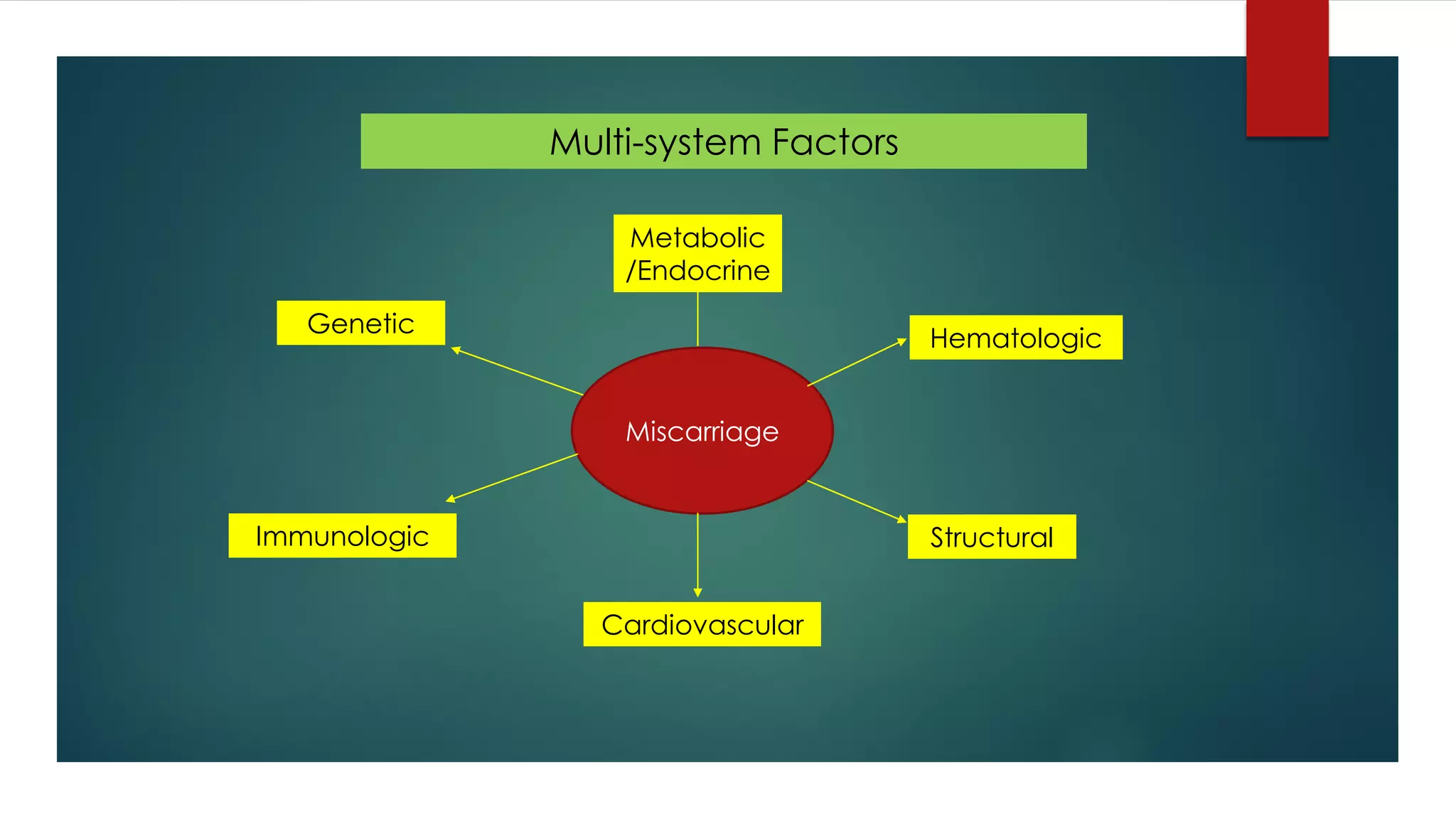
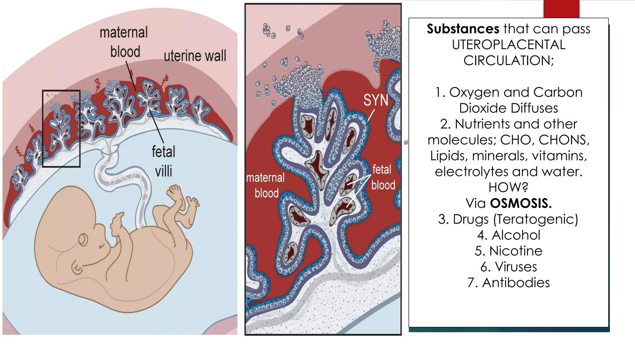
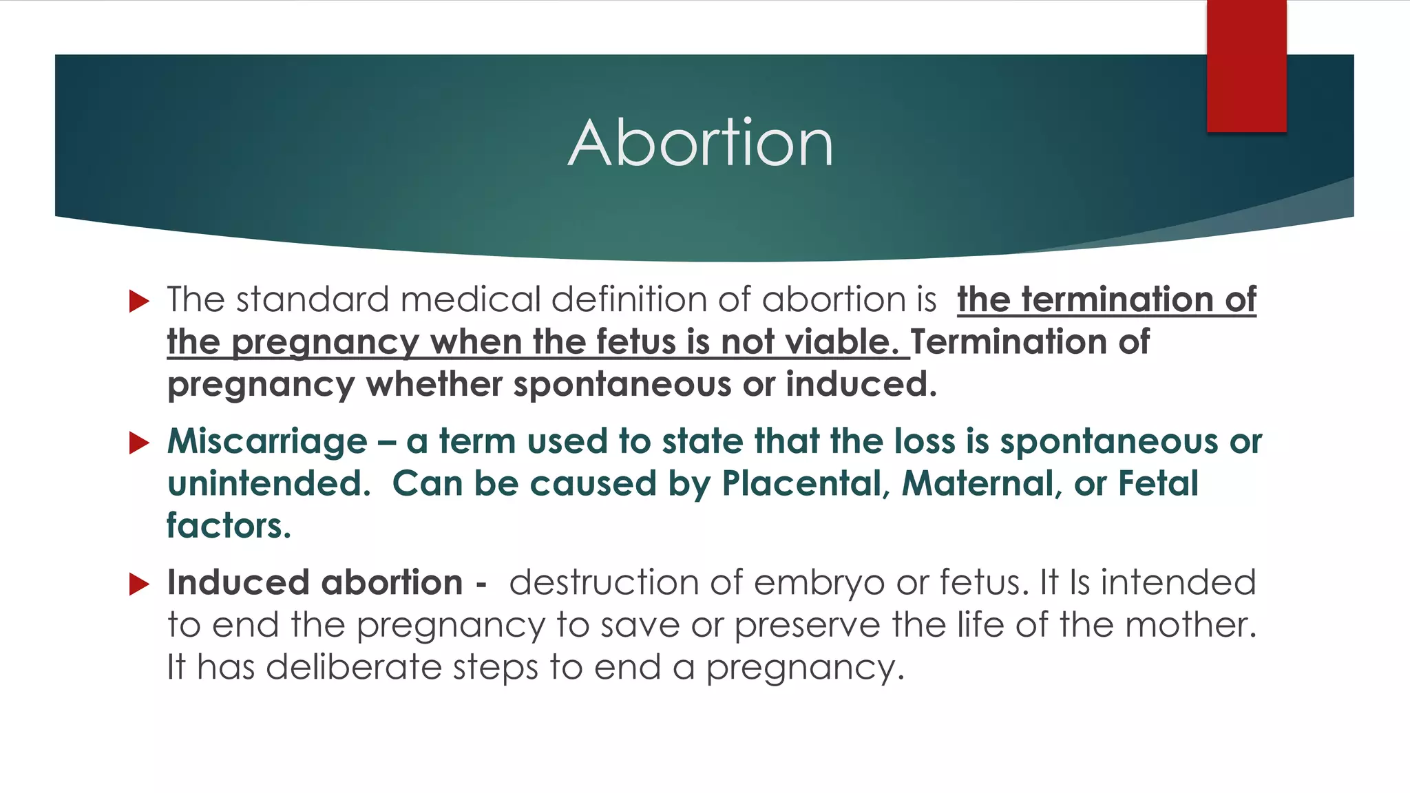
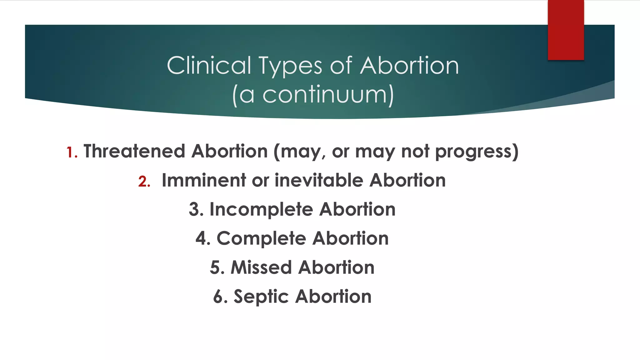
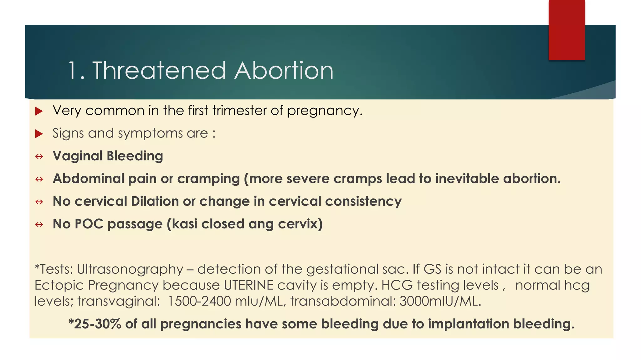
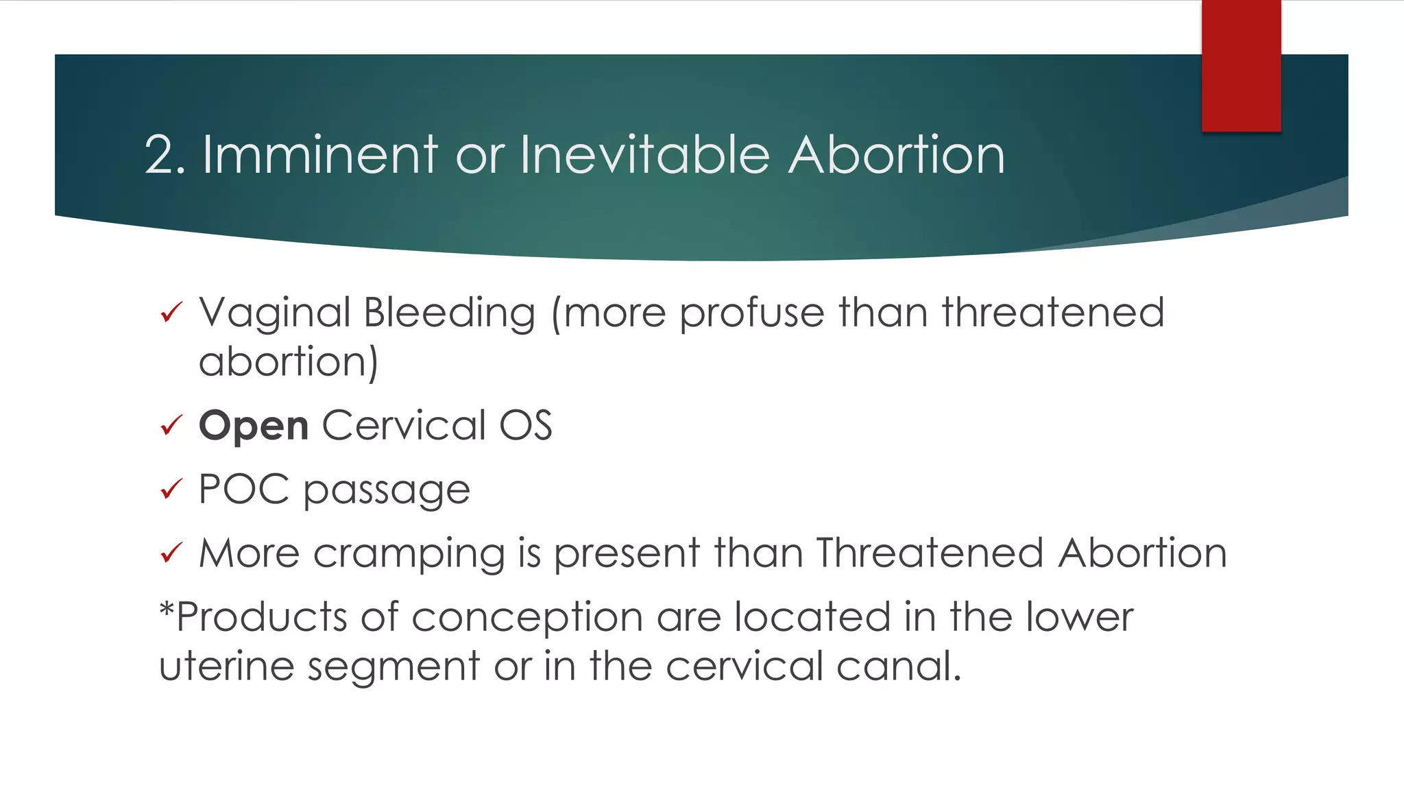
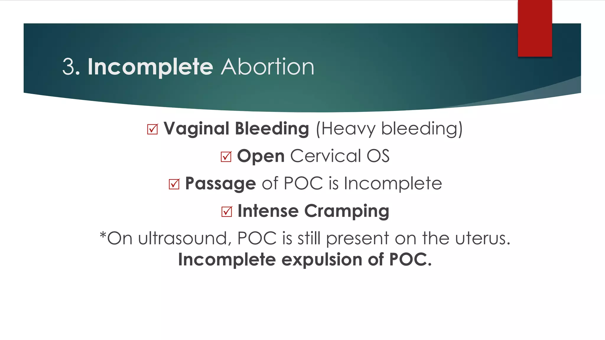
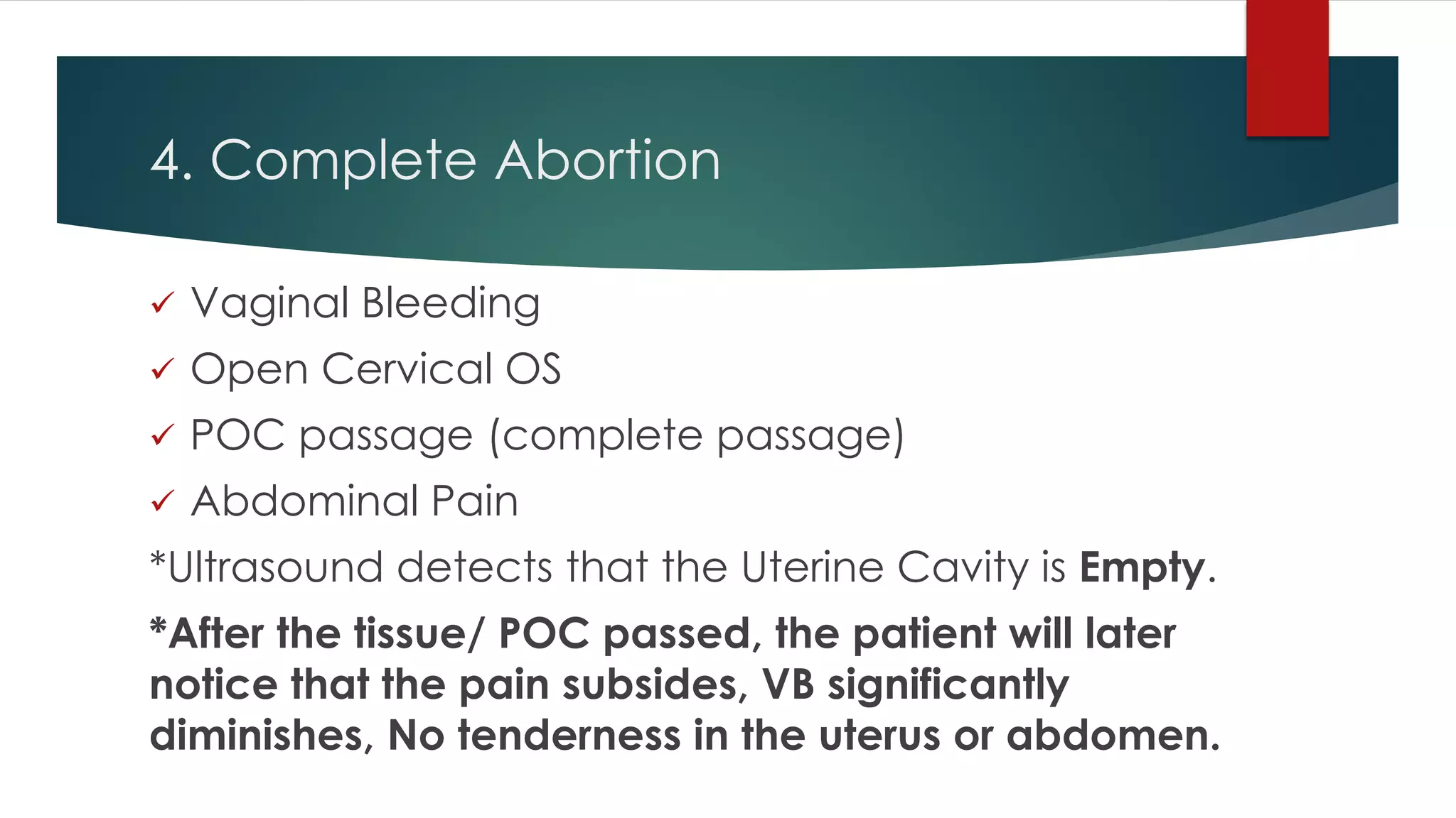
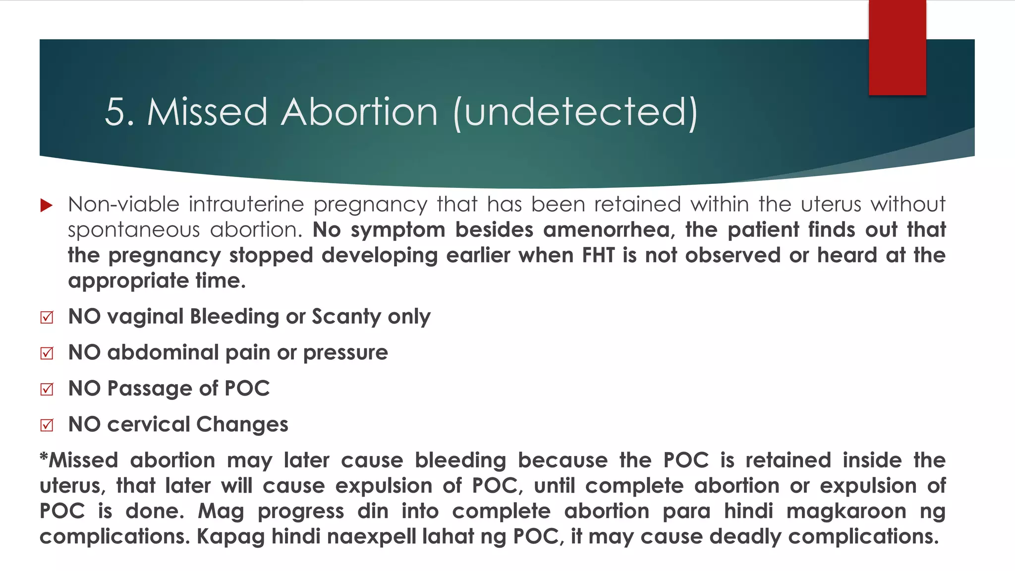
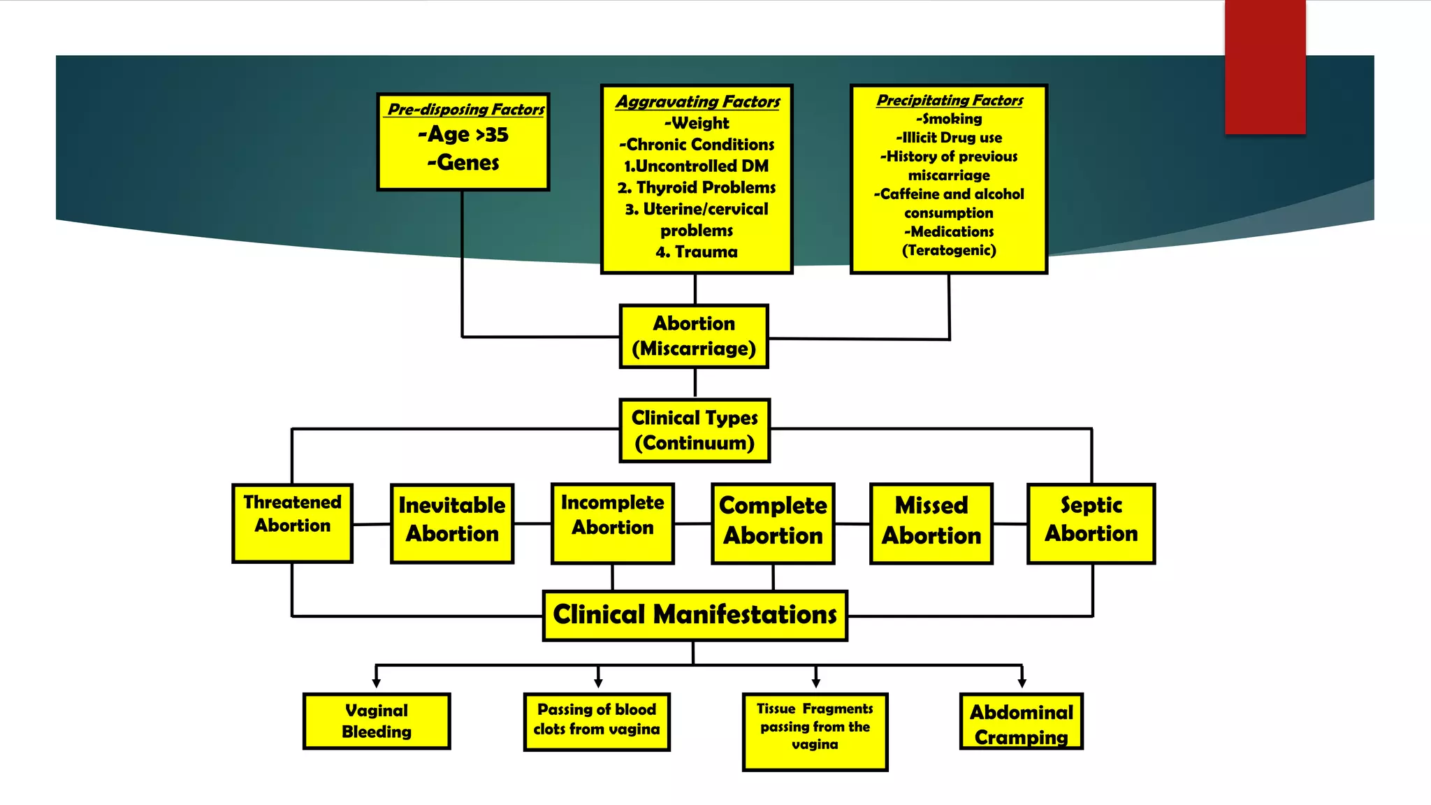
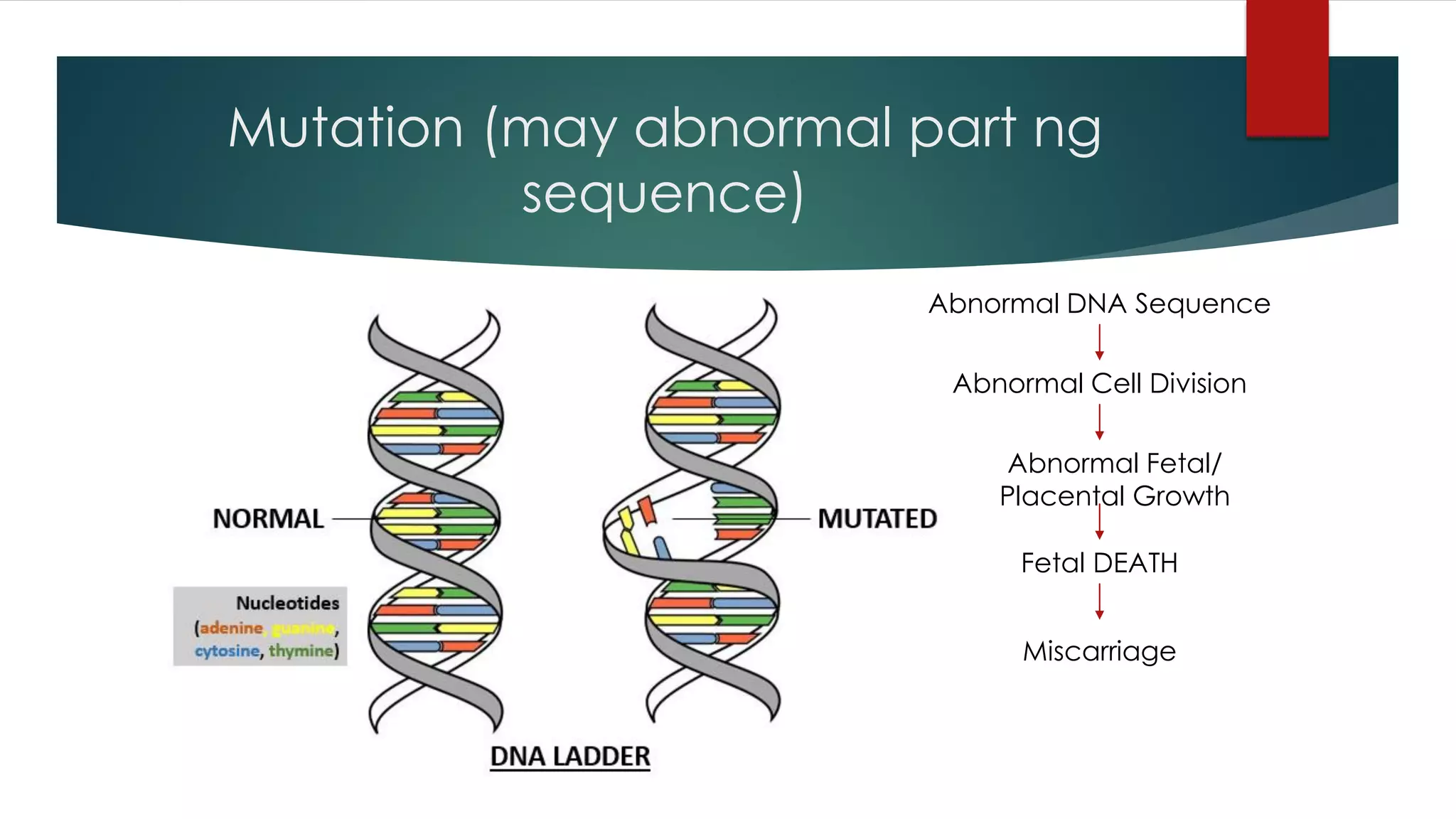
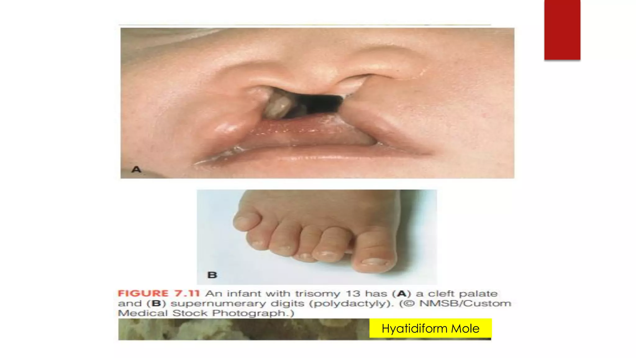
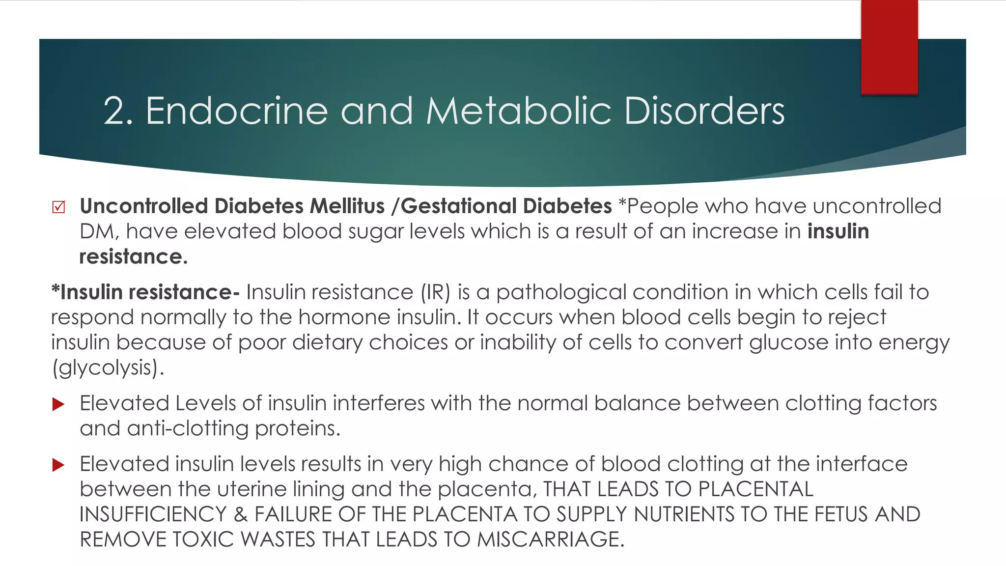
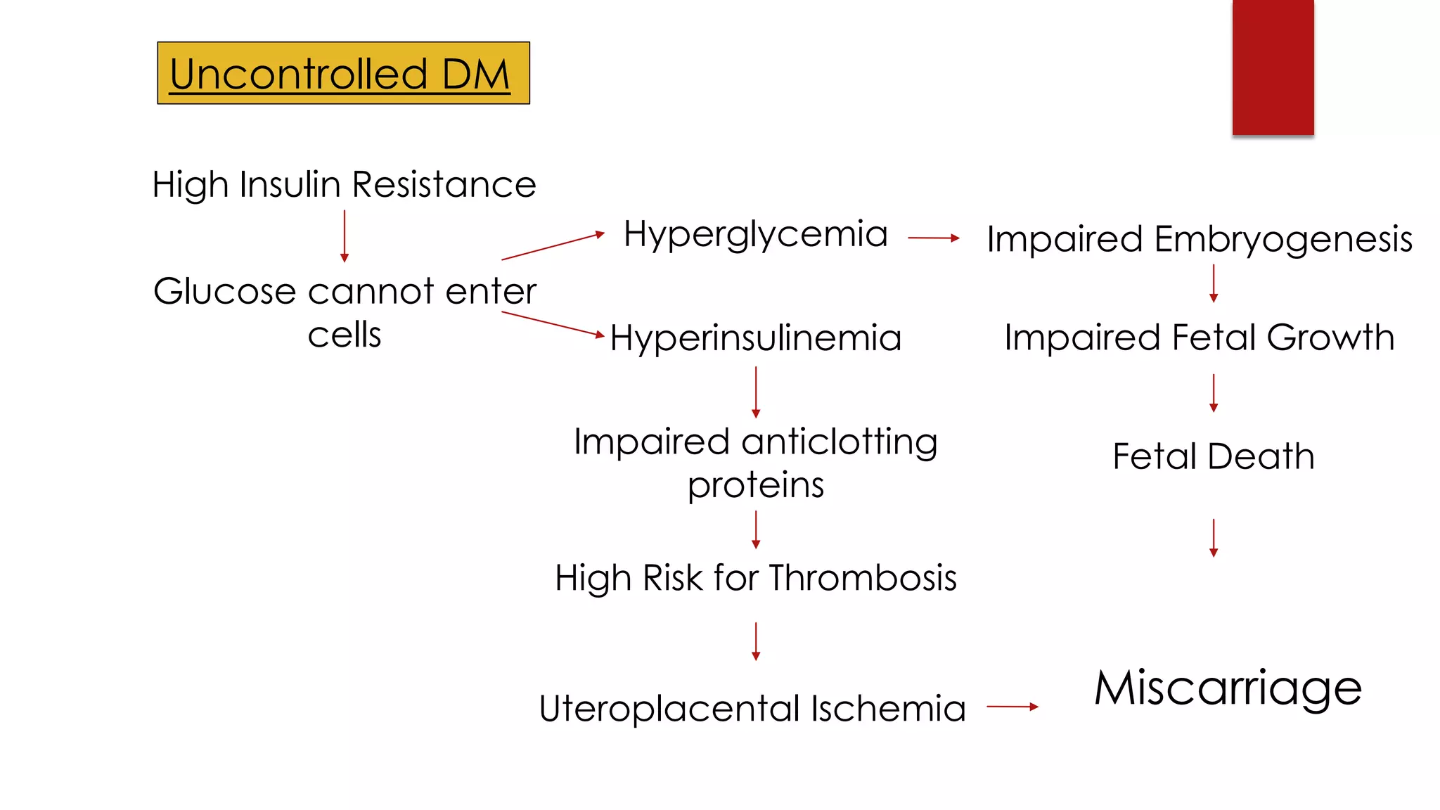
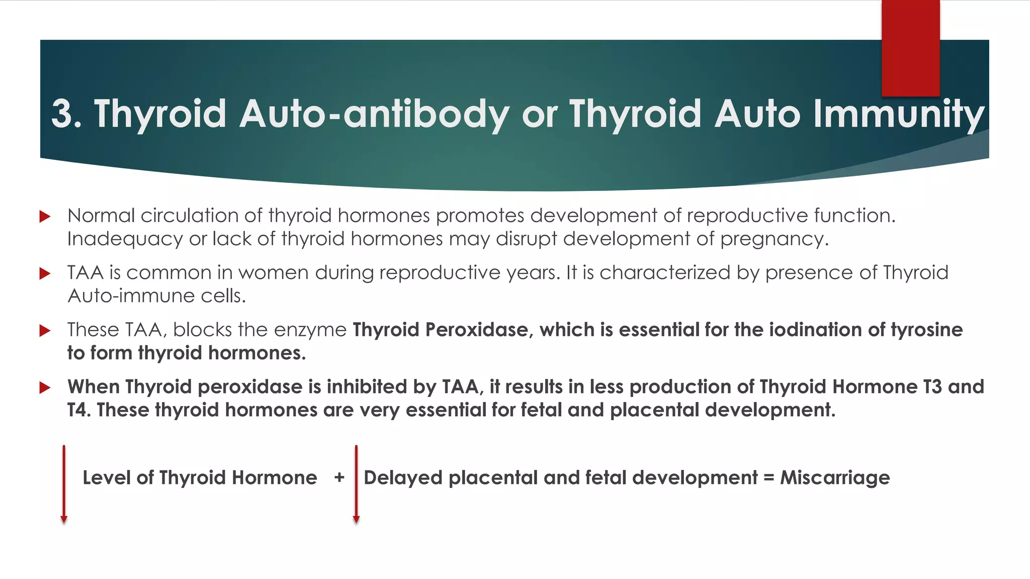
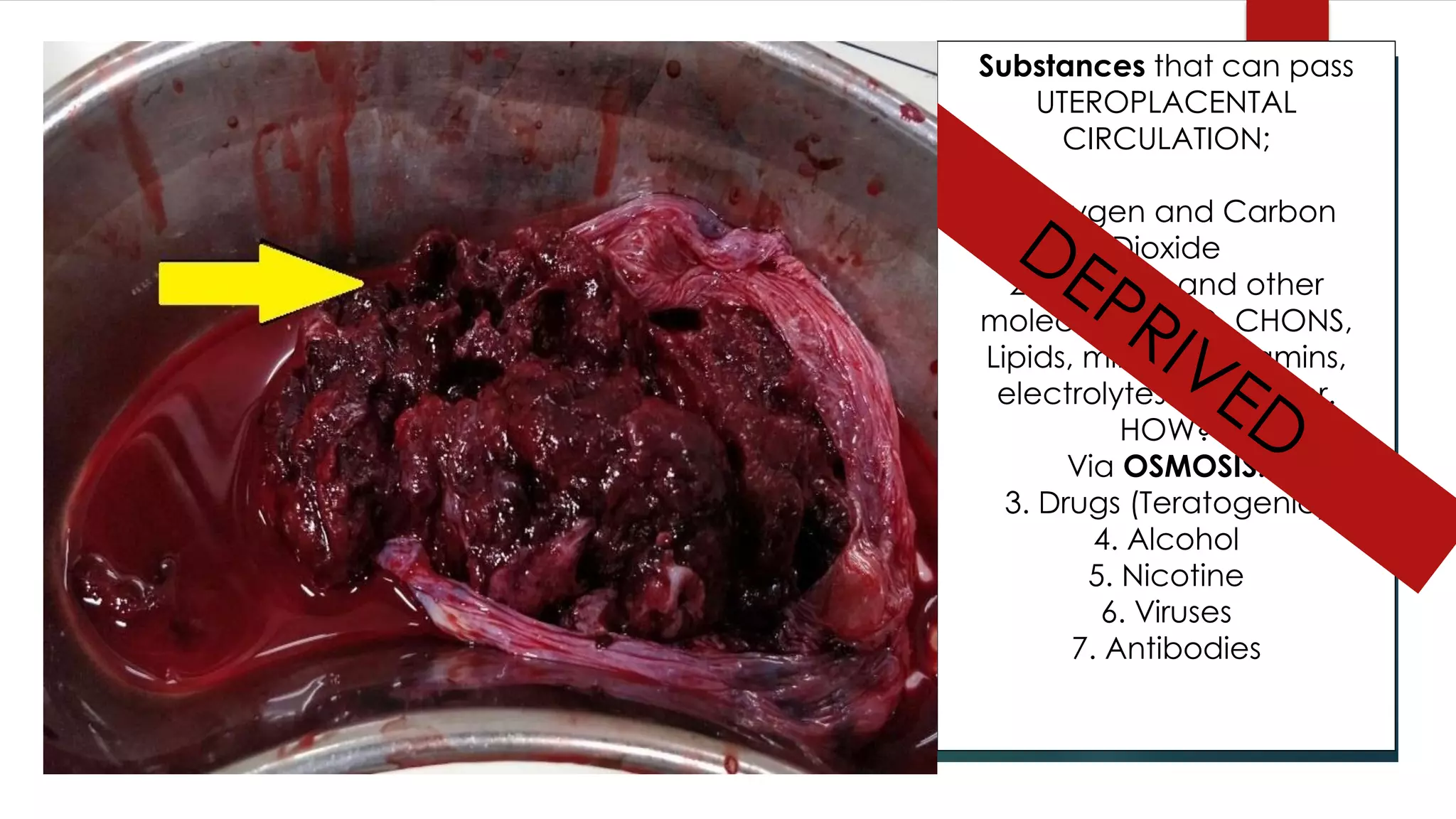
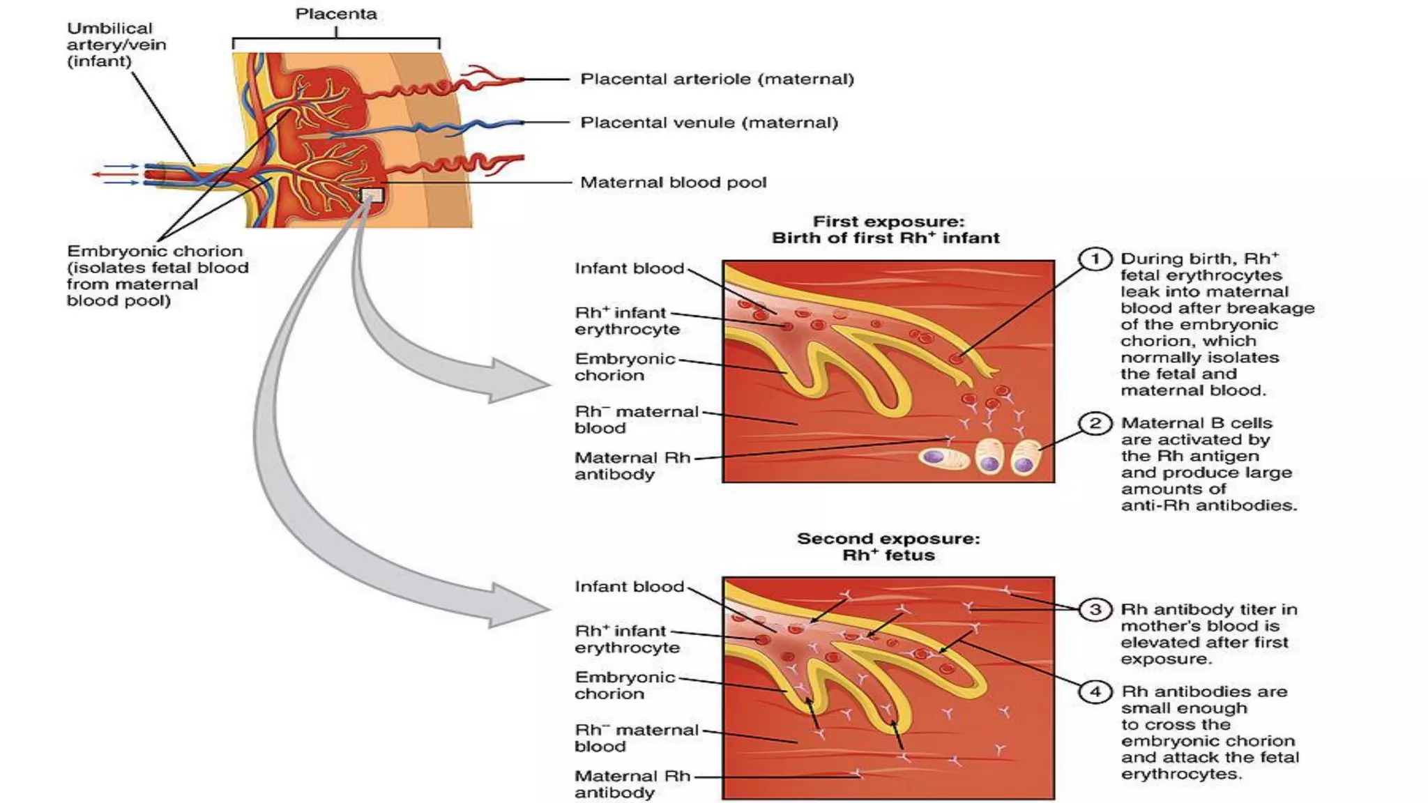
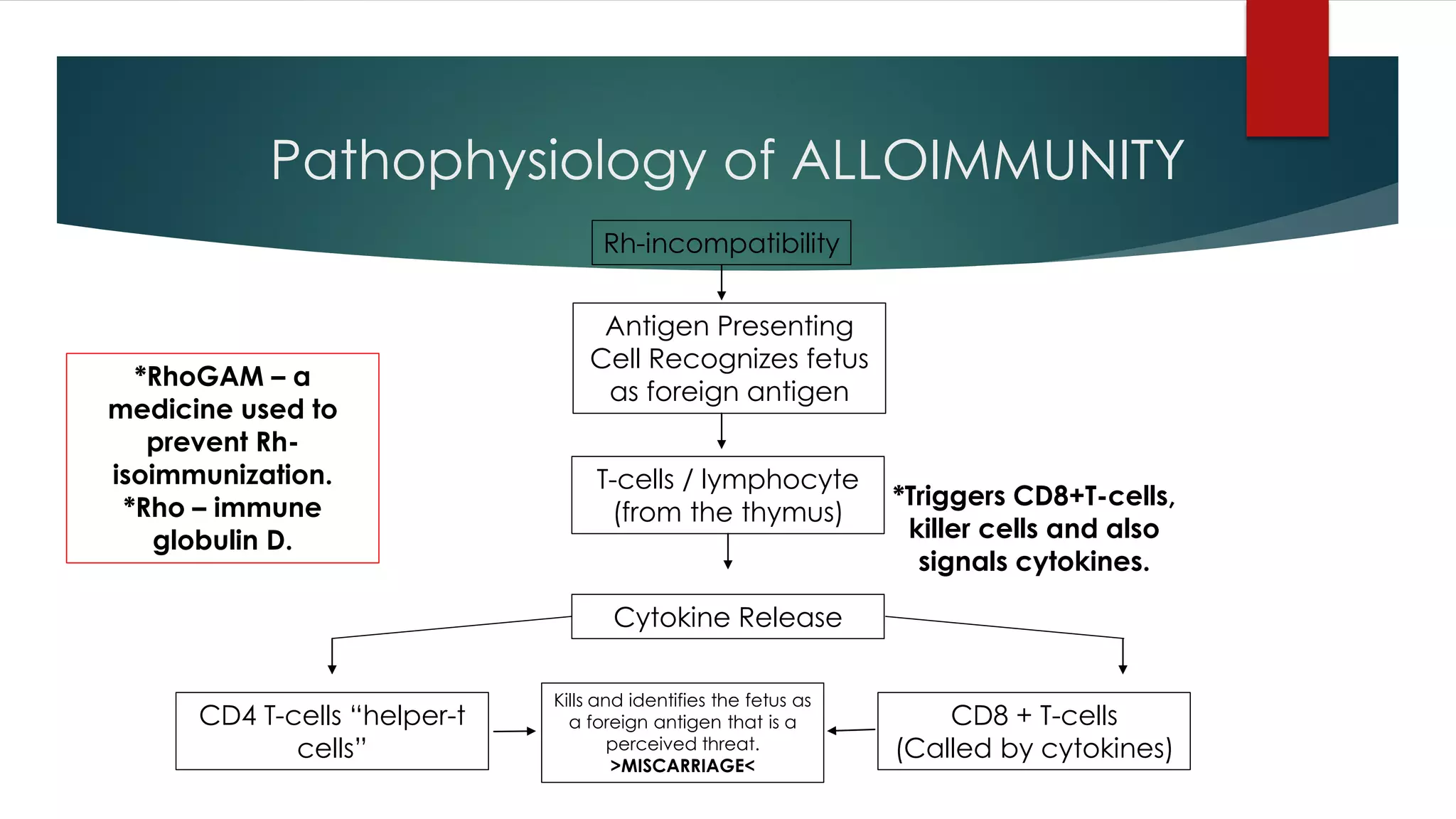
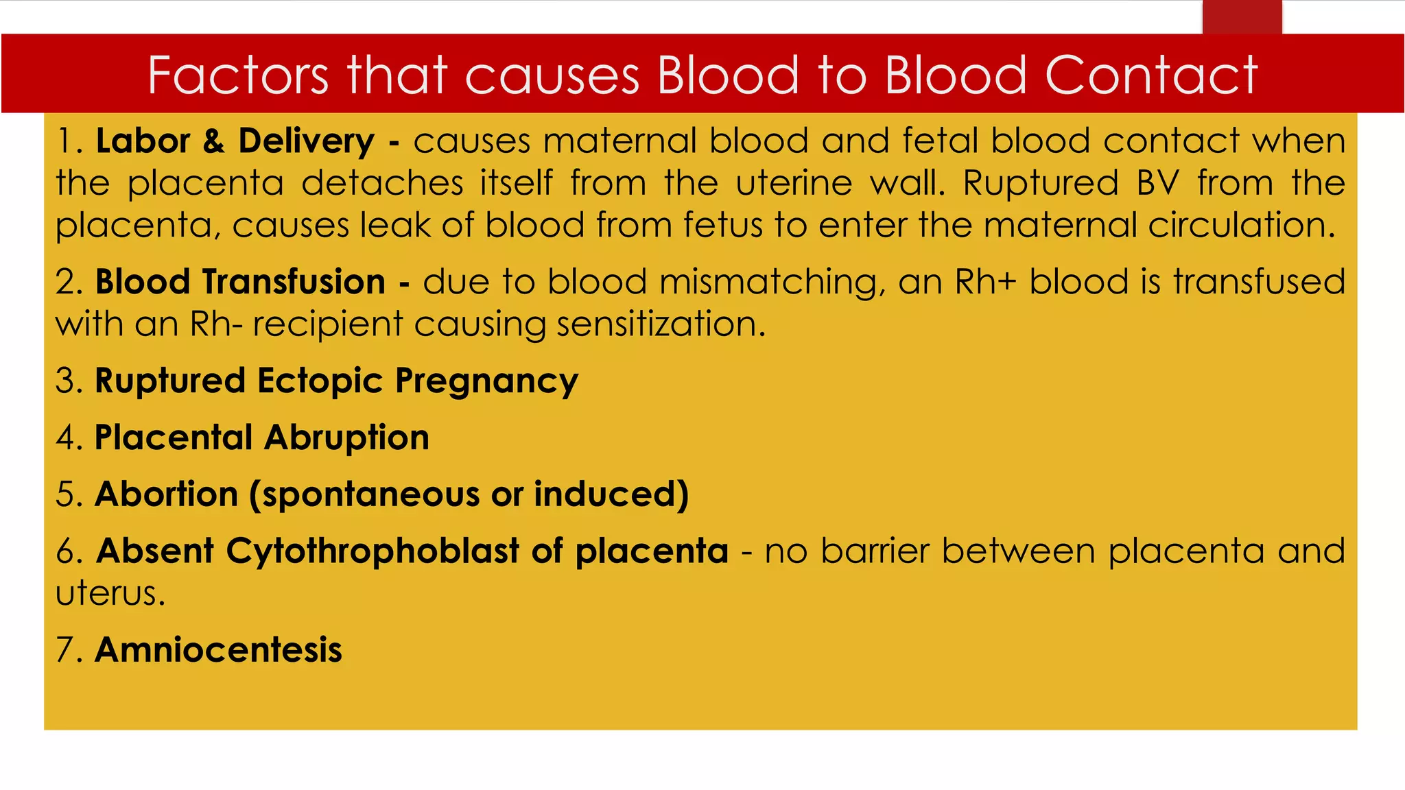
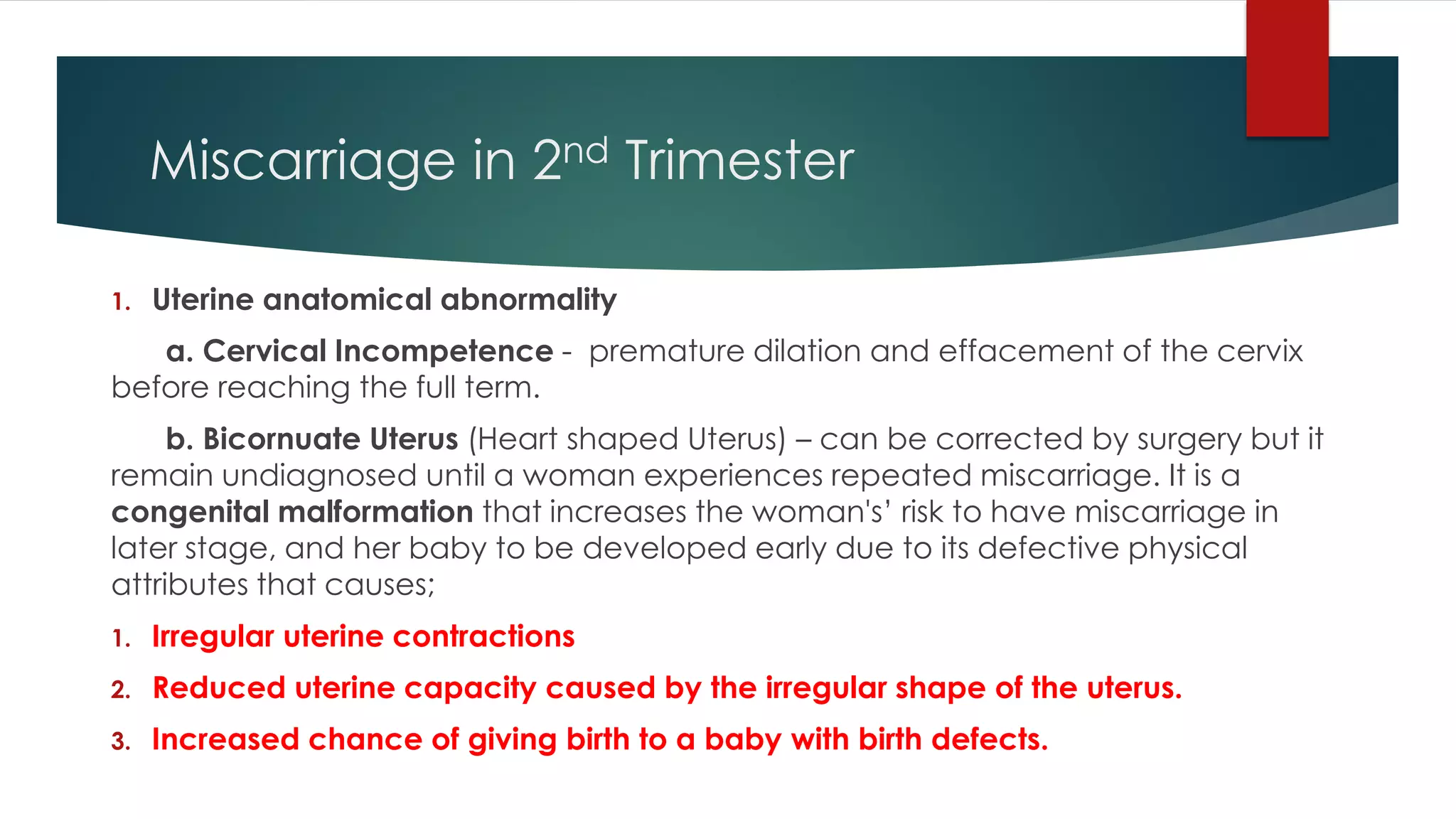
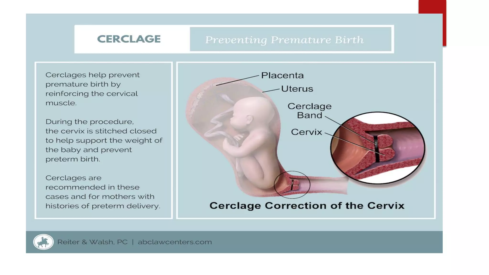
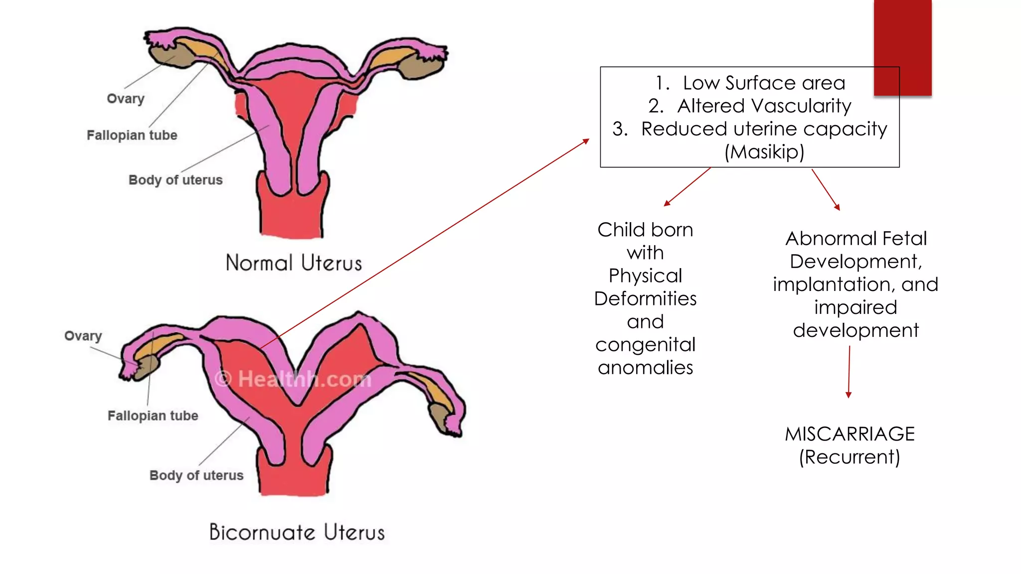
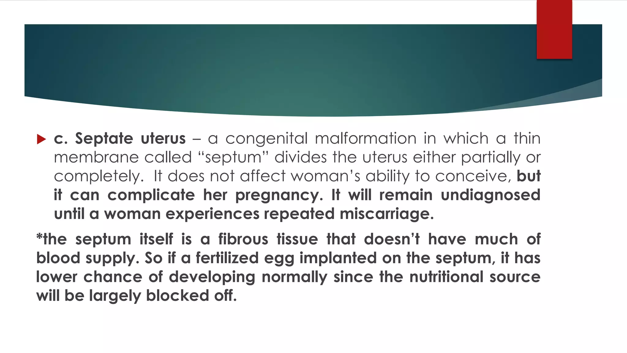
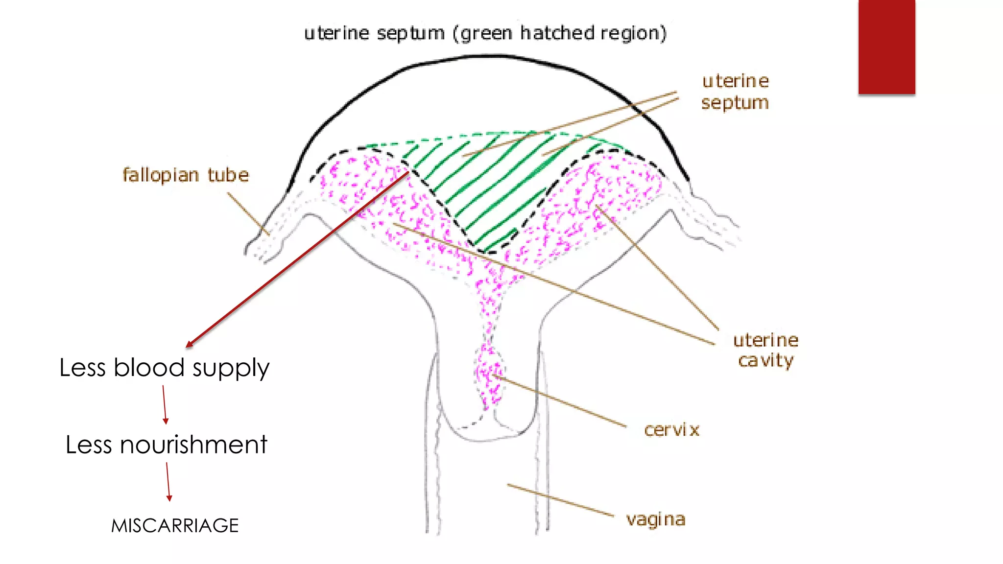
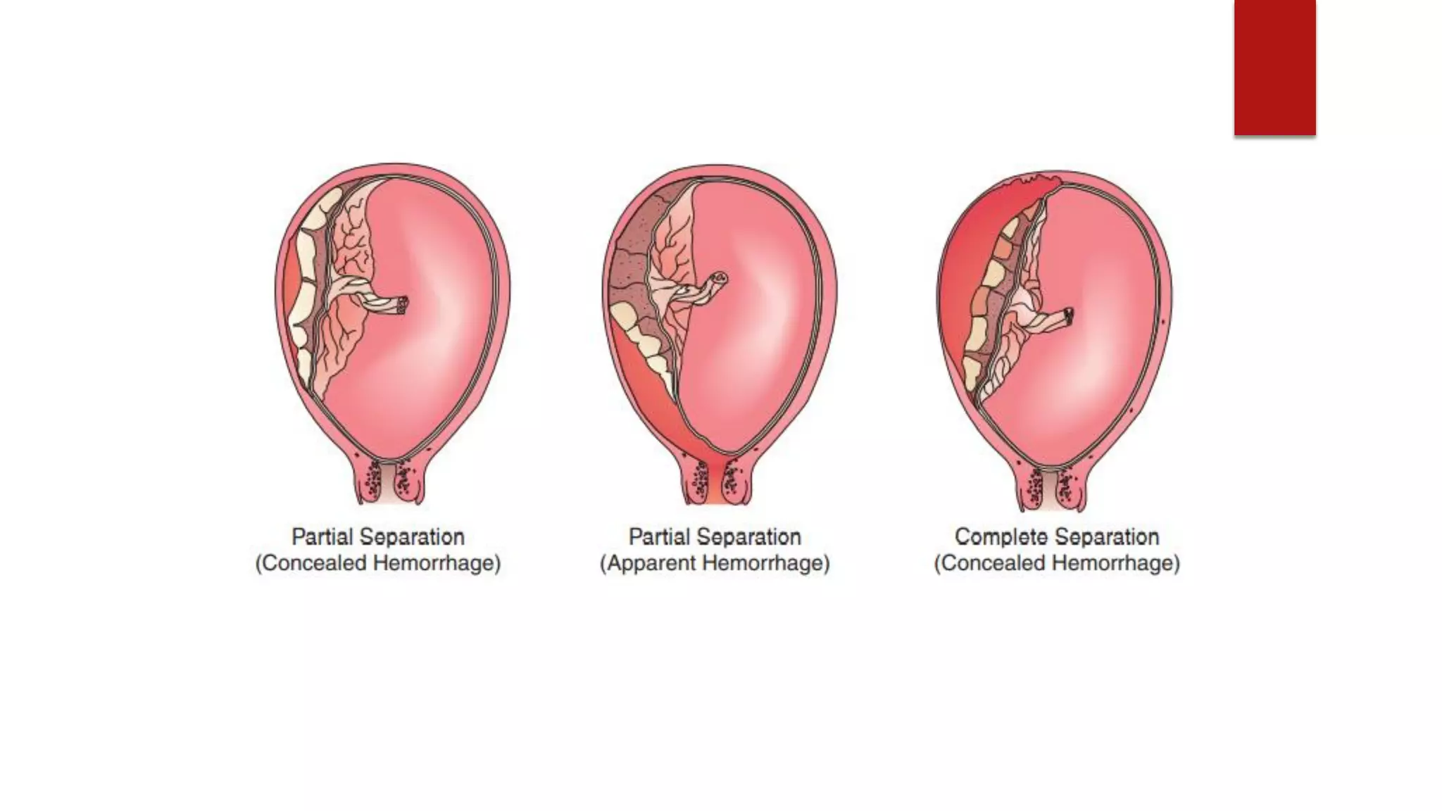
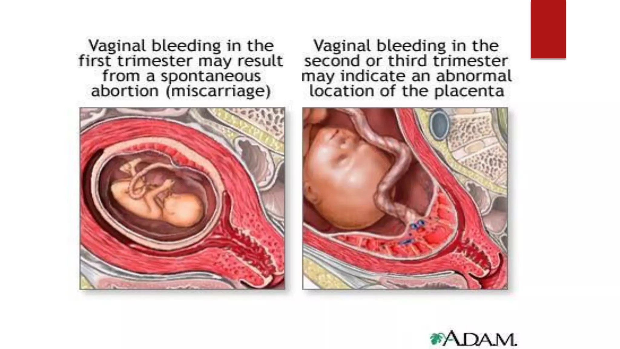
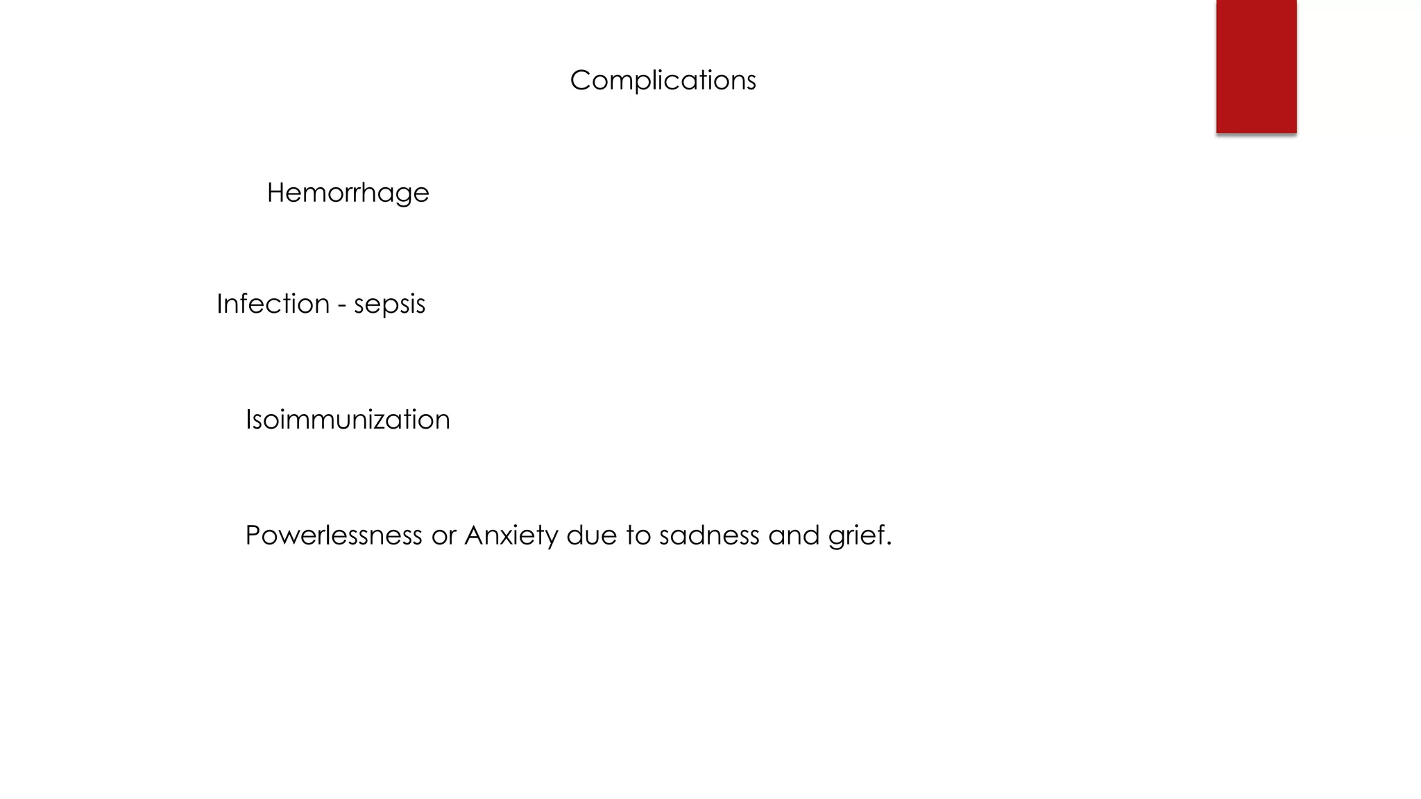
Introduction to
Pathophysiology
JERARD LLOYD B. DOMINGO
BSN 3A
Introduction to Pathophysiology
A. The functional changes associated with or resulting
from disease or injury.
B. The study of changes, in the way the body works that
result from disease.
C. Sa isang system na dating may normal function, tapos
nagkaroon ng abnormality or alterations (d/t some
reasons or factors), ano ang naging epekto nito sa
katawan at paano nagrerespond ang katawan dito.
Normal
ABNORMAL
Clinical Manifestations (Early)
“3PG”
Polyuria
Polyphagia
Polydipsia
Glycosuria
 Ihi ng ihi (Polyuria)
 Labis na Pagkauhaw (Polydipsia)
 Labis na Pagkagutom kahit kakatapos lang kumain (polyphagia)
 Nilalanggam ang ihi
 Madalas na pagkakaroon ng UTI at skin infections
 Pangangati ng ari lalo na sa kababaihan
 Labis na pagkapagod/walang energy
 Weight loss
 Paglabo ng paningin
Clinical Manifestations (Late symptoms up to onset
of Complications)
“Pathy”
Neuropathy
Nephropathy
Retinopathy
Infections
Dx
FBS
OGTT
HbA1C
Tx
 Diet
 Exercise
 Cessation of
smoking and
alcoholism
 Metformin
 Insulin
Madali ba?
Bat nagkaganun?
Palaging Stressed
Naninigarilyo, Lasenggo
May sugat na hindi gumagaling
RISK FACTOR - something
(CONDITION OR SITUATION)
that increases risk or
susceptibility for acquiring
or having the disease.
Risk Factors
Predisposing Precipitating Aggravating
Non-modifiable Risk Factor Cause or triggers the onset
of a disease, illness, or
incident.
Conditions that makes the
disease worse or
contributes to the
progression of a disease.
Age
Sex
Genes
Race
UV rays, smoking,
alcoholism, intake of
teratogenic medications,
history of previous
miscarriage, etc.
Anxiety, stress, history of
trauma, structural
abnormalities, etc.
*Note: Aggravating factors, sometimes CAN cause or
trigger the disease.
Precipitating and aggravating sometimes occurs in the
same time frame that makes the disease worse.
Mas pinadali
Non-modifiable Modifiable
Fixed Flexible
Genes
Race
Age
Sex
UV rays, smoking, stress,
alcoholism, intake of
teratogenic medications,
obesity, sedentary lifestyle,
etc.
Learning Objectives:
1. Define abortion and miscarriage
2. Differentiate abortion vs. miscarriage
3. Identify factors that caused the disease
4. Trace the pathophysiology of the disease
5. Trace the symptomatology of the disease
6. Identify the nursing and medical management for the
disease
7. Identify complications if not managed
Abortion
BY: JERARD LLOYD B. DOMINGO
BSN - 2A
Definition of Abortion
*A medical term for any interruption of a pregnancy before a fetus is
viable (Pillitteri, 2018)
Age of Viability: 20 -24th week (Pilliterri pg. 529, vol. 1).
1. The expulsion of the fetus and other products of conception from
the uterus before the fetus is capable of living outside the uterus.
2. A procedure to end a pregnancy.
3. A non-induced embryonic or fetal death or passage of POC before
20 weeks of gestation.
4. Expulsion of fetus or embryo from the uterus during the first half of
gestation or 20 weeks or less, or in absence of accurate dating
criteria, born weighing <500 grams.
ABORTION
INDUCED SPONTANEOUS
• Ipinalaglag / Induced/
Elective Termination/
Therapeutic
• INTENTIONAL (sinadya)
• Planned
• Nalaglag/ Spontaneous
• UNINTENTIONAL (di
sinadya)
• Unplanned
• Other term:
MISCARRIAGE
• Unskilled (HILOT)
• Medically Induced
Caused by numerous
factors; maternal, fetal,
and environmental factors.
Miscarriage
Genetic
Immunologic
Metabolic
/Endocrine
Cardiovascular
Hematologic
Structural
Multi-system Factors
Pre-disposing Factors
-Age >35
-Genes
Aggravating Factors
-Weight
-Chronic Conditions
1.Uncontrolled DM
2. Thyroid Problems
3. Uterine/cervical
problems
4. Trauma
Precipitating Factors
-Smoking
-Illicit Drug use
-History of previous
miscarriage
-Caffeine and alcohol
consumption
-Medications
(Teratogenic)
Abortion
(Miscarriage)
Clinical Types
(Continuum)
Septic
Abortion
Complete
Abortion
Missed
Abortion
Incomplete
Abortion
Clinical Manifestations
Threatened
Abortion
Inevitable
Abortion
Vaginal
Bleeding
Abdominal
Cramping
Passing of blood
clots from vagina
Tissue Fragments
passing from the
vagina
2nd
Trimester
1st
Trimester
Miscarriage
Bicornuate uterus &
Septate uterus
Syphilis &
Bacterial Vaginosis
Hypertension &
Heart disease
Cervical
Incompetence
Idiopathic
Infection
Chronic maternal
illness
Anatomic
abnormalities
Inherited
Thrombophilia
Infection Induced
Auto-immunity
Anti phospholipid
antibody syndrome
Protein C or Protein
S Deficiency
Uncontrolled
Diabetes mellitus
Chromosomal
translocation
Allo-immune factors
Thyroid auto antibody &
Luteal phase defect
Hyperhomocystenemia &
Prothrombin gene
mutation
Immune Factors
Genetics
Endocrine and
Metabolic
Substances that can pass
UTEROPLACENTAL
CIRCULATION;
1. Oxygen and Carbon
Dioxide Diffuses
2. Nutrients and other
molecules; CHO, CHONS,
Lipids, minerals, vitamins,
electrolytes and water.
HOW?
Via OSMOSIS.
3. Drugs (Teratogenic)
4. Alcohol
5. Nicotine
6. Viruses
7. Antibodies
Abortion
 The standard medical definition of abortion is the termination of
the pregnancy when the fetus is not viable. Termination of
pregnancy whether spontaneous or induced.
 Miscarriage – a term used to state that the loss is spontaneous or
unintended. Can be caused by Placental, Maternal, or Fetal
factors.
 Induced abortion - destruction of embryo or fetus. It Is intended
to end the pregnancy to save or preserve the life of the mother.
It has deliberate steps to end a pregnancy.
Clinical Types of Abortion
(a continuum)
1. Threatened Abortion (may, or may not progress)
2. Imminent or inevitable Abortion
3. Incomplete Abortion
4. Complete Abortion
5. Missed Abortion
6. Septic Abortion
1. Threatened Abortion
 Very common in the first trimester of pregnancy.
 Signs and symptoms are :
 Vaginal Bleeding
 Abdominal pain or cramping (more severe cramps lead to inevitable abortion.
 No cervical Dilation or change in cervical consistency
 No POC passage (kasi closed ang cervix)
*Tests: Ultrasonography – detection of the gestational sac. If GS is not intact it can be an
Ectopic Pregnancy because UTERINE cavity is empty. HCG testing levels , normal hcg
levels; transvaginal: 1500-2400 mIu/ML, transabdominal: 3000mIU/ML.
*25-30% of all pregnancies have some bleeding due to implantation bleeding.
2. Imminent or Inevitable Abortion
 Vaginal Bleeding (more profuse than threatened
abortion)
 Open Cervical OS
 POC passage
 More cramping is present than Threatened Abortion
*Products of conception are located in the lower
uterine segment or in the cervical canal.
3. Incomplete Abortion
 Vaginal Bleeding (Heavy bleeding)
 Open Cervical OS
 Passage of POC is Incomplete
 Intense Cramping
*On ultrasound, POC is still present on the uterus.
Incomplete expulsion of POC.
4. Complete Abortion
 Vaginal Bleeding
 Open Cervical OS
 POC passage (complete passage)
 Abdominal Pain
*Ultrasound detects that the Uterine Cavity is Empty.
*After the tissue/ POC passed, the patient will later
notice that the pain subsides, VB significantly
diminishes, No tenderness in the uterus or abdomen.
5. Missed Abortion (undetected)
 Non-viable intrauterine pregnancy that has been retained within the uterus without
spontaneous abortion. No symptom besides amenorrhea, the patient finds out that
the pregnancy stopped developing earlier when FHT is not observed or heard at the
appropriate time.
 NO vaginal Bleeding or Scanty only
 NO abdominal pain or pressure
 NO Passage of POC
 NO cervical Changes
*Missed abortion may later cause bleeding because the POC is retained inside the
uterus, that later will cause expulsion of POC, until complete abortion or expulsion of
POC is done. Mag progress din into complete abortion para hindi magkaroon ng
complications. Kapag hindi naexpell lahat ng POC, it may cause deadly complications.
Pre-disposing Factors
-Age >35
-Genes
Aggravating Factors
-Weight
-Chronic Conditions
1.Uncontrolled DM
2. Thyroid Problems
3. Uterine/cervical
problems
4. Trauma
Precipitating Factors
-Smoking
-Illicit Drug use
-History of previous
miscarriage
-Caffeine and alcohol
consumption
-Medications
(Teratogenic)
Abortion
(Miscarriage)
Clinical Types
(Continuum)
Septic
Abortion
Complete
Abortion
Missed
Abortion
Incomplete
Abortion
Clinical Manifestations
Threatened
Abortion
Inevitable
Abortion
Vaginal
Bleeding
Abdominal
Cramping
Passing of blood
clots from vagina
Tissue Fragments
passing from the
vagina
6. Septic Abortion (d/t/ self induced/hilot)
 Usually caused by an induced abortion done by unskilled
practitioners using a non-sterile technique.
 Typical causative organisms include Escherichia coli,
Enterobacter aerogenes, Proteus vulgaris, hemolytic
streptococci, staphylococci, and some anaerobic organisms
(eg, Clostridium perfringens). One or more organisms may be
involved.
 Symptoms and signs (e.g., chills, fever, vaginal discharge,
peritonitis, vaginal bleeding) typically appear within 24 to 48
hours after an abortion.
1.Defective spermatozoa or ova, genetic
mutation before embryogenesis.
2. Endocrine factors such as lowered levels of
protein-bound iodine (PBI), butanol-
extractable iodine (BEI), and globulin-bound
iodine (GBI); poor thyroid function; or luteal
phase defect
3.Deviations of the uterus, such as septate or
bicornuate uterus
4.Disruption with uteroplacental circulation
causing nutrient and oxygen deprivation to
the fetus.
Miscarriage in 1st Trimester
1. Chromosomal Aberrations (most common) - a condition in which part of a
chromosome has broken off and reattached to another location. Genetic
translocation has all the genetic material necessary for growth, but when the
parents’ cells divide to create an egg or sperm cells for reproduction, it usually ends
up with an extra genetic material or missing genetic material.
• Parents who has Translocation are normal people, but their gametes has defects on
its chromosomes resulting in abnormal development that causes abortion.
• 50-70% of miscarriages are caused by CHROMOSOMAL ABNORMALITIES.
• Chromosomal Abnormalities are associated with the increasing age of a woman.
Significant number of miscarriages are caused by a disorder where there are 3
copies of a chromosome instead of two copies or also called as TRISOMY.
Translocation (Nagkapalit palit)
Mutation (may abnormal part ng
sequence)
Abnormal DNA Sequence
Abnormal Cell Division
Abnormal Fetal/
Placental Growth
Fetal DEATH
Miscarriage
Hyatidiform Mole
2. Endocrine and Metabolic Disorders
 Uncontrolled Diabetes Mellitus /Gestational Diabetes *People who have uncontrolled
DM, have elevated blood sugar levels which is a result of an increase in insulin
resistance.
*Insulin resistance- Insulin resistance (IR) is a pathological condition in which cells fail to
respond normally to the hormone insulin. It occurs when blood cells begin to reject
insulin because of poor dietary choices or inability of cells to convert glucose into energy
(glycolysis).
 Elevated Levels of insulin interferes with the normal balance between clotting factors
and anti-clotting proteins.
 Elevated insulin levels results in very high chance of blood clotting at the interface
between the uterine lining and the placenta, THAT LEADS TO PLACENTAL
INSUFFICIENCY & FAILURE OF THE PLACENTA TO SUPPLY NUTRIENTS TO THE FETUS AND
REMOVE TOXIC WASTES THAT LEADS TO MISCARRIAGE.
Uncontrolled DM
High Insulin Resistance
Glucose cannot enter
cells
Hyperglycemia
Hyperinsulinemia
Impaired Embryogenesis
Impaired anticlotting
proteins
Impaired Fetal Growth
Fetal Death
High Risk for Thrombosis
Uteroplacental Ischemia Miscarriage
3. Thyroid Auto-antibody or Thyroid Auto Immunity
 Normal circulation of thyroid hormones promotes development of reproductive function.
Inadequacy or lack of thyroid hormones may disrupt development of pregnancy.
 TAA is common in women during reproductive years. It is characterized by presence of Thyroid
Auto-immune cells.
 These TAA, blocks the enzyme Thyroid Peroxidase, which is essential for the iodination of tyrosine
to form thyroid hormones.
 When Thyroid peroxidase is inhibited by TAA, it results in less production of Thyroid Hormone T3 and
T4. These thyroid hormones are very essential for fetal and placental development.
Level of Thyroid Hormone + Delayed placental and fetal development = Miscarriage
Tyrosine
Thyroid
Peroxidase
(iodination) Thyroid
Hormone (with
Iodine)
T3 and T4
Growth and
Development
(Normal Embryogenesis)
Thyroid
Autoantibodies
MISCARRIAGE
Thyroid
Autoantibodies
4. Luteal Phase Defect (LPD)
 Problem exists with the luteal phase that results in an incompetent uterine
lining that is essential for implantation for an embryo.
 The luteal phase is one stage of menstrual cycle. It occurs after ovulation
(when your ovaries release an egg) and before your period starts. During this
time, the lining of your uterus normally gets thicker to prepare for a possible
pregnancy.
 If you have a luteal phase defect, that lining doesn't grow properly each
month. This can make it difficult to become or remain pregnant.
 Luteal Phase Defect Results in absent or inadequate progesterone levels that
leads to incompetency of endometrial lining to support implantation or remain
pregnant. This may result to miscarriage and infertility.
5. Inherited Thrombophilia
 Group of inherited conditions that predispose to thrombosis.
*Thrombosis – formation of blood clot or thrombus within a blood vessel, that prevents
blood from flowing normally through the circulatory system.
*Thrombophilia – a state in which there is an increased tendency of the blood to clot.
When clotting occurs, suppression of nutrients, gas exchange, excretion of wastes was
diminished.
 Thrombophilia creates a HYPERCOAGULABLE STATE WHICH LEADS TO ARTERIAL OR
VENOUS THROMBOSIS AT THE SITE OF IMPLANTATION OR IN THE PLACENTAL BLOOD
VESSELS.
 ANTICOAGULANTS are effective treatment against recurrent pregnancy loss in women
with acquired thrombophilia due to anti-phospholipid antibody syndrome.
 Mechanism of action: Thrombophilia increases most clotting factors such as factor 8,,
Von Willebrand factor, platelets, fibrinogens and factor 7.
Coagulation State of A pregnant Woman
High
Estrogen
Antithrombin III
Natural Anticoagulant
Hypercoagulable State
Uteroplacental Thrombosis
MISCARRIAGE
5. Inherited Thrombophilia
 Factor V Leiden (RS6025) – “Leiden” a place in Europe where the mutation was
discovered.
- A mutated form of factor 5 – pro-accerin or AC-globulin that causes
hypercoagulability.
- This mutation leads to the inability of Factor 5 to bind with protein c.
*Protein C – an anticoagulant protein that that inhibits the pro-clotting activity of
Factor 5.
 Therefore there is an increased tendency for the patient to form abnormal and
potentially harmful blood clots
 Example is Deep Vein Thrombosis – may cause pulmonary embolism.
-Blockage of an artery in the lungs caused by an embolus (a
blood clot, air bubble, piece of fatty deposit, or other object
which has been carried in the bloodstream to lodge in a
vessel and cause an embolism.
Deep vein thrombosis (DVT) occurs when a blood clot (thrombus)
forms in one or more of the deep veins in your body, usually in your
legs. Deep vein thrombosis can cause leg pain or swelling, but also
can occur with no symptoms. Deep vein thrombosis can develop if
you have certain medical conditions that affect how your blood
clots.
Protein C and Protein S Deficiency
 Protein C - An anti-coagulant protein that inhibits the CF V and VII.
 Protein S – a vitamin K dependent physiological anticoagulant, act as activator of protein C to
activate clotting factors 5a and 7a. Protein S and C deficiency is a disorder of blood clotting.
People with this condition have an increased risk of developing abnormal blood clots.
 Protein S deficiency is caused by mutations in the PROS1 gene. This gene provides instructions
for making protein S, which is found in the bloodstream and is important for controlling blood
clotting. Protein S helps block the activity of (inactivate) certain proteins that promote the
formation of blood clots.
 Protein C deficiency is caused by mutations in the PROC gene. This gene provides instructions
for making protein C, which is found in the bloodstream and is important for controlling blood
clotting. Protein C blocks the activity of (inactivates) certain proteins that promote blood
clotting.
Prothrombin Gene Mutation
 The BODY MAKES OUT TOO MUCH PROTHROMBIN. Too much prothrombin
causes too much blood clotting.
 The prothrombin gene G20210A mutation differs from the gene for normal
prothrombin or factor II by a single nucleotide (nucleotides are the building
blocks of DNA). This mutation causes the body to produce excess amounts
of prothrombin. Consequently, in individuals with the prothrombin gene
mutation, prothrombin levels are higher, which in turn contributes to the
formation of blood clots.
 MORE CLOTS MORE CHANCES OF DISRUPTION OF BLOOD FLOW IN
UTEROPLACENTAL CIRCULATION LEADING TO INADEQUATE SUPPLY CAUSING
MISCARRIAGE OR EVEN DVT.
Protein C and Protein S Deficiency
 They are results of genetic mutations with PROS1 and PROC gene
responsible for the synthesis of protein s and protein c.
*These deficiencies can be acquired in certain conditions such as liver
damage or injury.
*Protein C and Protein S are synthesized by the liver together with the
clotting factors.
Prothrombin
Gene Mutation
Estrogen
Uteroplacental
Thrombosis
MISCARRIAGE
HYPERHOMOCYSTENEMIA D/T VITAMIN
DEFICIENCY
 Elevated levels of homocysteine.
*Homocysteine – an amino acid resulted from the conversion of cysteine by the Vitamin
B6, B12, and Folate enzymes to methionine (active form of protein).
 Hyperhomocystenemia resulted from the insufficiency of Vitamin B6, B12, and Folate
levels for the normal conversion of cysteine into methionine.
 Elevated cysteine levels (hyperhomocysteinemia) causes potential blood vessel
problems that may lead to stenosis or narrowing of BV and endothelial damage.
 Homocysteine is a common sulfur containing amino acid found in the body. It is
elevated during pregnancy due to vit. B6, B12, Folate insufficiency.
 Hypercysteinemia or hyperhomocysteinemia causes HYPERCOAGULABILITY & STENOSIS
OF blood vessels that causes large clots or thrombus in the uteroplacental circulation
that causes suppression of blood supply containing oxygen, nutrients and exchange of
waste products that may lead to miscarriage.
Homocysteine
Methionine
Protein
synthesis,
Amino Acid
Metabolism
Vitamin b6,
b12, folate
enzymes
Deficiency
Hyperhomocysteinemia
Thrombosis,
Narrowing,
hardening of
arteries
MISCARRIAGE
7. Anti-phospholipid Antibody Syndrome
 An autoimmune systemic disorder
*Phospholipids – found in all living cells and cell membranes including blood
cells and the lining of blood vessels.
*Antibodies – proteins that help the body fight against infections.
 Antiphospholipid Antibody Syndrome – antibodies mistakenly attacks
phospholipids that causes damage to cells. This also causes damage in the
endothelial layer of blood vessels that causes the formation of large clots,
that may interfere in the normal utero-placental circulation and delay or
stop the development of the growing fetus that may cause miscarriage. It
may also cause DVT, Pulmonary Embolism, MI or stroke. .
7. Anti-phospholipid Antibody Syndrome
 APAS causes destruction of Beta 2 Glycoprotein, that
causes a very hypercoagulable state.
 APAS may cause death, as large clots forms in the lungs
(pulmonary embolism), heart attack, brain (stroke).
 APAS was an abnormal result of Maternal Immune
Response.
Anti-phospholipid Antibody Syndrome
Abnormal Production
of Phospholipid
antibodies
B2 Glycoprotein
“Clot inhibitor”
Normal Clotting
Regulation
Endothelial Damage
Compensatory
Mechanism
Thromboplastin
Secretion
Thrombosis @UP
circulation
Abnormal Clotting
Activation
Attack!
Cells
MISCARRIAGE
CLOTS!!!
Substances that can pass
UTEROPLACENTAL
CIRCULATION;
1. Oxygen and Carbon
Dioxide
2. Nutrients and other
molecules; CHO, CHONS,
Lipids, minerals, vitamins,
electrolytes and water.
HOW?
Via OSMOSIS.
3. Drugs (Teratogenic)
4. Alcohol
5. Nicotine
6. Viruses
7. Antibodies
8. Allo-immune Factors
 Alloimunity or isoimmunity – another autoimmune response.
- It is an immune response to non-self antigens from members of the same species,
which are called allo antigens or iso antigens.
*Alloimmunity – refers to the immune response to the antigen of the baby that results in
deterioration of the fetus.
*Allo-immunity is a result of Maternal Immune Response that resulted in isoimmunization.
For example, when the mother is Rh- and the fetus is Rh+ isoimmunization occurs as
situations that causes mixing of maternal and fetal blood occurs such labour and
delivery. Isoimmunization is recognized by T-lymphocyte Activating System.
*Allo – other
*Auto – self
Rh Factor
 RhAg (antigen) are found on the
surface of the RBCs that may cause
isoimmunization.
 D Antigen is the most powerful Rh
Factor. if you possess it, you are Rh+,
and if not, you are Rh-.
 Exposure of Rh- people to even small
amounts of Rh+ blood, can result in
the production of anti-D allo-antibody
that may lead to Rh sensitization or
isoimmunization. Baby Mother
PHAGOCYTE
Antibodies
Key Terms
-Alloimmunity - (sometimes called isoimmunity) is an
immune response to nonself antigens from members of
the same species, which are called alloantigens or
isoantigens. Allo means other. Meaning, that this
alloimmunity is a reult of a non-self antigens.
-Rh Sensitization - when you’re Rh-negative, your body
treats Rh-positive blood that you come in contact with as
a foreign substance and builds antibodies to protect itself.
-Hemolysis - the destruction of red blood cells.
-Hemolytic anemia - refers to anemia caused by the
excess destruction of red blood cells.
Pathophysiology of ALLOIMMUNITY
Rh-incompatibility
*Triggers CD8+T-cells,
killer cells and also
signals cytokines.
T-cells / lymphocyte
(from the thymus)
Antigen Presenting
Cell Recognizes fetus
as foreign antigen
Cytokine Release
CD4 T-cells “helper-t
cells”
CD8 + T-cells
(Called by cytokines)
Kills and identifies the fetus as
a foreign antigen that is a
perceived threat.
>MISCARRIAGE<
*RhoGAM – a
medicine used to
prevent Rh-
isoimmunization.
*Rho – immune
globulin D.
Factors that causes Blood to Blood Contact
1. Labor & Delivery - causes maternal blood and fetal blood contact when
the placenta detaches itself from the uterine wall. Ruptured BV from the
placenta, causes leak of blood from fetus to enter the maternal circulation.
2. Blood Transfusion - due to blood mismatching, an Rh+ blood is transfused
with an Rh- recipient causing sensitization.
3. Ruptured Ectopic Pregnancy
4. Placental Abruption
5. Abortion (spontaneous or induced)
6. Absent Cytothrophoblast of placenta - no barrier between placenta and
uterus.
7. Amniocentesis
Miscarriage in 2nd Trimester
1. Uterine anatomical abnormality
a. Cervical Incompetence - premature dilation and effacement of the cervix
before reaching the full term.
b. Bicornuate Uterus (Heart shaped Uterus) – can be corrected by surgery but it
remain undiagnosed until a woman experiences repeated miscarriage. It is a
congenital malformation that increases the woman's’ risk to have miscarriage in
later stage, and her baby to be developed early due to its defective physical
attributes that causes;
1. Irregular uterine contractions
2. Reduced uterine capacity caused by the irregular shape of the uterus.
3. Increased chance of giving birth to a baby with birth defects.
1. Low Surface area
2. Altered Vascularity
3. Reduced uterine capacity
(Masikip)
Child born
with
Physical
Deformities
and
congenital
anomalies
Abnormal Fetal
Development,
implantation, and
impaired
development
MISCARRIAGE
(Recurrent)
 c. Septate uterus – a congenital malformation in which a thin
membrane called “septum” divides the uterus either partially or
completely. It does not affect woman’s ability to conceive, but
it can complicate her pregnancy. It will remain undiagnosed
until a woman experiences repeated miscarriage.
*the septum itself is a fibrous tissue that doesn’t have much of
blood supply. So if a fertilized egg implanted on the septum, it has
lower chance of developing normally since the nutritional source
will be largely blocked off.
Less blood supply
Less nourishment
MISCARRIAGE
INFECTION and MISCARRIAGE
 When a viral infection was present during pregnancy. Uteroplacental Transmission
may pass the virus from the mother to the growing fetus.
 Slow fetal growth may also arise due to maternal infection to microbes, parasites or
viruses (e.g. Toxoplasmosis, rubella, CMV, herpes and zika virus).
 There is a known mechanism that alters placental development.
 “THE INTERFERON EFFECT”
*Interferon – a molecule produced in response to infection, especially viral infection. It is
a substance produced by immune cells during infection to combat viruses and other
cellular microbes. High levels of interferon are observed in autoimmune or inflammatory
diseases such as lupus and other infections.
INTERFERON MECHANISM ACCDG. TO SCIENTISTS
 Interferons are responsible for placental abnormalities. How?
 Interferon prevents syncytiothropoblast formation. WHY?
 Interferon Induced Transmembrane Proteins blocks the
activity of syncytin 1.
*Syncytin 1 – a protein found in humans responsible for
placental development. Its molecular properties is an
endogenous retroviral element. It was derived from a retrovirus.
 Interferons are triggered by viral infections.
Viral INFECTION
Cytokine Signaling System
INTERFERON (IFN)
SIGNALLING GENES
(ISG)
Type 1
IFN
Type 2
IFN
Type 3
IFN
Syncytin 1
Congenital disorders, Fetal Death, Miscarriage
*Syncytin 1 – a protein
found in humans responsible
for placental development.
Its molecular properties is an
endogenous retroviral
element. It was derived from
a retrovirus.
Syncytyn 1 was
destroyed by interferons.
No placental
development, no fetal
growth, miscarriage.
Maternal Illness
 Pregnancy Induced Hypertension (Pre-eclampsia) – marked by
EDEMA, PROTEINURIA, & HIGH BLOOD PRESSURE. Marupok anf
tumubong blood vessels, masikip or stenosed, VERY RISKY FOR THE
MOTHER AND FETUS.
 Placental Problems – angiogenesis occurs in the placenta. The
newly formed blood vessels are narrower and stenosed that may
cause increase in Blood Pressure in the circulation.
 High blood pressure may cause Placental Abruption.
 Hypothyroidism – lacks in the circulating thyroid hormones
necessary for the growth and development of the pregnancy.
 Heart Diseases – may cause the inability of the heart to pump
enough blood for the maintaining the pregnancy.
Signs & Symptoms
Pre-disposing Factors
-Age >35
-Genes
Aggravating Factors
-Weight
-Chronic Conditions
1.Uncontrolled DM
2. Thyroid Problems
3. Uterine/cervical
problems
4. Trauma
Precipitating Factors
-Smoking
-Illicit Drug use
-History of previous
miscarriage
-Caffeine and alcohol
consumption
-Medications
(Teratogenic)
Abortion
(Miscarriage)
Clinical Types
(Continuum)
Septic
Abortion
Complete
Abortion
Missed
Abortion
Incomplete
Abortion
Clinical Manifestations
Threatened
Abortion
Inevitable
Abortion
Vaginal
Bleeding
Abdominal
Cramping
Passing of blood
clots from vagina
Tissue Fragments
passing from the
vagina
Physiology of Uterine Contractions
*Organs involved for Uterine Contractions
1. Hypothalamus – secretes the oxytocin and travels to the PPG.
2. Posterior Pituitary Gland (hormonal release)
2. Uterus (Myometrium)
Uterus has 3 layers; Endometrium, Myometrium, and Perimetrium.
*Most involved organ for uterine contractions is the myometrium.
Why?
- Because it has oxytocin receptors on its surface that affects
contractility. These Receptors are the target of many drugs used for
inducing labor.
- When Oxytocin is secreted by the PPG, it travels in the bloodstream
and bind to Oxytocin Receptors for initiating uterine contractions.
What causes vaginal bleeding?
Progesterone is an essential hormone for
maintaining the pregnancy. It has different
functions;
1. Suppresses Uterine Contractions
2. Quiescence of Uterine Activity.
3. Prevents Rejection of the Embryo.
When progesterone levels decline, it causes
UCs and detachment of the embryo to the
decidua, that results in the rupturation of blood
vessels that causes VAGINAL BLEEDING.
Oxytocin
Mechanical
Stretch Theory
Progesterone
Stimuli
PGE 1, PGE 2.
PAIN
Abdominal
Cramping
Bleeding
LABOR Onset by;
Mechanical Stretch, Release
of Cortisol from fetal blood,
LOW PROGESTERONE LEVELS.
Intense UC
Physiology of Uterine Contractions
1. Mechanical Stretch Theory
*Tension of myometrial cells as uterus distends. Physical stretching
causes the influx (pagpasok) of ions Ca2+ and Na to change action
potential to myometrial cells that facilitates the onset of UC.
- Also when the uterus enlarge as the pregnancy progress, it also
causes the myometrial cells to become over distended.
*Overdistention triggers the release of Inflammatory impulse that
activates the response of inflammatory mediators.
*Inflammatory mediators – e.g. cytokines and
prostaglandins.
Prostaglandin – increases in concentration when triggered
by mechanical stretch and low levels of progesterone.
Prostaglandins have 2 types : PGE1&PGE2.
1. Mechanical Stretch Theory
*PGE1 – Causes myometrial contractions to induce labor. These
intense uterine contractions causes PAIN.
Misoprostol is a synthetic version of PGE1.
*PGE2 – Causes the activation of PGE1 receptors for inducing a
potent uterine contraction. It also causes the release inflammatory
mediators 1- 8 and Tissue Necrosis factor alpha (TNF-α).
*TNF – α – is a member of cytokine family that has signaling
effects for inflammation.
*Inflammatory Mediators (1–8) – causes inflammation process
that causes pain. It activates the collagenases, and metallo-
proteinases that activates the SOFTENING OF THE CERVIX.
Physiology of Uterine Contractions
Placenta and amnion also produces the release of
prostaglandin that leads to the detachment of placenta
from the endometrial lining causing rupture of BV that
causes VAGINAL BLEEDING.
Oxytocin release is a positive feedback mechanism
(self-propagating). It will not stop stimulating the
uterine contraction until the fetus and POC’s are not
expelled. After expulsion, the (+) feedback turns into
a (-) feedback mechanism (self-limiting).
Physiology of Uterine Contractions
Oxytocin was released when mechanical
stretch occurs and the most important is
the declining levels of progesterone.
Vaginal
Bleeding
Dilatation and
Curettage
Dilatation and
Curettage
Prostaglandin Analogue
Complications
Hemorrhage
Infection - sepsis
Isoimmunization
Powerlessness or Anxiety due to sadness and grief.
Pre-disposing Factors
-Age >35
-Genes
Aggravating Factors
-Weight
-Chronic Conditions
1.Uncontrolled DM
2. Thyroid Problems
3. Uterine/cervical
problems
4. Trauma
Precipitating Factors
-Smoking
-Illicit Drug use
-History of previous
miscarriage
-Caffeine and alcohol
consumption
-Medications
(Teratogenic)
Abortion
(Miscarriage)
Clinical Types
(Continuum)
Septic
Abortion
Complete
Abortion
Missed
Abortion
Incomplete
Abortion
Clinical Manifestations
Threatened
Abortion
Inevitable
Abortion
Vaginal
Bleeding
Abdominal
Cramping
Passing of blood
clots from vagina
Tissue Fragments
passing from the
vagina
2nd
Trimester
1st
Trimester
Miscarriage
Bicornuate uterus &
Septate uterus
Syphilis &
Bacterial Vaginosis
Hypertension &
Heart disease
Cervical
Incompetence
Idiopathic
Infection
Chronic maternal
illness
Anatomic
abnormalities
Inherited
Thrombophilia
Infection Induced
Auto-immunity
Anti phospholipid
antibody syndrome
Protein C or Protein
S Deficiency
Uncontrolled
Diabetes mellitus
Chromosomal
translocation
Allo-immune factors
Thyroid auto antibody &
Luteal phase defect
Hyperhomocystenemia &
Prothrombin gene
mutation
Immune Factors
Genetics
Endocrine and
Metabolic
Thank You 
Ang presentation na ito ay hindi pa po tapos.
Madami pa ang additional risk factors for
abortion, and other relevant information ang
maaring maidagdag. Karamihan sa mechanisms
ay based on theory pa rin ng scientists.
PS: Review well. Pag may nakitang mali, inform
agad ako. Anjan LAHAT ng laman ng report 
-Jerardu

Pathophysiology ( Intro) and Abortion (patho)

  • 1.
  • 2.
    Introduction to Pathophysiology A.The functional changes associated with or resulting from disease or injury. B. The study of changes, in the way the body works that result from disease. C. Sa isang system na dating may normal function, tapos nagkaroon ng abnormality or alterations (d/t some reasons or factors), ano ang naging epekto nito sa katawan at paano nagrerespond ang katawan dito.
  • 3.
  • 4.
  • 6.
    Clinical Manifestations (Early) “3PG” Polyuria Polyphagia Polydipsia Glycosuria Ihi ng ihi (Polyuria)  Labis na Pagkauhaw (Polydipsia)  Labis na Pagkagutom kahit kakatapos lang kumain (polyphagia)  Nilalanggam ang ihi  Madalas na pagkakaroon ng UTI at skin infections  Pangangati ng ari lalo na sa kababaihan  Labis na pagkapagod/walang energy  Weight loss  Paglabo ng paningin
  • 7.
    Clinical Manifestations (Latesymptoms up to onset of Complications) “Pathy” Neuropathy Nephropathy Retinopathy Infections Dx FBS OGTT HbA1C Tx  Diet  Exercise  Cessation of smoking and alcoholism  Metformin  Insulin
  • 8.
  • 9.
  • 10.
  • 11.
    RISK FACTOR -something (CONDITION OR SITUATION) that increases risk or susceptibility for acquiring or having the disease.
  • 12.
    Risk Factors Predisposing PrecipitatingAggravating Non-modifiable Risk Factor Cause or triggers the onset of a disease, illness, or incident. Conditions that makes the disease worse or contributes to the progression of a disease. Age Sex Genes Race UV rays, smoking, alcoholism, intake of teratogenic medications, history of previous miscarriage, etc. Anxiety, stress, history of trauma, structural abnormalities, etc. *Note: Aggravating factors, sometimes CAN cause or trigger the disease. Precipitating and aggravating sometimes occurs in the same time frame that makes the disease worse.
  • 13.
    Mas pinadali Non-modifiable Modifiable FixedFlexible Genes Race Age Sex UV rays, smoking, stress, alcoholism, intake of teratogenic medications, obesity, sedentary lifestyle, etc.
  • 14.
    Learning Objectives: 1. Defineabortion and miscarriage 2. Differentiate abortion vs. miscarriage 3. Identify factors that caused the disease 4. Trace the pathophysiology of the disease 5. Trace the symptomatology of the disease 6. Identify the nursing and medical management for the disease 7. Identify complications if not managed
  • 15.
    Abortion BY: JERARD LLOYDB. DOMINGO BSN - 2A
  • 16.
    Definition of Abortion *Amedical term for any interruption of a pregnancy before a fetus is viable (Pillitteri, 2018) Age of Viability: 20 -24th week (Pilliterri pg. 529, vol. 1). 1. The expulsion of the fetus and other products of conception from the uterus before the fetus is capable of living outside the uterus. 2. A procedure to end a pregnancy. 3. A non-induced embryonic or fetal death or passage of POC before 20 weeks of gestation. 4. Expulsion of fetus or embryo from the uterus during the first half of gestation or 20 weeks or less, or in absence of accurate dating criteria, born weighing <500 grams.
  • 17.
    ABORTION INDUCED SPONTANEOUS • Ipinalaglag/ Induced/ Elective Termination/ Therapeutic • INTENTIONAL (sinadya) • Planned • Nalaglag/ Spontaneous • UNINTENTIONAL (di sinadya) • Unplanned • Other term: MISCARRIAGE • Unskilled (HILOT) • Medically Induced Caused by numerous factors; maternal, fetal, and environmental factors.
  • 18.
  • 19.
    Pre-disposing Factors -Age >35 -Genes AggravatingFactors -Weight -Chronic Conditions 1.Uncontrolled DM 2. Thyroid Problems 3. Uterine/cervical problems 4. Trauma Precipitating Factors -Smoking -Illicit Drug use -History of previous miscarriage -Caffeine and alcohol consumption -Medications (Teratogenic) Abortion (Miscarriage) Clinical Types (Continuum) Septic Abortion Complete Abortion Missed Abortion Incomplete Abortion Clinical Manifestations Threatened Abortion Inevitable Abortion Vaginal Bleeding Abdominal Cramping Passing of blood clots from vagina Tissue Fragments passing from the vagina
  • 20.
    2nd Trimester 1st Trimester Miscarriage Bicornuate uterus & Septateuterus Syphilis & Bacterial Vaginosis Hypertension & Heart disease Cervical Incompetence Idiopathic Infection Chronic maternal illness Anatomic abnormalities Inherited Thrombophilia Infection Induced Auto-immunity Anti phospholipid antibody syndrome Protein C or Protein S Deficiency Uncontrolled Diabetes mellitus Chromosomal translocation Allo-immune factors Thyroid auto antibody & Luteal phase defect Hyperhomocystenemia & Prothrombin gene mutation Immune Factors Genetics Endocrine and Metabolic
  • 21.
    Substances that canpass UTEROPLACENTAL CIRCULATION; 1. Oxygen and Carbon Dioxide Diffuses 2. Nutrients and other molecules; CHO, CHONS, Lipids, minerals, vitamins, electrolytes and water. HOW? Via OSMOSIS. 3. Drugs (Teratogenic) 4. Alcohol 5. Nicotine 6. Viruses 7. Antibodies
  • 22.
    Abortion  The standardmedical definition of abortion is the termination of the pregnancy when the fetus is not viable. Termination of pregnancy whether spontaneous or induced.  Miscarriage – a term used to state that the loss is spontaneous or unintended. Can be caused by Placental, Maternal, or Fetal factors.  Induced abortion - destruction of embryo or fetus. It Is intended to end the pregnancy to save or preserve the life of the mother. It has deliberate steps to end a pregnancy.
  • 23.
    Clinical Types ofAbortion (a continuum) 1. Threatened Abortion (may, or may not progress) 2. Imminent or inevitable Abortion 3. Incomplete Abortion 4. Complete Abortion 5. Missed Abortion 6. Septic Abortion
  • 24.
    1. Threatened Abortion Very common in the first trimester of pregnancy.  Signs and symptoms are :  Vaginal Bleeding  Abdominal pain or cramping (more severe cramps lead to inevitable abortion.  No cervical Dilation or change in cervical consistency  No POC passage (kasi closed ang cervix) *Tests: Ultrasonography – detection of the gestational sac. If GS is not intact it can be an Ectopic Pregnancy because UTERINE cavity is empty. HCG testing levels , normal hcg levels; transvaginal: 1500-2400 mIu/ML, transabdominal: 3000mIU/ML. *25-30% of all pregnancies have some bleeding due to implantation bleeding.
  • 25.
    2. Imminent orInevitable Abortion  Vaginal Bleeding (more profuse than threatened abortion)  Open Cervical OS  POC passage  More cramping is present than Threatened Abortion *Products of conception are located in the lower uterine segment or in the cervical canal.
  • 26.
    3. Incomplete Abortion Vaginal Bleeding (Heavy bleeding)  Open Cervical OS  Passage of POC is Incomplete  Intense Cramping *On ultrasound, POC is still present on the uterus. Incomplete expulsion of POC.
  • 27.
    4. Complete Abortion Vaginal Bleeding  Open Cervical OS  POC passage (complete passage)  Abdominal Pain *Ultrasound detects that the Uterine Cavity is Empty. *After the tissue/ POC passed, the patient will later notice that the pain subsides, VB significantly diminishes, No tenderness in the uterus or abdomen.
  • 28.
    5. Missed Abortion(undetected)  Non-viable intrauterine pregnancy that has been retained within the uterus without spontaneous abortion. No symptom besides amenorrhea, the patient finds out that the pregnancy stopped developing earlier when FHT is not observed or heard at the appropriate time.  NO vaginal Bleeding or Scanty only  NO abdominal pain or pressure  NO Passage of POC  NO cervical Changes *Missed abortion may later cause bleeding because the POC is retained inside the uterus, that later will cause expulsion of POC, until complete abortion or expulsion of POC is done. Mag progress din into complete abortion para hindi magkaroon ng complications. Kapag hindi naexpell lahat ng POC, it may cause deadly complications.
  • 29.
    Pre-disposing Factors -Age >35 -Genes AggravatingFactors -Weight -Chronic Conditions 1.Uncontrolled DM 2. Thyroid Problems 3. Uterine/cervical problems 4. Trauma Precipitating Factors -Smoking -Illicit Drug use -History of previous miscarriage -Caffeine and alcohol consumption -Medications (Teratogenic) Abortion (Miscarriage) Clinical Types (Continuum) Septic Abortion Complete Abortion Missed Abortion Incomplete Abortion Clinical Manifestations Threatened Abortion Inevitable Abortion Vaginal Bleeding Abdominal Cramping Passing of blood clots from vagina Tissue Fragments passing from the vagina
  • 30.
    6. Septic Abortion(d/t/ self induced/hilot)  Usually caused by an induced abortion done by unskilled practitioners using a non-sterile technique.  Typical causative organisms include Escherichia coli, Enterobacter aerogenes, Proteus vulgaris, hemolytic streptococci, staphylococci, and some anaerobic organisms (eg, Clostridium perfringens). One or more organisms may be involved.  Symptoms and signs (e.g., chills, fever, vaginal discharge, peritonitis, vaginal bleeding) typically appear within 24 to 48 hours after an abortion.
  • 31.
    1.Defective spermatozoa orova, genetic mutation before embryogenesis. 2. Endocrine factors such as lowered levels of protein-bound iodine (PBI), butanol- extractable iodine (BEI), and globulin-bound iodine (GBI); poor thyroid function; or luteal phase defect 3.Deviations of the uterus, such as septate or bicornuate uterus 4.Disruption with uteroplacental circulation causing nutrient and oxygen deprivation to the fetus.
  • 32.
    Miscarriage in 1stTrimester 1. Chromosomal Aberrations (most common) - a condition in which part of a chromosome has broken off and reattached to another location. Genetic translocation has all the genetic material necessary for growth, but when the parents’ cells divide to create an egg or sperm cells for reproduction, it usually ends up with an extra genetic material or missing genetic material. • Parents who has Translocation are normal people, but their gametes has defects on its chromosomes resulting in abnormal development that causes abortion. • 50-70% of miscarriages are caused by CHROMOSOMAL ABNORMALITIES. • Chromosomal Abnormalities are associated with the increasing age of a woman. Significant number of miscarriages are caused by a disorder where there are 3 copies of a chromosome instead of two copies or also called as TRISOMY.
  • 33.
  • 34.
    Mutation (may abnormalpart ng sequence) Abnormal DNA Sequence Abnormal Cell Division Abnormal Fetal/ Placental Growth Fetal DEATH Miscarriage
  • 35.
  • 36.
    2. Endocrine andMetabolic Disorders  Uncontrolled Diabetes Mellitus /Gestational Diabetes *People who have uncontrolled DM, have elevated blood sugar levels which is a result of an increase in insulin resistance. *Insulin resistance- Insulin resistance (IR) is a pathological condition in which cells fail to respond normally to the hormone insulin. It occurs when blood cells begin to reject insulin because of poor dietary choices or inability of cells to convert glucose into energy (glycolysis).  Elevated Levels of insulin interferes with the normal balance between clotting factors and anti-clotting proteins.  Elevated insulin levels results in very high chance of blood clotting at the interface between the uterine lining and the placenta, THAT LEADS TO PLACENTAL INSUFFICIENCY & FAILURE OF THE PLACENTA TO SUPPLY NUTRIENTS TO THE FETUS AND REMOVE TOXIC WASTES THAT LEADS TO MISCARRIAGE.
  • 37.
    Uncontrolled DM High InsulinResistance Glucose cannot enter cells Hyperglycemia Hyperinsulinemia Impaired Embryogenesis Impaired anticlotting proteins Impaired Fetal Growth Fetal Death High Risk for Thrombosis Uteroplacental Ischemia Miscarriage
  • 38.
    3. Thyroid Auto-antibodyor Thyroid Auto Immunity  Normal circulation of thyroid hormones promotes development of reproductive function. Inadequacy or lack of thyroid hormones may disrupt development of pregnancy.  TAA is common in women during reproductive years. It is characterized by presence of Thyroid Auto-immune cells.  These TAA, blocks the enzyme Thyroid Peroxidase, which is essential for the iodination of tyrosine to form thyroid hormones.  When Thyroid peroxidase is inhibited by TAA, it results in less production of Thyroid Hormone T3 and T4. These thyroid hormones are very essential for fetal and placental development. Level of Thyroid Hormone + Delayed placental and fetal development = Miscarriage
  • 39.
    Tyrosine Thyroid Peroxidase (iodination) Thyroid Hormone (with Iodine) T3and T4 Growth and Development (Normal Embryogenesis) Thyroid Autoantibodies MISCARRIAGE Thyroid Autoantibodies
  • 40.
    4. Luteal PhaseDefect (LPD)  Problem exists with the luteal phase that results in an incompetent uterine lining that is essential for implantation for an embryo.  The luteal phase is one stage of menstrual cycle. It occurs after ovulation (when your ovaries release an egg) and before your period starts. During this time, the lining of your uterus normally gets thicker to prepare for a possible pregnancy.  If you have a luteal phase defect, that lining doesn't grow properly each month. This can make it difficult to become or remain pregnant.  Luteal Phase Defect Results in absent or inadequate progesterone levels that leads to incompetency of endometrial lining to support implantation or remain pregnant. This may result to miscarriage and infertility.
  • 43.
    5. Inherited Thrombophilia Group of inherited conditions that predispose to thrombosis. *Thrombosis – formation of blood clot or thrombus within a blood vessel, that prevents blood from flowing normally through the circulatory system. *Thrombophilia – a state in which there is an increased tendency of the blood to clot. When clotting occurs, suppression of nutrients, gas exchange, excretion of wastes was diminished.  Thrombophilia creates a HYPERCOAGULABLE STATE WHICH LEADS TO ARTERIAL OR VENOUS THROMBOSIS AT THE SITE OF IMPLANTATION OR IN THE PLACENTAL BLOOD VESSELS.  ANTICOAGULANTS are effective treatment against recurrent pregnancy loss in women with acquired thrombophilia due to anti-phospholipid antibody syndrome.  Mechanism of action: Thrombophilia increases most clotting factors such as factor 8,, Von Willebrand factor, platelets, fibrinogens and factor 7.
  • 44.
    Coagulation State ofA pregnant Woman High Estrogen Antithrombin III Natural Anticoagulant Hypercoagulable State Uteroplacental Thrombosis MISCARRIAGE
  • 45.
    5. Inherited Thrombophilia Factor V Leiden (RS6025) – “Leiden” a place in Europe where the mutation was discovered. - A mutated form of factor 5 – pro-accerin or AC-globulin that causes hypercoagulability. - This mutation leads to the inability of Factor 5 to bind with protein c. *Protein C – an anticoagulant protein that that inhibits the pro-clotting activity of Factor 5.  Therefore there is an increased tendency for the patient to form abnormal and potentially harmful blood clots  Example is Deep Vein Thrombosis – may cause pulmonary embolism. -Blockage of an artery in the lungs caused by an embolus (a blood clot, air bubble, piece of fatty deposit, or other object which has been carried in the bloodstream to lodge in a vessel and cause an embolism. Deep vein thrombosis (DVT) occurs when a blood clot (thrombus) forms in one or more of the deep veins in your body, usually in your legs. Deep vein thrombosis can cause leg pain or swelling, but also can occur with no symptoms. Deep vein thrombosis can develop if you have certain medical conditions that affect how your blood clots.
  • 46.
    Protein C andProtein S Deficiency  Protein C - An anti-coagulant protein that inhibits the CF V and VII.  Protein S – a vitamin K dependent physiological anticoagulant, act as activator of protein C to activate clotting factors 5a and 7a. Protein S and C deficiency is a disorder of blood clotting. People with this condition have an increased risk of developing abnormal blood clots.  Protein S deficiency is caused by mutations in the PROS1 gene. This gene provides instructions for making protein S, which is found in the bloodstream and is important for controlling blood clotting. Protein S helps block the activity of (inactivate) certain proteins that promote the formation of blood clots.  Protein C deficiency is caused by mutations in the PROC gene. This gene provides instructions for making protein C, which is found in the bloodstream and is important for controlling blood clotting. Protein C blocks the activity of (inactivates) certain proteins that promote blood clotting.
  • 47.
    Prothrombin Gene Mutation The BODY MAKES OUT TOO MUCH PROTHROMBIN. Too much prothrombin causes too much blood clotting.  The prothrombin gene G20210A mutation differs from the gene for normal prothrombin or factor II by a single nucleotide (nucleotides are the building blocks of DNA). This mutation causes the body to produce excess amounts of prothrombin. Consequently, in individuals with the prothrombin gene mutation, prothrombin levels are higher, which in turn contributes to the formation of blood clots.  MORE CLOTS MORE CHANCES OF DISRUPTION OF BLOOD FLOW IN UTEROPLACENTAL CIRCULATION LEADING TO INADEQUATE SUPPLY CAUSING MISCARRIAGE OR EVEN DVT.
  • 48.
    Protein C andProtein S Deficiency  They are results of genetic mutations with PROS1 and PROC gene responsible for the synthesis of protein s and protein c. *These deficiencies can be acquired in certain conditions such as liver damage or injury. *Protein C and Protein S are synthesized by the liver together with the clotting factors.
  • 49.
  • 50.
    HYPERHOMOCYSTENEMIA D/T VITAMIN DEFICIENCY Elevated levels of homocysteine. *Homocysteine – an amino acid resulted from the conversion of cysteine by the Vitamin B6, B12, and Folate enzymes to methionine (active form of protein).  Hyperhomocystenemia resulted from the insufficiency of Vitamin B6, B12, and Folate levels for the normal conversion of cysteine into methionine.  Elevated cysteine levels (hyperhomocysteinemia) causes potential blood vessel problems that may lead to stenosis or narrowing of BV and endothelial damage.  Homocysteine is a common sulfur containing amino acid found in the body. It is elevated during pregnancy due to vit. B6, B12, Folate insufficiency.  Hypercysteinemia or hyperhomocysteinemia causes HYPERCOAGULABILITY & STENOSIS OF blood vessels that causes large clots or thrombus in the uteroplacental circulation that causes suppression of blood supply containing oxygen, nutrients and exchange of waste products that may lead to miscarriage.
  • 51.
    Homocysteine Methionine Protein synthesis, Amino Acid Metabolism Vitamin b6, b12,folate enzymes Deficiency Hyperhomocysteinemia Thrombosis, Narrowing, hardening of arteries MISCARRIAGE
  • 52.
    7. Anti-phospholipid AntibodySyndrome  An autoimmune systemic disorder *Phospholipids – found in all living cells and cell membranes including blood cells and the lining of blood vessels. *Antibodies – proteins that help the body fight against infections.  Antiphospholipid Antibody Syndrome – antibodies mistakenly attacks phospholipids that causes damage to cells. This also causes damage in the endothelial layer of blood vessels that causes the formation of large clots, that may interfere in the normal utero-placental circulation and delay or stop the development of the growing fetus that may cause miscarriage. It may also cause DVT, Pulmonary Embolism, MI or stroke. .
  • 53.
    7. Anti-phospholipid AntibodySyndrome  APAS causes destruction of Beta 2 Glycoprotein, that causes a very hypercoagulable state.  APAS may cause death, as large clots forms in the lungs (pulmonary embolism), heart attack, brain (stroke).  APAS was an abnormal result of Maternal Immune Response.
  • 54.
    Anti-phospholipid Antibody Syndrome AbnormalProduction of Phospholipid antibodies B2 Glycoprotein “Clot inhibitor” Normal Clotting Regulation Endothelial Damage Compensatory Mechanism Thromboplastin Secretion Thrombosis @UP circulation Abnormal Clotting Activation Attack! Cells MISCARRIAGE
  • 55.
  • 56.
    Substances that canpass UTEROPLACENTAL CIRCULATION; 1. Oxygen and Carbon Dioxide 2. Nutrients and other molecules; CHO, CHONS, Lipids, minerals, vitamins, electrolytes and water. HOW? Via OSMOSIS. 3. Drugs (Teratogenic) 4. Alcohol 5. Nicotine 6. Viruses 7. Antibodies
  • 57.
    8. Allo-immune Factors Alloimunity or isoimmunity – another autoimmune response. - It is an immune response to non-self antigens from members of the same species, which are called allo antigens or iso antigens. *Alloimmunity – refers to the immune response to the antigen of the baby that results in deterioration of the fetus. *Allo-immunity is a result of Maternal Immune Response that resulted in isoimmunization. For example, when the mother is Rh- and the fetus is Rh+ isoimmunization occurs as situations that causes mixing of maternal and fetal blood occurs such labour and delivery. Isoimmunization is recognized by T-lymphocyte Activating System. *Allo – other *Auto – self
  • 58.
    Rh Factor  RhAg(antigen) are found on the surface of the RBCs that may cause isoimmunization.  D Antigen is the most powerful Rh Factor. if you possess it, you are Rh+, and if not, you are Rh-.  Exposure of Rh- people to even small amounts of Rh+ blood, can result in the production of anti-D allo-antibody that may lead to Rh sensitization or isoimmunization. Baby Mother PHAGOCYTE Antibodies
  • 59.
    Key Terms -Alloimmunity -(sometimes called isoimmunity) is an immune response to nonself antigens from members of the same species, which are called alloantigens or isoantigens. Allo means other. Meaning, that this alloimmunity is a reult of a non-self antigens. -Rh Sensitization - when you’re Rh-negative, your body treats Rh-positive blood that you come in contact with as a foreign substance and builds antibodies to protect itself. -Hemolysis - the destruction of red blood cells. -Hemolytic anemia - refers to anemia caused by the excess destruction of red blood cells.
  • 61.
    Pathophysiology of ALLOIMMUNITY Rh-incompatibility *TriggersCD8+T-cells, killer cells and also signals cytokines. T-cells / lymphocyte (from the thymus) Antigen Presenting Cell Recognizes fetus as foreign antigen Cytokine Release CD4 T-cells “helper-t cells” CD8 + T-cells (Called by cytokines) Kills and identifies the fetus as a foreign antigen that is a perceived threat. >MISCARRIAGE< *RhoGAM – a medicine used to prevent Rh- isoimmunization. *Rho – immune globulin D.
  • 62.
    Factors that causesBlood to Blood Contact 1. Labor & Delivery - causes maternal blood and fetal blood contact when the placenta detaches itself from the uterine wall. Ruptured BV from the placenta, causes leak of blood from fetus to enter the maternal circulation. 2. Blood Transfusion - due to blood mismatching, an Rh+ blood is transfused with an Rh- recipient causing sensitization. 3. Ruptured Ectopic Pregnancy 4. Placental Abruption 5. Abortion (spontaneous or induced) 6. Absent Cytothrophoblast of placenta - no barrier between placenta and uterus. 7. Amniocentesis
  • 63.
    Miscarriage in 2ndTrimester 1. Uterine anatomical abnormality a. Cervical Incompetence - premature dilation and effacement of the cervix before reaching the full term. b. Bicornuate Uterus (Heart shaped Uterus) – can be corrected by surgery but it remain undiagnosed until a woman experiences repeated miscarriage. It is a congenital malformation that increases the woman's’ risk to have miscarriage in later stage, and her baby to be developed early due to its defective physical attributes that causes; 1. Irregular uterine contractions 2. Reduced uterine capacity caused by the irregular shape of the uterus. 3. Increased chance of giving birth to a baby with birth defects.
  • 65.
    1. Low Surfacearea 2. Altered Vascularity 3. Reduced uterine capacity (Masikip) Child born with Physical Deformities and congenital anomalies Abnormal Fetal Development, implantation, and impaired development MISCARRIAGE (Recurrent)
  • 66.
     c. Septateuterus – a congenital malformation in which a thin membrane called “septum” divides the uterus either partially or completely. It does not affect woman’s ability to conceive, but it can complicate her pregnancy. It will remain undiagnosed until a woman experiences repeated miscarriage. *the septum itself is a fibrous tissue that doesn’t have much of blood supply. So if a fertilized egg implanted on the septum, it has lower chance of developing normally since the nutritional source will be largely blocked off.
  • 67.
    Less blood supply Lessnourishment MISCARRIAGE
  • 68.
    INFECTION and MISCARRIAGE When a viral infection was present during pregnancy. Uteroplacental Transmission may pass the virus from the mother to the growing fetus.  Slow fetal growth may also arise due to maternal infection to microbes, parasites or viruses (e.g. Toxoplasmosis, rubella, CMV, herpes and zika virus).  There is a known mechanism that alters placental development.  “THE INTERFERON EFFECT” *Interferon – a molecule produced in response to infection, especially viral infection. It is a substance produced by immune cells during infection to combat viruses and other cellular microbes. High levels of interferon are observed in autoimmune or inflammatory diseases such as lupus and other infections.
  • 69.
    INTERFERON MECHANISM ACCDG.TO SCIENTISTS  Interferons are responsible for placental abnormalities. How?  Interferon prevents syncytiothropoblast formation. WHY?  Interferon Induced Transmembrane Proteins blocks the activity of syncytin 1. *Syncytin 1 – a protein found in humans responsible for placental development. Its molecular properties is an endogenous retroviral element. It was derived from a retrovirus.  Interferons are triggered by viral infections.
  • 70.
    Viral INFECTION Cytokine SignalingSystem INTERFERON (IFN) SIGNALLING GENES (ISG) Type 1 IFN Type 2 IFN Type 3 IFN Syncytin 1 Congenital disorders, Fetal Death, Miscarriage *Syncytin 1 – a protein found in humans responsible for placental development. Its molecular properties is an endogenous retroviral element. It was derived from a retrovirus. Syncytyn 1 was destroyed by interferons. No placental development, no fetal growth, miscarriage.
  • 71.
    Maternal Illness  PregnancyInduced Hypertension (Pre-eclampsia) – marked by EDEMA, PROTEINURIA, & HIGH BLOOD PRESSURE. Marupok anf tumubong blood vessels, masikip or stenosed, VERY RISKY FOR THE MOTHER AND FETUS.  Placental Problems – angiogenesis occurs in the placenta. The newly formed blood vessels are narrower and stenosed that may cause increase in Blood Pressure in the circulation.  High blood pressure may cause Placental Abruption.  Hypothyroidism – lacks in the circulating thyroid hormones necessary for the growth and development of the pregnancy.  Heart Diseases – may cause the inability of the heart to pump enough blood for the maintaining the pregnancy.
  • 73.
  • 74.
    Pre-disposing Factors -Age >35 -Genes AggravatingFactors -Weight -Chronic Conditions 1.Uncontrolled DM 2. Thyroid Problems 3. Uterine/cervical problems 4. Trauma Precipitating Factors -Smoking -Illicit Drug use -History of previous miscarriage -Caffeine and alcohol consumption -Medications (Teratogenic) Abortion (Miscarriage) Clinical Types (Continuum) Septic Abortion Complete Abortion Missed Abortion Incomplete Abortion Clinical Manifestations Threatened Abortion Inevitable Abortion Vaginal Bleeding Abdominal Cramping Passing of blood clots from vagina Tissue Fragments passing from the vagina
  • 76.
    Physiology of UterineContractions *Organs involved for Uterine Contractions 1. Hypothalamus – secretes the oxytocin and travels to the PPG. 2. Posterior Pituitary Gland (hormonal release) 2. Uterus (Myometrium) Uterus has 3 layers; Endometrium, Myometrium, and Perimetrium. *Most involved organ for uterine contractions is the myometrium. Why? - Because it has oxytocin receptors on its surface that affects contractility. These Receptors are the target of many drugs used for inducing labor. - When Oxytocin is secreted by the PPG, it travels in the bloodstream and bind to Oxytocin Receptors for initiating uterine contractions.
  • 77.
    What causes vaginalbleeding? Progesterone is an essential hormone for maintaining the pregnancy. It has different functions; 1. Suppresses Uterine Contractions 2. Quiescence of Uterine Activity. 3. Prevents Rejection of the Embryo. When progesterone levels decline, it causes UCs and detachment of the embryo to the decidua, that results in the rupturation of blood vessels that causes VAGINAL BLEEDING.
  • 78.
    Oxytocin Mechanical Stretch Theory Progesterone Stimuli PGE 1,PGE 2. PAIN Abdominal Cramping Bleeding LABOR Onset by; Mechanical Stretch, Release of Cortisol from fetal blood, LOW PROGESTERONE LEVELS. Intense UC
  • 79.
    Physiology of UterineContractions 1. Mechanical Stretch Theory *Tension of myometrial cells as uterus distends. Physical stretching causes the influx (pagpasok) of ions Ca2+ and Na to change action potential to myometrial cells that facilitates the onset of UC. - Also when the uterus enlarge as the pregnancy progress, it also causes the myometrial cells to become over distended. *Overdistention triggers the release of Inflammatory impulse that activates the response of inflammatory mediators. *Inflammatory mediators – e.g. cytokines and prostaglandins. Prostaglandin – increases in concentration when triggered by mechanical stretch and low levels of progesterone. Prostaglandins have 2 types : PGE1&PGE2.
  • 80.
    1. Mechanical StretchTheory *PGE1 – Causes myometrial contractions to induce labor. These intense uterine contractions causes PAIN. Misoprostol is a synthetic version of PGE1. *PGE2 – Causes the activation of PGE1 receptors for inducing a potent uterine contraction. It also causes the release inflammatory mediators 1- 8 and Tissue Necrosis factor alpha (TNF-α). *TNF – α – is a member of cytokine family that has signaling effects for inflammation. *Inflammatory Mediators (1–8) – causes inflammation process that causes pain. It activates the collagenases, and metallo- proteinases that activates the SOFTENING OF THE CERVIX. Physiology of Uterine Contractions
  • 81.
    Placenta and amnionalso produces the release of prostaglandin that leads to the detachment of placenta from the endometrial lining causing rupture of BV that causes VAGINAL BLEEDING. Oxytocin release is a positive feedback mechanism (self-propagating). It will not stop stimulating the uterine contraction until the fetus and POC’s are not expelled. After expulsion, the (+) feedback turns into a (-) feedback mechanism (self-limiting). Physiology of Uterine Contractions Oxytocin was released when mechanical stretch occurs and the most important is the declining levels of progesterone. Vaginal Bleeding
  • 83.
  • 84.
  • 86.
  • 88.
  • 89.
    Pre-disposing Factors -Age >35 -Genes AggravatingFactors -Weight -Chronic Conditions 1.Uncontrolled DM 2. Thyroid Problems 3. Uterine/cervical problems 4. Trauma Precipitating Factors -Smoking -Illicit Drug use -History of previous miscarriage -Caffeine and alcohol consumption -Medications (Teratogenic) Abortion (Miscarriage) Clinical Types (Continuum) Septic Abortion Complete Abortion Missed Abortion Incomplete Abortion Clinical Manifestations Threatened Abortion Inevitable Abortion Vaginal Bleeding Abdominal Cramping Passing of blood clots from vagina Tissue Fragments passing from the vagina
  • 90.
    2nd Trimester 1st Trimester Miscarriage Bicornuate uterus & Septateuterus Syphilis & Bacterial Vaginosis Hypertension & Heart disease Cervical Incompetence Idiopathic Infection Chronic maternal illness Anatomic abnormalities Inherited Thrombophilia Infection Induced Auto-immunity Anti phospholipid antibody syndrome Protein C or Protein S Deficiency Uncontrolled Diabetes mellitus Chromosomal translocation Allo-immune factors Thyroid auto antibody & Luteal phase defect Hyperhomocystenemia & Prothrombin gene mutation Immune Factors Genetics Endocrine and Metabolic
  • 91.
  • 92.
    Ang presentation naito ay hindi pa po tapos. Madami pa ang additional risk factors for abortion, and other relevant information ang maaring maidagdag. Karamihan sa mechanisms ay based on theory pa rin ng scientists. PS: Review well. Pag may nakitang mali, inform agad ako. Anjan LAHAT ng laman ng report  -Jerardu