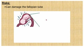
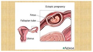
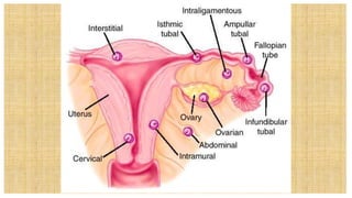
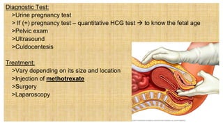
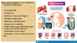
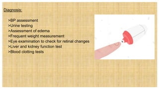
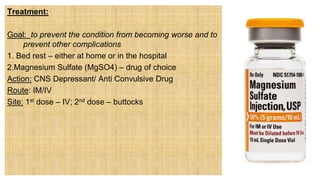
NCM 109- Care of Mother
and Child at Risk or with
Problems
(Acute and Chronic)-LECTURE
Wesleyan University –Philippines
Cabanatuan City
CONAMS
Jhonee Balmeo
Instructor
II. Care given to a mother experiencing a sudden pregnancy
complication utilizing the nursing care plan.
II. Care given to a mother experiencing a sudden
pregnancy complication utilizing the nursing care
plan.
1.Assessment
 Always ask women during prenatal visit about
any symptoms that may indicate a
complication such as pain or vaginal
symptoms (leaking of fluid, or bleeding)
 Other symptoms: head ache, blurring of
vision, back pains
 Review the danger signs of pregnancy
2.Nursing Diagnosis: should reflect both the physical problem and the woman’s or
family’s concern
Examples:
 Anxiety related to guarded pregnancy outcome
 Fear of preterm labor ending the pregnancy
 Anticipatory grieving related to uncertain pregnancy outcome
 Deficient knowledge related to signs and symptoms of possible complications
 Risk for infection related to incomplete miscarriage
 Deficient fluid volume related to third-trimester bleeding
 Risk for ineffective tissue perfusion related to gestational hypertension
3.Outcome Identification and Planning:
Outcomes addresses both fetal and maternal (mother) welfare as well as family
welfare
Treatment and management should be regularly updated and maintained
Referrals can be included for counseling
4.Implementation:
Interventions require an interpersonal
approach that speaks to several different
areas:
• Continued both healthy maternal and fetal
physical growth
• A woman’s and family’s psychological health
• Continuation of the pregnancy for as long
as possible
5. Outcome Evaluation
Patient’s blood pressure is maintained within acceptable parameters for
remainder of pregnancy
Couple states they feel able to cope with anxiety associated with the pregnancy
complication
Patient’s signs and symptoms of hypertension of pregnancy do not progress to
eclampsia
Patient accurately verbalizes crucial signs and symptoms she should
immediately report to her primary health care provider
Couple expresses feelings of sadness over pregnancy loss.
Patient is able to adhere to the medical treatment regimen and experiences no
adverse effects from the treatment
A. Bleeding Disorder During the First Trimester of Pregnancy
A.Bleeding Disorder During the First Trimester of
Pregnancy
1.Abortion
 Is a medical term for any interruption of a pregnancy
before a fetus is viable (able to survive outside the
uterus if born at that time)
 termination of pregnancy before the age of viability (20
weeks or 5 mos)
 a procedure, either surgical or medical, to end a
pregnancy by removing the fetus and placenta from the
uterus
• A fetus born before this point is considered a
miscarriage or is termed as premature or immature
birth
Types:
1. a. Spontaneous Abortion/Miscarriage – is an early miscarriage if it occurs
before week 16 of pregnancy and a late miscarriage if it occurs before weeks 16
and 20
Signs and symptoms:
 > Low back pain or abdominal pain
that is dull, sharp, or cramping
 > Vaginal bleeding, with or without
abdominal cramps
 > Tissue or clot – like material that
passes from the vagina
> For the first 6 weeks (between 1st to 2nd
month) of pregnancy, the developing placenta is
tentatively attached to the decidua of the
uterus.
During weeks 6 to 12 (2nd to 3rd month) of
pregnancy, the placenta is moderately attached
After week 12, the attachment is penetrating
and deep
Bleeding before week 6 is rarely severe
Bleeding after week 12 can be profuse because
the placenta is implanted so deeply.
Common causes:
1. Abnormal fetal development due either to
teratogenic factor (Any agent that can disturb
the development of an embryo or fetus.) or
chromosomal aberration (changes
in chromosome number:gains or losses)
2.Immunologic factors
3.Implantation abnormalities
Common causes:
4.Failure of the corpus luteum on the ovary to produce enough progesterone to
maintain the decidua basalis
5. Ingestion of alcoholic beverages during pregnancy
6. Urinary tract infection
7. Systemic infections such as rubella, syphylis, poliomyelitis, cytomegalovirus and
toxoplasmosis
Signs and Test
 1. Pelvic Exam – thinning of cervix (effacement)
* Increased cervical dilatation
* Evidence of rupture membranes
Signs and Test
 1. Pelvic Exam – thinning of cervix (effacement)
* Increased cervical dilatation
* Evidence of rupture membranes
Signs and Test
 2. HCG – (qualitative and quantitative urine and blood) – urine HCG test is a
common method of determining if a woman is pregnant; detectable in the blood or
urine 1 to 2 days after implantation of the fertilized egg ( that is 10 days after
ovulation)
Normal Values: Qualitative Urine and Blood
> the test is negative if client is not pregnant
> the test is positive if client is pregnant
Treatment and Management
 > Tissue passed from the vagina should be examined to determine the source (fetal
V/S H-Mole)
 > If remaining tissues are present – surgery or D & C
It is (usually) defined as a fetus of more than
20 to 24 weeks of gestation or one that
weighs at least 500 grams
• A. AOG
• B. Viable fetus
• C. Vailability
• D. Normal Birth Weight
The following are s/sx of miscarriage, but
one.
 > Low back pain
 > Vaginal bleeding,
 > Abdominal Pain
 > Decrease O2 Saturation
 > Tissue or clot – like material passes from the vagina
 AOTA
Classification Of Spontaneous Abortion:
a.1. Threatened Abortion – pregnancy is jeopardized by bleeding and cramping but
the cervix is closed.
Signs and Symptoms
 > Vaginal bleeding during the first 20 weeks of pregnancy
 > Abdominal cramps may or may not accompany vaginal bleeding
Treatment/Management
 > Complete Bed Rest (CBR) or pelvic rest for 24 to 48 hours-key intervention
 > Abstaining from intercourse
 > Avoid douching
 > Avoid using tampons
a.2. Imminent/ Inevitable Abortion – moderate bleeding,
cramping, tissue protrudes from the cervix (cervical dilatation)
Signs and Symptoms
> low back pain or abdominal pain that is dull, sharp, or
cramping
> vaginal bleeding, with or without abdominal cramps
> tissue or clot – like material that passes from the
vagina
TYPES
1. A.2.1.Complete Abortion – all products
of conception are expelled
2. A.2.2. Incomplete Abortion – placenta
and membranes are retained
Complication:
 > Infection-may also occur after a
complete abortion
 * Escherrichia coli- organism responsible
after miscarriage
 >spread from the rectum forward into the
vagina
 *Group A streptococcus
Management:
> For complete abortion – emotional support
for incomplete abortion – D & C – dilating the cervix and scraping the lining of the
uterus with an instrument called a curette
b. Habitual Abortion – three or more consecutive pregnancies result in abortion
usually related to incompetent cervix
Other Possible Causes:
> Defective spermatozoa
> endocrine factors such as lowered levels of protein bound iodine (PBI), butanol-
extractable iodine (BEI), and globulin-bound iodine (GBI), poor thyroid functions or a
luteal phase defect
>deviations of the uterus such as separate or bicornuate uterus
>resistance to uterine artery blood flow
>chorioamnionitis or uterine infection
>autoimmune disorders such as those involving lupus anticoagulant and
antiphospholipid antibodies
b. Habitual Abortion – three or more consecutive pregnancies result in abortion
usually related to incompetent cervix
Other Possible Causes:
> Defective spermatozoa
> endocrine factors such as lowered levels of protein bound iodine (PBI), butanol-
extractable iodine (BEI), and globulin-bound iodine (GBI), poor thyroid functions or a
luteal phase defect
>deviations of the uterus such as separate or bicornuate uterus
>resistance to uterine artery blood flow
>chorioamnionitis or uterine infection
>autoimmune disorders such as those involving lupus anticoagulant and
antiphospholipid antibodies  both causes clotting problem
b. Habitual Abortion – three or more consecutive pregnancies result in abortion
usually related to incompetent cervix
Other Possible Causes:
> Defective spermatozoa
> endocrine factors such as lowered levels of protein bound iodine (PBI), butanol-
extractable iodine (BEI), and globulin-bound iodine (GBI), poor thyroid functions or a
luteal phase defect
>deviations of the uterus such as separate or bicornuate uterus
>resistance to uterine artery blood flow
>chorioamnionitis or uterine infection
>autoimmune disorders such as those involving lupus anticoagulant and
antiphospholipid antibodies
Test to detect the cause:
1.X-ray or ultrasound of the uterus
2. Transvaginal ultrasound
3.Blood test:
 >Thrombophilia and thyroid function test
 >Karyogram
 >CBC to determine the degree of blood
loss
 >WBC and differential to rule out
infection
A karyogram allows a geneticist to determine a
person's karyotype - a written description of their
chromosomes including anything out of the ordinary
Management:
 > Surgery for Habitual Abortion, if the cause is incompetent cervix
Temporary
> McDonald Procedure
> Temporary Cerclage
> Delivery: Normal Delivery
Permanent
> Shirodkar procedure
> Delivery: C/S
Nursing Management:
 >Check for signs of infection
 >Check for signs of labor
 >Check for normal bleeding
Identification:
this is a type of abortion/miscarriage where
some fragments has been retained from the
womb?
• ____________________?
refers to a variety of procedures that use
sutures or synthetic tape to reinforce the
cervix during pregnancy in women with a
history of a short cervix.
A. Cervical cerclage.
B. Vaginal suturing
C. Mc Donald’s Procedure
D. Shirodcar Procedure
A client of yours asked you regarding the procedure that will be
conducted to her due to her incompetent cervix. The OB
mentioned about “McDonalds’ Proc. What is the best response
for this question?
A. This procedure uses a sutures that pass through the walls of
the cervix so they're not exposed. It is a permanent stitch
around the cervix which will not be removed and therefore
a Caesarean section will be necessary to deliver the baby.
B. It uses a pursestring stitch to cinch the cervix shut; the
cervix stitching involves a band of suture at the upper part
of the cervix while the lower part has already started to
efface
C. It is the stitching of the uterus to make a closure in the
cervix, so that the fetus may reach the age of viability.
c. Missed abortion – fetus dies; product of conception remain
in uterus 4 weeks or longer; signs of pregnancy cease
Signs and Symptoms
 >Scanty dark bleeding
 >Negative pregnancy test
Management
 >Induced labor – oxytocin/vaginal suppositories with
prostaglandin hormone
 >Vacuum extraction
d. Infected/Septic Abortion – abortion associated with an infection inside a pregnant
woman’s uterus.
an abortion that is complicated by infection (Uzelac & Garmel, 2007). Infection can
occur after a spontaneous miscarriage, but more frequently it occurs in women who
have tried to self-abort or were aborted illegally using a nonsterile instrument such
as a knitting needle.
 >Abortion may be spontaneous, which is referred to as miscarriage.
d. Infected/Septic Abortion
 >May also be an elective surgical or medical abortion, meaning, the woman chose
to terminate her pregnancy.
Pathophysiology: A septic abortion can occur when
Bacteria enter the uterus through the mucus plug.
These bacteria can be introduced by unclean tools used during an elective abortion.
The bacteria may also be those that normally live in a woman’s vagina.
If the woman has a sexually transmitted disease (STD) such as chlamydia, the
bacteria causing the STD can infect the uterus.
If the infection reaches the bloodstream, it is called sepsis
Signs and Symptoms:
 >High fever, usually above 101 F
 >Chills
 >Severe abdominal pain or cramping
 >Prolonged or heavy vaginal bleeding
 >Foul-smelling vaginal discharge
 >backache
If condition becomes serious, signs of shock may appear: These include:
 >Low blood pressure
 >Low body temperature
 >Little or no urine output
 >Troubled breathing
Causes of Septic Abortion:
> The membranes surrounding the fetus have ruptured
sometimes without detected
 >STD
 >IUD left in place during pregnancy
 >Tissue from the fetus or placenta is left inside the uterus
after a miscarriage or abortion
 >Attempts were made to end the pregnancy, often
illegally, by inserting tools, chemicals, or soaps into the
uterus
Long Term effects : Infertility Treatment: D & C
Risks: Death of the fetus
One of your patients asks you what are the
different classifications of Spontaneous Abortion.
Enumerating those is the best response to your
patient. SATA
A. threatened abortion,
B. inevitable abortion,
C. incomplete abortion,
D. missed abortion,
E. septic abortion,
F. complete abortion, and
G. recurrent spontaneous abortion
Dudong? Ano-ano ang mga dahilan
bakit nagkakaroon ng septic abortion?
Magbigay ng isa.
b.1. Septicemia – is the presence of bacteria in the blood (bacteremia) and is often
associated with severe disease.
Causes: Septicemia is a serious, life-threatening infection that get worse very quickly.
It can arise from infections throughout the body, including infections in the lungs,
abdomen, and urinary tract
 Septicemia can rapidly lead to septic shock and death. Septicemia associated with
some organisms (germs) such as meningogococci can lead to shock, adrenal
collapse, and disseminated intravascular coagulopathy, a condition called
Waterhouse-Friderichsen syndrome
Signs and Symptoms:
 >Fever (sudden onset, often spiking)
 >Chills
 >Toxic looking (looks acutely ill)
 >Changes in mental state
• >Irritable
• >Lethargic
• >Anxious
• >Agitated
• >Unresponsive
• >comatose
 >Shock
• >cold, clammy skin
• >Pale
• >Cyanotic
• >Skin signs associated with clotting abnormalities
 >Petechiae
 >Ecchymosis (often large, flat, purplish lesions that do not blanch when pressed)
 >Gangrene (early changes in the extremities suggesting decresed or absent blood
flow)
 >Decreased or no urine output
Test that can confirm infection:
 >Blood culture
 >Urine culture
 >CSF culture
 >CBC
 >Platelet count
 >Clotting studies – Pt, PTT, fibrinogen levels
Complications:
 >Irreversible shock
 >Waterhouse-Friderichesen syndrome
 >Adult respiratory distress syndrome (ARDS)
End of 1st session
Session 2
2. Ectopic Pregnancy
An ectopic pregnancy occurs when a fertilized egg implants and
grows outside the main cavity of the uterus. An ectopic
pregnancy most often occurs in a fallopian tube, which carries
eggs from the ovaries to the uterus.
> occurs when gestation is located outside the uterine cavity/tubal
pregnancies
Causes:
 >Fallopian tube damage often from infection-can block the fertilized
egg’s path to the uterus causing it to implant and grow in the tube
 >Surgery
 >Endometriosis
 >Smoking
 >Previous ectopic pregnancy
 >Pelvic infection – chlamydia or gonorrhea
 >Fertility drugs that increase egg production
 >Pelvic or abdominal surgery
Risks:
 >Can damage the fallopian tube
Signs and Symptoms:
 Normal signs of pregnancy
 Pain- first red flag sign
Other Signs and Symptoms:
 Vaginal spotting or bleeding
 Dizziness or fainting (caused by blood loss)
 Low blood pressure (caused by blood loss)
 Lower back pain
Unruptured Tubal Ruptured
>missed period >sudden sharp severe pain
> abdominal pain within >shoulder pain (indicative
3-5 weeks of intraperitoneal bleeding that
> scant, dark brown extends to diaphragm and
vaginal bleeding Phrenic nerve)
> vague discomfort > + Cullen’s sign – bluish tinged
umbilicus
Diagnostic Test:
 >Urine pregnancy test
 > If (+) pregnancy test – quantitative HCG test  to know the fetal age
 >Pelvic exam
 >Ultrasound
 >Culdocentesis
Treatment:
 >Vary depending on its size and location
 >Injection of methotrexate
 >Surgery
 >Laparoscopy
Future Pregnancies:
 >30% who have had ectopic pregnancy will have difficulty becoming
pregnant again.
 >If the fallopian tube has been spared, the chances of a future successful
pregnancy are 60%. Even if one fallopian tube has been removed, the
chances of having a successful pregnancy with the other tube can be greater
than 40%.
High Risk Women:
 >Age – 35 and 44 y/o
 >With PID – Pelvic Inflammatory Disease
 >Previous Ectopic Pregnancy
 >Surgery on fallopian tube
 >Infertility problems or medication to stimulate ovulation
Nursing Care:
 >Vital Signs
 >Administer IVF
 >Monitor vaginal bleeding
 >Monitor I&O
 > Prepare for Culdocentesis-
> Culdocentesis- is a procedure in which peritoneal fluid is obtained from the cul de
sac of a female patient. It involves the introduction of a spinal needle through the
vaginal wall into the peritoneal space of the pouch of Douglas
Result: to determine if clotting
or non clotting
 >If clotting – negative for
ectopic pregnancy
 >If non – clotting – positive
for ectopic pregnancy
Where do you think are the other sites for
ectopic pregnancy?
A. ampulla
B. Infundibular
C. isthmic segments of the fallopian tube
D. AOTA
You were assigned on the OB ward when one of
your clients asks you regarding the causes of EP,
you will respond by enumerating the following:
SATA
A. Smoking
B. Previous ectopic pregnancy
C. Pelvic infection – chlamydia or
gonorrhea
D. Damaged fallopian tube
E. Chromosomal disorder
It is a procedure used to diagnose the presence of
ruptured ectopic pregnancy by evaluating for
hemoperitoneum by inserting a needle and drawing back
fluid from the pouch of Douglas
B. BLEEDING DISORDER DURING THE SECOND TRIMESTER OF BLEEDING
1.Hydatidiform Mole ( H-Mole)-an abnormal proliferation and degeneration of the
trophoblastic villi
 >Molar pregnancy
 >Gestational Trophoblastic Disease
 >Bunch of Grapes
 >Hydatid – means drop of water; mole – means spot
B. BLEEDING DISORDER DURING THE
SECOND TRIMESTER OF
BLEEDING
1.Hydatidiform Mole ( H-Mole)-an
abnormal proliferation and
degeneration of the trophoblastic villi
 >Molar pregnancy
 >Gestational Trophoblastic Disease
 >Bunch of Grapes
 >Hydatid – means drop of water; mole
– means spot
Types:
 a.Partial Molar – pregnancy that includes an abnormal embryo (a fertilized egg that
has begun to grow) but does not survive
 b.Complete Molar –pregnancy in which there is small cluster of clear blisters or
pouches that don’t contain an embryo
Drug of Choice: Methotrexate
Etiology: Unknown
Other Causes:
 >Problems with the chromosome
 >Problem with the nutrition – low protein intake
 >Problem with the ovaries and uterus
 >Mole sometimes can develop from a placental tissue that is left behind in the
uterus after a miscarriage or childbirth
Signs and Symptoms
 >(+) pregnancy test
 >Symptoms for the first 3-4 months
 >Uterus grow abnormally fast
 >End of 3rd month-woman will experience vaginal bleeding ranging from scant
spotting to excessive bleeding
May predispose the:
 >Presence of hyperthyroidism (overproduction of thyroid hormone) leads to:
• >Weight loss
• >Increase appetite
• >Intolerance to heat
 >Grapelike cluster of cells itself will be shed with the blood during this time
 >Nausea and vomiting due to increase HCG and progesterone
 >(-) fetal movement
 >(-)fetal heart rate
Early Signs:
 >Vesicles passed thru the vagina
 >Hyperemesis gravidarum
 >Fundal height – rapidly increases
 >Vaginal bleeding (scant or profuse)
 > Pre-eclampsia at about 12 weeks
Late Signs
 >HPN before 20th week
 >Vesicles look like a ‘snowstorm” on sonogram
 >Anemia
 >Abdominal cramping
Serious Late Complications
 >Hyperthyroidism
 >Pulmonary embolus
Diagnosis:
> suspect until 3rd month or later if fetal heartbeat is present with bleeding and
severe nausea and vomiting
 >Physician will examine the woman’s abdomen feeling for any strange humps or
abnormalities in the uterus
 >Tubal pregnancy will be ruled out
 >Abnormally increased HCG level with vaginal bleeding;
 >(-) FHB
 >unusually large uterus will indicate a molar pregnancy
 >Ultrasound – confirm no living fetus
Treatment
 >often, the tissue is naturally expelled by the fourth month of pregnancy.
 In some instances, the physician will give the woman a drug called oxytocin to
trigger the release of the mole that is not spontaneously aborted
 >If this does not happen, a vacuum aspiration can be performed to remove the mole
Treatment
 D&C
* woman is given anesthetic
* Cervix is dilated and the contents of the uterus is gently suctioned out.
* After the mole has been mostly removed, gentle scraping of the uterus lining
is usually performed.
* If the woman is older and does not want any more children, the uterus can be
surgically removed (hysterectomy) instead of a vacuum aspiration because of the
higher risk of cancerous moles in this age group
* Monitoring the patient for at least 2 months after the end of a molar
pregnancy for HCG level
 >Hcg level will be checked every 2 weeks – if don’t return to normal by that time,
the mole may have become cancerous
 >If HCG level is normal, the woman’s HCG will be tested each month for 6 months
and every 2 months for a year
 >If mole become cancerous, treatment includes removal of the cancerous tissue
and chemotherapy
 >If cancer spread to other parts of the body, radiation will be added
 >Woman should not be pregnant within a year after HCG levels have returned to
normal
 >If woman got pregnant within that time, it is difficult to tell whether the resulting
high levels of HCG were caused by the pregnancy or as a cancer from the mole
Why would the doctor recommend the oxytocin for
patient with H-Mole?
a. Pitocin induces the labor, helping the
woman to expel the products
b. Increases the oxygen supply to the
woman
c. Helps in destroying the growing hmole
d. Suppresses the Oxytocin production
of the body
Psssst!!! Thru or fols?
Methotrexate is a drug of choice for Hmole?
C. BLEEDING DISORDERS DURING THE THIRD TRIMESTER OF PREGNANCY
1. 1.Placenta Previa – occurs when the placenta is improperly implanted in the
lower uterine segment, sometimes covering the cervical os.
Signs and Symptoms
> Frank, bright red, painless vaginal bleeding
>Engagement (usually has not occurred)
>Fetal distress
>Presentation (usually abnormal) – baby is
breech or in transverse position
>Uterus measures larger than it should
according to gestational age
Types:
a. a.Partial Placenta Previa – a portion of the cervix is covered by the placenta
b. b.Complete Placental Previa/Total – cervical opening is completely covered
c. c.Marginal Placenta Previa – extends just to the edge of the cervix
Management
1.Bed Rest
 >If the patient presents with mild bleeding before the fetal lungs are mature
2.Depending on the gestational age; steroid shots may be given to help mature the
baby’s lungs
3.If the bleeding cannot be controlled, an immediate cesarian delivery is usually done
regardless of the length of pregnancy
4.Near term, fetal lung maturity may be assessed by amniocentesis and the
preferred method is C/S
 >Some marginal previas can be delivered vaginally
 >Complete or partial previous would require a C/S
2. Avoid intercourse
3. Limit or no travelling
4. Avoid pelvic exams/internal exams – can cause profuse bleeding
Predisposing Factors
>Old Age
>Smoking
>intake of alcoholic beverages
>history of placenta previa in the past
pregnancy
Surgical Management:C/S with
blood transfusion based on blood
loss
It is a medical term often referred to as “baby dropping.”
This means that the infant's head or buttocks have
settled into the pelvis prior to labor.
A. Engagement
B. Presentation
C. Dropping form
D. Fetal implantation
2. Abruptio Placenta
 > Premature separation of the placenta from the implantation site. It usually occurs
after the 20th week of pregnancy
 > Bleeding into the deciduas basalis (the layer between the placenta and
myometrium) compresses and compromises the function of adjacent placenta
Other Names:
 >Premature Separation of Placenta
 >Accidental Hemorrhage
 >Ablatio Placenta
 >Placental Abruption
Signs and Symptoms:
 >Painful vaginal bleeding
 >Severe abdominal pain
 >Concealed bleeding (retroplacental)
 >Rigid abdomen
 >Couvelaire uterus (caused by bleeding into the myometrium)
 >Dropping Coagulation factor ( a potential for DIC)
Signs and Symptoms:
 >Painful vaginal bleeding
 >Severe abdominal pain
 >Concealed bleeding (retroplacental)
 >Rigid abdomen
 >Couvelaire uterus (caused by bleeding into the myometrium)
 >Dropping Coagulation factor ( a potential for DIC)
 Couvelaire uterus (also known as uteroplacental apoplexy) is
a life-threatening condition in which loosening of the
placenta (abruptio placentae) causes bleeding that penetrates into
the uterine myometrium forcing its way into the peritoneal cavity.
Other Signs and Symptoms:
 >Uterine hypertonous  contractions with a duration lasting more than 2
minutes
 >Back pain
 >Preterm labor
 >Hypovolemic shock
 >Non reassuring fetal heart tracking and fetal demise
Severe Cases
 >Maternal hypotension
 >Uterine hypertonicity
 >Fetal distress
 >Death
 >Clotting abnormalities
Cause: unknown
Predisposing Factors:
 >Mechanical factors such as: abdominal trauma – car accident of fall
 >Sudden loss in uterine volume as occurs with rapid loss of amniotic fluid or the
delivery of a first twin
 >Abnormally short umbilical cord
 >Hypertension
 >Pre-eclampsia
 >Multiparity
 >Rupture of membranes more than 24H
Signs and Tests:
 >During a physical examination, uterine tenderness and or increased uterine tone
may be noticed
 >CBC – decreased hematocrit and hemoglobin and platelets
 >Prothrombin time test
 >Partial thromboplastin time test
 >Fibrinogen level test
 >Ultrasound
Treatment and Management:
 >IVF
 >Blood Transfusion
 >Check for presence of shock and fetal distress
 >Emergency C/S – for fetal distress or maternal bleeding
 Immature fetus with small placental separation – hospitalization – for observation –
release after several days if no evidence of progressing abruption occurs
 If mature fetus – vaginal delivery if maternal and fetal distress is minimal
 C/S – to protect the mother and child
General Nursing Care
 >Infuse IVF, prepare to administer blood
 >Type and cross match blood components (PRBC)
 >Monitor FHR
 >Insert foley catheter
 >Measure blood loss – count pads
 >Report signs and symptoms of DIC
 >Monitor V/S
 >Strict I & O
Abruptio placentae (or placental
abruption) is the _____ of the placenta and
the uterine wall.
A. Adherence
B. Connecting
C. Separating
D. reattachment
The most common cause of placental
abruption is:
A. Diabetes
B.Trauma
C.High blood pressure
D.drugs
The biggest risk to the mother after a placental
abruption is:
A. hemorrhaging
B.seizures
C.Oxygen deprivation
D.Pain
D. Pregnancy Induced Hypertension (PIH)
 > A form of increased blood pressure in pregnancy
 > Also called toxemia or pre – eclampsia
 > Eclampsia is a severe form of PIH accompanied with seizures
 > HELLP Syndrome – is a pregnancy complication that affects the blood and
liver.
 Hemolysis with Elevated Liver Enzymes and Decreased/Low Platelet Counts- is a
complication of severe pre-eclampsia or eclampsia.
Signs and Symptoms of HELLP Syndrome:
 > Breakdown of RBC
 > Changes in the liver
 > Decreased platelets (cells found in the blood that are needed to help the blood to
clot in order to control bleeding)
THREE PRIMARY CHARACTERISTICS:
 >Increased blood pressure, reading greater than 140/90 mmHg or a significant
increased in one or both pressures
 >Protein in the urine – proteinuria
 >Edema – swelling of face and fingers
Cause : unknown
Predisposing Factors:
 >Pre – existing HPN (increased BP) >PIH with previous pregnancy
 >Kidney disease >Mother’s age younger than 20 or
>Diabetes older than 40
 >Multiple gestation (twins/triplets)
Note:
* PIH should be treated immediately since with increased BP, there is also an
increased in the resistance of blood vessels. This may hinder blood flow in many
different organ systems in an expectant
Other Problems:
 >Occurrence of placental abruptio
 > Fetal problems such as intrauterine growth restriction (poor fetal growth) and
stillbirth
Signs and Symptoms: may experience
symptoms differently:
 >Increased BP
 >Proteinuria
 >Edema on face and fingers
 >Sudden weight gain
 >Blurring or double vision
 >Nausea and vomiting
 >Right sided upper abdominal pain
or pain around the stomach
 >Decreased urine output
 >Changes in liver or kidney function
test
Diagnosis:
 >BP assessment
 >Urine testing
 >Assessment of edema
 >Frequent weight measurement
 >Eye examination to check for retinal changes
 >Liver and kidney function test
 >Blood clotting tests
Treatment:
Goal: to prevent the condition from becoming worse and to
prevent other complications
1. Bed rest – either at home or in the hospital
2.Magnesium Sulfate (MgSO4) – drug of choice
Action: CNS Depressant/ Anti Convulsive Drug
Route: IM/IV
Site: 1st dose – IV; 2nd dose – buttocks
Nursing Considerations:
 >Consider the rights in giving medications
 >Check the expiration date of the medication
 >Check for proper color of the medication
> Check the patient’s BP before and after giving of
medication
 > Insert Foley catheter as per doctor’s order
Before giving the 2nd dose: check for the following:
 >BP – increased or decreased
 >Urine output – 30 cc/H; if less than 30 cc/H, hold the 2nd dose, notify the physician at once
and document the findings
 >Check for knee jerk – (+) or (-), if (-), hold the 2nd dose, notify the physician at once
 *(-) knee jerk is a sign of MgSo4 toxicity
 >Give antidote: Calcium Gluconate
3. Fetal Monitoring – to check for the health of the fetus when the mother has PIH
Include the following
 >Fetal movement counting – increased or decreased – fetal distress
 >Non stress testing test – tests that measure the fetal HR in response to fetus’
movements
 >Biophysical profile – test that combine nonstress test with ultrsound to observe the
fetus
 >Doppler waves – to measure the flow of blood through a blood vessel
4. Continued laboratory testing of urine and blood
5. Medications called corticosteroids that may help mature the lungs of the fetus
6. Delivery of the baby ( if treatment do not control the PIH, if the fetus or the mother is
in danger), C/S is recommended
For The General Nursing Care, remember this acronym:
P
E
A
C
E
GENERAL NURSING CARE:
P – PROMOTE BED REST
 > Prevent convulsion by nursing measures: seizure precautions
• *Quiet and calm environment
• *Minimal handling
• *Avoid jarring the bed
• *Provide tongue guard – to prevent biting the tongue in case of seizure attack
• *All side rails up (at all times) – to ensure safety of the client
• *Prepare the following at bed side:
 >Suction machine
 >Oxygen
 >Suction tip
 >NSS
 Note: make sure all machine and equipment are functioning well and in good
status, this is considered as one of the nurse’s responsibilities
• >During seizure attack – stay with the patient; do not restrict movements of
extremities to prevent contracture deformity; ensure patient’s safety (prevent patient
from falling)
• > After the attack – turn patient to side.
E - NSURE HIGH PROTEIN INTAKE ( 1 G/KG/DAY)
A – NTIHYPERTENSIVE DRUG : HYDRALAZINE
C - NS DEPRESSANT (MGSO4) ANTICONVULSANT DRUG
E – VALUATE PHYSICAL PARAMETERS FOR MAGNESIUM SULFATE TOXICITY
B – BP decreased
U – urine output decreased
R – RR less than 12/min
P – patellar reflex absent
Note: if one of these is present, hold the 2nd dose, report the findings to the physician,
document the findings and actions taken
Your Patient in the Gyne Ward had developed
severe pre-eclampsia, what would be the drug you
would expect to administer?
• a. Magnesium sulfate.
• b. Ranitidine (Zantac).
• c. A nonsteroidal anti-inflammatory agent.
• d. A loop diuretic.
Huy!! Nakikinig ka ba? Kung Oo,
What is HELLP syndrome?
A. A life-threatening condition
B. A hypertensive disorder of pregnancy
C. A condition involving H - hemolysis EL -
elevated liver enzymes and P - low platelets
D. All answers are correct
Eh ito? Sige nga? Subukan mo sagutin.
What is preeclampsia?
1.A life-threatening condition
2.A hypertensive disorder of pregnancy
3.Seizures during pregnancy
4.No answers are correct
End of session 2
E.Oligohydramnios
Refers to a pregnancy with less than the
average amount of amniotic fluid.
Part of the volume of amniotic fluid is
formed by the addition of fetal urine, this
reduced amount of fluid is usually caused by
a bladder or renal disorder in the fetus that is
interfering with voiding
Another cause: due to growth restriction of
the fetus, he/she is not voiding as much as
usual
Diagnosis:
1.Physical Assessment: Inspection: suspected during pregnancy if the uterus fails
to meet its expected growth rate
2.Ultrasound – pockets of amniotic fluid are less than average
Effects on the fetus after birth:
Muscles are weak due to cramped space during pregnancy
Lungs fail to develop that can lead to hypoplastic lungs- difficulty of breathing
Potter syndrome- distorted features of the face
Potter syndrome
Nursing Consideration:
> Careful inspection among infants at birth to rule out kidney disease and
compromised lung development
Potter syndrome
Nursing Consideration:
> Careful inspection among infants at birth to rule out kidney disease and
compromised lung development
F.Polyhydramnios
Occurs when there is excess fluid of more than 2,000ml or an amniotic fluid
index above 24 cm.
Normal volume at term: 500 to 1000 ml
Effects on Pregnancy:
Can cause fetal malpresentation due to the additional uterine space and can
allow the fetus to turn on a transverse lie
Can lead to premature rupture of the membranes from the increased
pressure that can lead to risks for infection, prolapsed cord and preterm birth
Normal Process during Pregnancy:
> Amniotic fluid is formed by a combination of the cells of the amniotic
membrane and from fetal urine
It is evacuated by being swallowed by the fetus, absorbed across the intestinal membrane
into the fetal bloodstream and transferred across the placenta
With polyhydramnios, accumulation of amniotic fluid suggests difficulty with the fetus’s
ability to swallow or absorb fluid
Causes of inability to swallow fluids:
Anencephalic
Fetus with tracheoesophageal fistula with stenosis
Fetus with intestinal obstruction
Occurs among infant with diabetic mother
Assessment/Diagnosis:
1. Physical Assessment: Inspection- rapid enlargement of the uterus
2. Difficulty to palpate fetal parts because the uterus is unusually tense
3. Difficulty in auscultating the FHR due to the depth of the increased amount of
fluid surrounding the fetus
4. Woman may have extreme shortness of breath due to pushing up of the
uterus against her diaphragm
5. Presence of varicosities and hemorrhoids due to blockage of venous return
from the lower extremities by extensive uterine pressure
6.Increased weight gain due to increased amount of amniotic fluid
7. Ultrasound
Therapeutic Management:
1. Hospitalization or home care
Goal:
a.For adequate rest
b.For further evaluation
c.To maintain adequate uteroplacental circulation
d.To reduce pressure on the cervix and prevent preterm labor
2.Advice woman to report any sign of ruptured membranes or uterine
contractions
3.Advice woman to have high fiber diet and consult her doctor for stool softener
if diet is ineffective-to prevent constipation and straining during defecation to
prevent uterine pressure
4.Monitor vital signs if in the hospital
5.Monitor presence of edema in the lower extremities
6.Amniocentesis – to remove excess some of the extra fluid
7.Tocolytics- to prevent or halt preterm labor
If preterm rupture of the membranes occurs:
Membranes can be “Needled” (insertion of a thin needle vaginally to pierce
them) to slow, control the release of fluid and to prevent prolapsed cord during
labor
Assess infant after birth for gastrointestinal blockage.
Basic knowledge check tayo 
A fetus ______.
A. develops in the uterus
B. is surrounded by amniotic fluid
C. is enveloped inside the amniotic sac
D. All answers are correct
Stock knowledge? How about this one?
Which is not a function of amniotic fluid?
A. To help practice using the digestive system
and aid in its development
B. To keep fragile, growing body parts lubricated
C. To protect the fetus from blows
D. To provide the fetus with nutrients
G.Postterm Pregnancy
> A term pregnancy is 38 to 42 weeks
Any pregnancy that extends this period is postterm
Causes:
1. Women who have long menstrual cycle
40 to 45 days: they do not ovulate on day 14 which is the normal period
They ovulate 14 days from the end of their cycle, or on day 26 or 31, children
will be considered “late” by 12 to 17 days
2. Women who are receiving high dose of salicylates for their severe sinus
headaches or rheumatoid arthritis- this interferes with the synthesis of
prostaglandin
3.Myometrial quiescence-uterus that does not respond to normal labor
stimulation
Danger to the fetus:
1.Meconium aspiration
2.Macrosomia
3.Lack of growth-placenta is functioning for only 40-42 weeks-exposes the fetus
to decreased blood perfusion, oxygen, fluid and nutrients
Management:
1. Biophysical profile- to evaluate the placental perfusion and amount of
amniotic fluid present;
if normal, it is assumed miscalculation occurs
If abnormal result or physical examination or biparietal diameter on
ultrasound result the fetus is in term size, labor will be induced
How to induce labor:
1.Prostaglandin gel or misoprostol (Cytotec) –applied to the vagina to initiate
uterine contraction followed by an oxytocin infusion
2. If oxytocin is ineffective, C/S is performed
Nursing Consideration during the labor process:
1.Monitor FHR, V/S
After Birth:
1.Assess newborn for meconium aspiration
>Establish and maintain patent airway
2.Assess for polycythemia – due to decreased oxygenation in the final weeks
>hematocrit may be elevated due to polycythemia and dehydration that leads to
lowered circulating plasma level.
3.Asess for hypoglycemia-because fetus had to use stores of glycogen for
nourishment in the final weeks of intrauterine life
4.Maintain an adequate temperature
Newborn has low subcutaneous fats levels
5. Follow up care until at least school age to track their developmental abilities
Care of the Woman:
Allow woman to stay a longer period of time with her newborn and let her or
assist her in providing appropriate interventions to her newborn
H. Preterm Labor
Labor that occurs before the end of week 37
of gestation
Danger:
Infant is immature
Assessment:
> Any pregnant woman having persistent
uterine contractions, mild and widely spaced
should be considered to be in labor, if
contractions have caused cervical effacement
or dilatation over 1 cm
Measures on How to Prevent Preterm Labor:
1.Remain on bed rest except to use on bathroom.
2.Drink 8 to 10 glasses of fluids daily
3.Keep mentally active by reading or working on a project to prevent boredom
4.Avoid activities that could stimulate labor
5. Consult your primary care giver whether sexual relations should be restricted
6.Immediately report signs of ruptured membranes and sudden gush of fluid
from the vagina) or vaginal bleeding
7.Report signs of urinary tract infections or vaginal infection (burning or
frequency of urination, vaginal itching or pain)
8.Keep appointments for prenatal care.
9.Empty bladder to prevent pressure on the uterus
10.Lie down on your left or right side to encourage blood return to the fetus
Signs and Symptoms:
persistent, dull, and low backache
vaginal spotting
Feeling of pelvic pressure
Abdominal tightening
Menstrual-like cramping
Increased vaginal discharge
Uterine contractions
Intestinal cramping
Diagnosis:
Analyzing changes in the length of the cervix by ultrasound
Analysis of vaginal mucus for the presence of fetal fibronectin, a protein
produced by trophoblast cells
* If this is present in vaginal mucus, preterm labor occurs, labor will not occur if
the protein is absent for at least 14 days
Therapeutic Management:
1.Medical attempts can be made to stop preterm labor if:
a.The fetal membranes have not ruptures
b.No fetal distress
c.No evidence of bleeding
d.Cervix is not dilated more than 4 to 5 cm
e.Effacement is not more than 50%
2.If in preterm labor:
a.Admission in the hospital
b.Bed rest – to relieve the pressure of the fetus in the cervix
c.Monitoring the uterine contractions
d.IVF therapy-to keep the woman well hydrated
e.Vaginal and cervical cultures and a clean catch urine sample- to rule out infection
Drug Administration:
1.Tocolytic drugs- an agent to halt labor
>Terbutaline-drug of choice
*carries a “black box” warning- should not be used for over 48
to 72 hours
*Reason: could cause serious maternal heart problems and
death
*should not be used in out patient or home setting-requires
constant professional assessment
Drug Administration:
2.Magnesium Sulfate-used traditionally to treat
pre eclampsia and prevent eclamptic seizures,
can also be used
*recent research does not support this as
tocolytic agent
3.Corticosteroid
> bethamethasone- to promote the formation of
lung surfactant to prevent respiratory distress
syndrome among newborn
Fetal Assessment: if woman is sent home:
1.Advice woman to keep herself well hydrated
2.Maintain adequate nutrition
3.Mainatin bed rest and avoid strenuous activities
4.Advice the woman to have a record of daily” kick” count or “count to 10” test of her baby’s
movements inside her womb.
LABOR THAT CANNOT BE HALTED
Membranes have ruptured-point of no return
Effacement is more than 50%
Cervical dilatation is more than 3 to 4 cm
Management:
> If fetus is very immature, C/S – to reduce pressure on the fetal head and reduce the
possibility of subdural or intrventricular hemorrhage from a vaginal birth
What to expect:
First stage of labor-the longest stage
Second stage of labor- maybe shorter
Artificial rupture of the membranes is not done because of the risk for
prolapse of the cord around a small head
Analgesics are administered with caution- immature infant have difficulty of
breathing at birth
Epidural anesthesia is preferred if the woman wants pharmacological pain
management
Subukan ulit natin galling mo 
Kangaroo care is an important method to increase intimacy and bonding between the mother and
her new child in those first moments after birth.
• A.True
B.False
A birth is considered premature when the
child is born before ____ weeks gestation.
• A. 35
• B. 36
• C. 37
• D. 38
NICU is the abbreviation for:
• A.Neonatal intensive care unit
• B.Newborn infant child unit
• C.Narnia in closet unleashed
• D. No I Cant Unsee
I.Intrauterine Fetal Death
Fetal death is determined by the point of gestation when death occurs
Missed abortion – when the fetus dies before 20 weeks of gestation and is not
aborted spontaneously
Fetal death – occurs after 20 weeks of gestation and may be used when labor does
not begin within 48H of death
Signs and Symptoms:
Painless spotting
Uterine contractions with cervical effacement and dilatation
Fetus is born lifeless and emaciated
Dx:
 > (-) fetal movement
 > (-) FHB
 > Uterine growth ceases
 > Uterine size decrease
 > Fetal heart movement cannot be visualized by UTZ
 > X-ray detected by the appearance of intravascular or intra abdominal fetal gas
(Robert’s sign)
Management:
1.Induced labor- combination of
misoprostol(Cytotec) applied to the vagina to
effect cervical ripening and oxytocin
administration to begin uterine contraction
2.Bllod studies: test for DIC
Rh Incompatibility
J. Rh Incompatibility
> Occurs when an Rh-negative mother (one negative for a D antigen or one with a dd
genotype carries a fetus with an Rh positive blood type (DD or Dd genotype)
For this to happen:
> The father of the child must either be homozygous (DD) or heterozygous (Dd) Rh
positive
If the father of the child is homozygous (DD) for the factor, 100% of the couple’s children will
be Rh positive (Dd)
If the father is heterozygous for the trait, 50% of their children can be Rh positive (Dd)
People who have Rh-positive blood have a protein factor (the D antigen) that Rh – negative
people do not, when an Rh –positive fetus begins to grow inside an Rh-negative mother who
is sensitized, her body reacts by forming antibodies against the invading substance-the
fetus.
> The Rh factor exists as a portion of the red blood cells so these maternal antibodies cross
the placenta and cause destruction
A fetus can become so deficient in RBC from this that a sufficient oxygen transport to body
cells cannot be maintained
This condition is termed as hemolytic disease of the newborn or erythroblastosis fetalis
Assessment:
1.Antibody titer
>if results are normal: 0, a ratio below 1:8 is minimal, the test is repeated at week 28 of
pregnancy, if the result is normal, no therapy is needed
If the woman anti-D antibody is elevated (1:16 or greater, showing Rh sensitization- the
fetus will be monitored for 2 weeks or more by Doppler velocity - a technique that can
predict when anemia is present or fetal red cells are being destroyed
If the results are high-fetus is not developing anemia and mostly an Rh-negative fetus
If the results are low – fetus is in danger, immediate birth will be carried out if near term; if
not near term, efforts to rdeucecthe numbe rof antibodies in the woman will be made or
replacing damaged red cells in the fetus began
THERAPEUTIC MANAGEMENT:
1. RhIG, a commercial prperation of passive Rh (D) antibodies against the Rh factor is
administered to women who are Rh negative at 28 weeks of pregnancy
It cannot cross the placenta and destroy fetal red cells because the antibodies are not the IgG
class which is the only type that crosses the placenta
RhIG (Rhogam)-given again by injection to the woman in the first 72 hours after birth of an
Rh-positive child- to further prevent the woman from forming natural antibodies
Nursing consideration after birth:
1.Determine the infant’s blood type
>if Rh positive, the mother will receive the RhIG injection
If Rh negative, no antibodies have been formed in the mother’s circulation during pregnancy
and none will form-no need for RhoGAM injection
Nakakapagod?
Pero worth it naman diba?
Expect for a quiz and requirement next week!

NCM 109 WEEK 3

  • 1.
    NCM 109- Careof Mother and Child at Risk or with Problems (Acute and Chronic)-LECTURE Wesleyan University –Philippines Cabanatuan City CONAMS Jhonee Balmeo Instructor
  • 2.
    II. Care givento a mother experiencing a sudden pregnancy complication utilizing the nursing care plan.
  • 3.
    II. Care givento a mother experiencing a sudden pregnancy complication utilizing the nursing care plan. 1.Assessment  Always ask women during prenatal visit about any symptoms that may indicate a complication such as pain or vaginal symptoms (leaking of fluid, or bleeding)  Other symptoms: head ache, blurring of vision, back pains  Review the danger signs of pregnancy
  • 4.
    2.Nursing Diagnosis: shouldreflect both the physical problem and the woman’s or family’s concern Examples:  Anxiety related to guarded pregnancy outcome  Fear of preterm labor ending the pregnancy  Anticipatory grieving related to uncertain pregnancy outcome  Deficient knowledge related to signs and symptoms of possible complications  Risk for infection related to incomplete miscarriage  Deficient fluid volume related to third-trimester bleeding  Risk for ineffective tissue perfusion related to gestational hypertension
  • 5.
    3.Outcome Identification andPlanning: Outcomes addresses both fetal and maternal (mother) welfare as well as family welfare Treatment and management should be regularly updated and maintained Referrals can be included for counseling
  • 6.
    4.Implementation: Interventions require aninterpersonal approach that speaks to several different areas: • Continued both healthy maternal and fetal physical growth • A woman’s and family’s psychological health • Continuation of the pregnancy for as long as possible
  • 7.
    5. Outcome Evaluation Patient’sblood pressure is maintained within acceptable parameters for remainder of pregnancy Couple states they feel able to cope with anxiety associated with the pregnancy complication Patient’s signs and symptoms of hypertension of pregnancy do not progress to eclampsia Patient accurately verbalizes crucial signs and symptoms she should immediately report to her primary health care provider Couple expresses feelings of sadness over pregnancy loss. Patient is able to adhere to the medical treatment regimen and experiences no adverse effects from the treatment
  • 8.
    A. Bleeding DisorderDuring the First Trimester of Pregnancy
  • 9.
    A.Bleeding Disorder Duringthe First Trimester of Pregnancy 1.Abortion  Is a medical term for any interruption of a pregnancy before a fetus is viable (able to survive outside the uterus if born at that time)  termination of pregnancy before the age of viability (20 weeks or 5 mos)  a procedure, either surgical or medical, to end a pregnancy by removing the fetus and placenta from the uterus • A fetus born before this point is considered a miscarriage or is termed as premature or immature birth
  • 10.
    Types: 1. a. SpontaneousAbortion/Miscarriage – is an early miscarriage if it occurs before week 16 of pregnancy and a late miscarriage if it occurs before weeks 16 and 20 Signs and symptoms:  > Low back pain or abdominal pain that is dull, sharp, or cramping  > Vaginal bleeding, with or without abdominal cramps  > Tissue or clot – like material that passes from the vagina
  • 11.
    > For thefirst 6 weeks (between 1st to 2nd month) of pregnancy, the developing placenta is tentatively attached to the decidua of the uterus. During weeks 6 to 12 (2nd to 3rd month) of pregnancy, the placenta is moderately attached After week 12, the attachment is penetrating and deep Bleeding before week 6 is rarely severe Bleeding after week 12 can be profuse because the placenta is implanted so deeply.
  • 12.
    Common causes: 1. Abnormalfetal development due either to teratogenic factor (Any agent that can disturb the development of an embryo or fetus.) or chromosomal aberration (changes in chromosome number:gains or losses) 2.Immunologic factors 3.Implantation abnormalities
  • 13.
    Common causes: 4.Failure ofthe corpus luteum on the ovary to produce enough progesterone to maintain the decidua basalis 5. Ingestion of alcoholic beverages during pregnancy 6. Urinary tract infection 7. Systemic infections such as rubella, syphylis, poliomyelitis, cytomegalovirus and toxoplasmosis
  • 14.
    Signs and Test 1. Pelvic Exam – thinning of cervix (effacement) * Increased cervical dilatation * Evidence of rupture membranes
  • 15.
    Signs and Test 1. Pelvic Exam – thinning of cervix (effacement) * Increased cervical dilatation * Evidence of rupture membranes
  • 16.
    Signs and Test 2. HCG – (qualitative and quantitative urine and blood) – urine HCG test is a common method of determining if a woman is pregnant; detectable in the blood or urine 1 to 2 days after implantation of the fertilized egg ( that is 10 days after ovulation) Normal Values: Qualitative Urine and Blood > the test is negative if client is not pregnant > the test is positive if client is pregnant
  • 17.
    Treatment and Management > Tissue passed from the vagina should be examined to determine the source (fetal V/S H-Mole)  > If remaining tissues are present – surgery or D & C
  • 18.
    It is (usually)defined as a fetus of more than 20 to 24 weeks of gestation or one that weighs at least 500 grams • A. AOG • B. Viable fetus • C. Vailability • D. Normal Birth Weight
  • 19.
    The following ares/sx of miscarriage, but one.  > Low back pain  > Vaginal bleeding,  > Abdominal Pain  > Decrease O2 Saturation  > Tissue or clot – like material passes from the vagina  AOTA
  • 20.
    Classification Of SpontaneousAbortion: a.1. Threatened Abortion – pregnancy is jeopardized by bleeding and cramping but the cervix is closed. Signs and Symptoms  > Vaginal bleeding during the first 20 weeks of pregnancy  > Abdominal cramps may or may not accompany vaginal bleeding
  • 21.
    Treatment/Management  > CompleteBed Rest (CBR) or pelvic rest for 24 to 48 hours-key intervention  > Abstaining from intercourse  > Avoid douching  > Avoid using tampons
  • 22.
    a.2. Imminent/ InevitableAbortion – moderate bleeding, cramping, tissue protrudes from the cervix (cervical dilatation) Signs and Symptoms > low back pain or abdominal pain that is dull, sharp, or cramping > vaginal bleeding, with or without abdominal cramps > tissue or clot – like material that passes from the vagina
  • 23.
    TYPES 1. A.2.1.Complete Abortion– all products of conception are expelled 2. A.2.2. Incomplete Abortion – placenta and membranes are retained Complication:  > Infection-may also occur after a complete abortion  * Escherrichia coli- organism responsible after miscarriage  >spread from the rectum forward into the vagina  *Group A streptococcus
  • 24.
    Management: > For completeabortion – emotional support for incomplete abortion – D & C – dilating the cervix and scraping the lining of the uterus with an instrument called a curette
  • 25.
    b. Habitual Abortion– three or more consecutive pregnancies result in abortion usually related to incompetent cervix Other Possible Causes: > Defective spermatozoa > endocrine factors such as lowered levels of protein bound iodine (PBI), butanol- extractable iodine (BEI), and globulin-bound iodine (GBI), poor thyroid functions or a luteal phase defect >deviations of the uterus such as separate or bicornuate uterus >resistance to uterine artery blood flow >chorioamnionitis or uterine infection >autoimmune disorders such as those involving lupus anticoagulant and antiphospholipid antibodies
  • 26.
    b. Habitual Abortion– three or more consecutive pregnancies result in abortion usually related to incompetent cervix Other Possible Causes: > Defective spermatozoa > endocrine factors such as lowered levels of protein bound iodine (PBI), butanol- extractable iodine (BEI), and globulin-bound iodine (GBI), poor thyroid functions or a luteal phase defect >deviations of the uterus such as separate or bicornuate uterus >resistance to uterine artery blood flow >chorioamnionitis or uterine infection >autoimmune disorders such as those involving lupus anticoagulant and antiphospholipid antibodies
  • 27.
    b. Habitual Abortion– three or more consecutive pregnancies result in abortion usually related to incompetent cervix Other Possible Causes: > Defective spermatozoa > endocrine factors such as lowered levels of protein bound iodine (PBI), butanol- extractable iodine (BEI), and globulin-bound iodine (GBI), poor thyroid functions or a luteal phase defect >deviations of the uterus such as separate or bicornuate uterus >resistance to uterine artery blood flow >chorioamnionitis or uterine infection >autoimmune disorders such as those involving lupus anticoagulant and antiphospholipid antibodies  both causes clotting problem
  • 28.
    b. Habitual Abortion– three or more consecutive pregnancies result in abortion usually related to incompetent cervix Other Possible Causes: > Defective spermatozoa > endocrine factors such as lowered levels of protein bound iodine (PBI), butanol- extractable iodine (BEI), and globulin-bound iodine (GBI), poor thyroid functions or a luteal phase defect >deviations of the uterus such as separate or bicornuate uterus >resistance to uterine artery blood flow >chorioamnionitis or uterine infection >autoimmune disorders such as those involving lupus anticoagulant and antiphospholipid antibodies
  • 29.
    Test to detectthe cause: 1.X-ray or ultrasound of the uterus 2. Transvaginal ultrasound 3.Blood test:  >Thrombophilia and thyroid function test  >Karyogram  >CBC to determine the degree of blood loss  >WBC and differential to rule out infection A karyogram allows a geneticist to determine a person's karyotype - a written description of their chromosomes including anything out of the ordinary
  • 30.
    Management:  > Surgeryfor Habitual Abortion, if the cause is incompetent cervix Temporary > McDonald Procedure > Temporary Cerclage > Delivery: Normal Delivery
  • 31.
    Permanent > Shirodkar procedure >Delivery: C/S Nursing Management:  >Check for signs of infection  >Check for signs of labor  >Check for normal bleeding
  • 32.
    Identification: this is atype of abortion/miscarriage where some fragments has been retained from the womb? • ____________________?
  • 33.
    refers to avariety of procedures that use sutures or synthetic tape to reinforce the cervix during pregnancy in women with a history of a short cervix. A. Cervical cerclage. B. Vaginal suturing C. Mc Donald’s Procedure D. Shirodcar Procedure
  • 34.
    A client ofyours asked you regarding the procedure that will be conducted to her due to her incompetent cervix. The OB mentioned about “McDonalds’ Proc. What is the best response for this question? A. This procedure uses a sutures that pass through the walls of the cervix so they're not exposed. It is a permanent stitch around the cervix which will not be removed and therefore a Caesarean section will be necessary to deliver the baby. B. It uses a pursestring stitch to cinch the cervix shut; the cervix stitching involves a band of suture at the upper part of the cervix while the lower part has already started to efface C. It is the stitching of the uterus to make a closure in the cervix, so that the fetus may reach the age of viability.
  • 35.
    c. Missed abortion– fetus dies; product of conception remain in uterus 4 weeks or longer; signs of pregnancy cease Signs and Symptoms  >Scanty dark bleeding  >Negative pregnancy test Management  >Induced labor – oxytocin/vaginal suppositories with prostaglandin hormone  >Vacuum extraction
  • 38.
    d. Infected/Septic Abortion– abortion associated with an infection inside a pregnant woman’s uterus. an abortion that is complicated by infection (Uzelac & Garmel, 2007). Infection can occur after a spontaneous miscarriage, but more frequently it occurs in women who have tried to self-abort or were aborted illegally using a nonsterile instrument such as a knitting needle.  >Abortion may be spontaneous, which is referred to as miscarriage.
  • 39.
    d. Infected/Septic Abortion >May also be an elective surgical or medical abortion, meaning, the woman chose to terminate her pregnancy.
  • 40.
    Pathophysiology: A septicabortion can occur when Bacteria enter the uterus through the mucus plug. These bacteria can be introduced by unclean tools used during an elective abortion. The bacteria may also be those that normally live in a woman’s vagina. If the woman has a sexually transmitted disease (STD) such as chlamydia, the bacteria causing the STD can infect the uterus. If the infection reaches the bloodstream, it is called sepsis
  • 41.
    Signs and Symptoms: >High fever, usually above 101 F  >Chills  >Severe abdominal pain or cramping  >Prolonged or heavy vaginal bleeding  >Foul-smelling vaginal discharge  >backache If condition becomes serious, signs of shock may appear: These include:  >Low blood pressure  >Low body temperature  >Little or no urine output  >Troubled breathing
  • 42.
    Causes of SepticAbortion: > The membranes surrounding the fetus have ruptured sometimes without detected  >STD  >IUD left in place during pregnancy  >Tissue from the fetus or placenta is left inside the uterus after a miscarriage or abortion  >Attempts were made to end the pregnancy, often illegally, by inserting tools, chemicals, or soaps into the uterus Long Term effects : Infertility Treatment: D & C Risks: Death of the fetus
  • 43.
    One of yourpatients asks you what are the different classifications of Spontaneous Abortion. Enumerating those is the best response to your patient. SATA A. threatened abortion, B. inevitable abortion, C. incomplete abortion, D. missed abortion, E. septic abortion, F. complete abortion, and G. recurrent spontaneous abortion
  • 44.
    Dudong? Ano-ano angmga dahilan bakit nagkakaroon ng septic abortion? Magbigay ng isa.
  • 45.
    b.1. Septicemia –is the presence of bacteria in the blood (bacteremia) and is often associated with severe disease. Causes: Septicemia is a serious, life-threatening infection that get worse very quickly. It can arise from infections throughout the body, including infections in the lungs, abdomen, and urinary tract  Septicemia can rapidly lead to septic shock and death. Septicemia associated with some organisms (germs) such as meningogococci can lead to shock, adrenal collapse, and disseminated intravascular coagulopathy, a condition called Waterhouse-Friderichsen syndrome
  • 46.
    b.1. Septicemia –is the presence of bacteria in the blood (bacteremia) and is often associated with severe disease. Causes: Septicemia is a serious, life-threatening infection that get worse very quickly. It can arise from infections throughout the body, including infections in the lungs, abdomen, and urinary tract  Septicemia can rapidly lead to septic shock and death. Septicemia associated with some organisms (germs) such as meningogococci can lead to shock, adrenal collapse, and disseminated intravascular coagulopathy, a condition called Waterhouse-Friderichsen syndrome
  • 47.
    Signs and Symptoms: >Fever (sudden onset, often spiking)  >Chills  >Toxic looking (looks acutely ill)  >Changes in mental state • >Irritable • >Lethargic • >Anxious • >Agitated • >Unresponsive • >comatose
  • 48.
     >Shock • >cold,clammy skin • >Pale • >Cyanotic • >Skin signs associated with clotting abnormalities  >Petechiae  >Ecchymosis (often large, flat, purplish lesions that do not blanch when pressed)  >Gangrene (early changes in the extremities suggesting decresed or absent blood flow)  >Decreased or no urine output
  • 49.
    Test that canconfirm infection:  >Blood culture  >Urine culture  >CSF culture  >CBC  >Platelet count  >Clotting studies – Pt, PTT, fibrinogen levels Complications:  >Irreversible shock  >Waterhouse-Friderichesen syndrome  >Adult respiratory distress syndrome (ARDS)
  • 50.
    End of 1stsession
  • 51.
  • 52.
    2. Ectopic Pregnancy Anectopic pregnancy occurs when a fertilized egg implants and grows outside the main cavity of the uterus. An ectopic pregnancy most often occurs in a fallopian tube, which carries eggs from the ovaries to the uterus.
  • 53.
    > occurs whengestation is located outside the uterine cavity/tubal pregnancies Causes:  >Fallopian tube damage often from infection-can block the fertilized egg’s path to the uterus causing it to implant and grow in the tube  >Surgery  >Endometriosis  >Smoking  >Previous ectopic pregnancy  >Pelvic infection – chlamydia or gonorrhea  >Fertility drugs that increase egg production  >Pelvic or abdominal surgery
  • 54.
    Risks:  >Can damagethe fallopian tube
  • 57.
    Signs and Symptoms: Normal signs of pregnancy  Pain- first red flag sign Other Signs and Symptoms:  Vaginal spotting or bleeding  Dizziness or fainting (caused by blood loss)  Low blood pressure (caused by blood loss)  Lower back pain
  • 58.
    Unruptured Tubal Ruptured >missedperiod >sudden sharp severe pain > abdominal pain within >shoulder pain (indicative 3-5 weeks of intraperitoneal bleeding that > scant, dark brown extends to diaphragm and vaginal bleeding Phrenic nerve) > vague discomfort > + Cullen’s sign – bluish tinged umbilicus
  • 59.
    Diagnostic Test:  >Urinepregnancy test  > If (+) pregnancy test – quantitative HCG test  to know the fetal age  >Pelvic exam  >Ultrasound  >Culdocentesis Treatment:  >Vary depending on its size and location  >Injection of methotrexate  >Surgery  >Laparoscopy
  • 60.
    Future Pregnancies:  >30%who have had ectopic pregnancy will have difficulty becoming pregnant again.  >If the fallopian tube has been spared, the chances of a future successful pregnancy are 60%. Even if one fallopian tube has been removed, the chances of having a successful pregnancy with the other tube can be greater than 40%.
  • 61.
    High Risk Women: >Age – 35 and 44 y/o  >With PID – Pelvic Inflammatory Disease  >Previous Ectopic Pregnancy  >Surgery on fallopian tube  >Infertility problems or medication to stimulate ovulation
  • 62.
    Nursing Care:  >VitalSigns  >Administer IVF  >Monitor vaginal bleeding  >Monitor I&O  > Prepare for Culdocentesis-
  • 63.
    > Culdocentesis- isa procedure in which peritoneal fluid is obtained from the cul de sac of a female patient. It involves the introduction of a spinal needle through the vaginal wall into the peritoneal space of the pouch of Douglas Result: to determine if clotting or non clotting  >If clotting – negative for ectopic pregnancy  >If non – clotting – positive for ectopic pregnancy
  • 64.
    Where do youthink are the other sites for ectopic pregnancy? A. ampulla B. Infundibular C. isthmic segments of the fallopian tube D. AOTA
  • 65.
    You were assignedon the OB ward when one of your clients asks you regarding the causes of EP, you will respond by enumerating the following: SATA A. Smoking B. Previous ectopic pregnancy C. Pelvic infection – chlamydia or gonorrhea D. Damaged fallopian tube E. Chromosomal disorder
  • 66.
    It is aprocedure used to diagnose the presence of ruptured ectopic pregnancy by evaluating for hemoperitoneum by inserting a needle and drawing back fluid from the pouch of Douglas
  • 67.
    B. BLEEDING DISORDERDURING THE SECOND TRIMESTER OF BLEEDING 1.Hydatidiform Mole ( H-Mole)-an abnormal proliferation and degeneration of the trophoblastic villi  >Molar pregnancy  >Gestational Trophoblastic Disease  >Bunch of Grapes  >Hydatid – means drop of water; mole – means spot
  • 68.
    B. BLEEDING DISORDERDURING THE SECOND TRIMESTER OF BLEEDING 1.Hydatidiform Mole ( H-Mole)-an abnormal proliferation and degeneration of the trophoblastic villi  >Molar pregnancy  >Gestational Trophoblastic Disease  >Bunch of Grapes  >Hydatid – means drop of water; mole – means spot
  • 69.
    Types:  a.Partial Molar– pregnancy that includes an abnormal embryo (a fertilized egg that has begun to grow) but does not survive  b.Complete Molar –pregnancy in which there is small cluster of clear blisters or pouches that don’t contain an embryo
  • 71.
    Drug of Choice:Methotrexate
  • 72.
    Etiology: Unknown Other Causes: >Problems with the chromosome  >Problem with the nutrition – low protein intake  >Problem with the ovaries and uterus  >Mole sometimes can develop from a placental tissue that is left behind in the uterus after a miscarriage or childbirth
  • 73.
    Signs and Symptoms >(+) pregnancy test  >Symptoms for the first 3-4 months  >Uterus grow abnormally fast  >End of 3rd month-woman will experience vaginal bleeding ranging from scant spotting to excessive bleeding May predispose the:  >Presence of hyperthyroidism (overproduction of thyroid hormone) leads to: • >Weight loss • >Increase appetite • >Intolerance to heat
  • 74.
     >Grapelike clusterof cells itself will be shed with the blood during this time  >Nausea and vomiting due to increase HCG and progesterone  >(-) fetal movement  >(-)fetal heart rate Early Signs:  >Vesicles passed thru the vagina  >Hyperemesis gravidarum  >Fundal height – rapidly increases  >Vaginal bleeding (scant or profuse)  > Pre-eclampsia at about 12 weeks
  • 75.
    Late Signs  >HPNbefore 20th week  >Vesicles look like a ‘snowstorm” on sonogram  >Anemia  >Abdominal cramping Serious Late Complications  >Hyperthyroidism  >Pulmonary embolus
  • 76.
    Diagnosis: > suspect until3rd month or later if fetal heartbeat is present with bleeding and severe nausea and vomiting  >Physician will examine the woman’s abdomen feeling for any strange humps or abnormalities in the uterus  >Tubal pregnancy will be ruled out  >Abnormally increased HCG level with vaginal bleeding;  >(-) FHB  >unusually large uterus will indicate a molar pregnancy  >Ultrasound – confirm no living fetus
  • 77.
    Treatment  >often, thetissue is naturally expelled by the fourth month of pregnancy.  In some instances, the physician will give the woman a drug called oxytocin to trigger the release of the mole that is not spontaneously aborted  >If this does not happen, a vacuum aspiration can be performed to remove the mole
  • 78.
    Treatment  D&C * womanis given anesthetic * Cervix is dilated and the contents of the uterus is gently suctioned out. * After the mole has been mostly removed, gentle scraping of the uterus lining is usually performed. * If the woman is older and does not want any more children, the uterus can be surgically removed (hysterectomy) instead of a vacuum aspiration because of the higher risk of cancerous moles in this age group * Monitoring the patient for at least 2 months after the end of a molar pregnancy for HCG level
  • 79.
     >Hcg levelwill be checked every 2 weeks – if don’t return to normal by that time, the mole may have become cancerous  >If HCG level is normal, the woman’s HCG will be tested each month for 6 months and every 2 months for a year  >If mole become cancerous, treatment includes removal of the cancerous tissue and chemotherapy  >If cancer spread to other parts of the body, radiation will be added  >Woman should not be pregnant within a year after HCG levels have returned to normal  >If woman got pregnant within that time, it is difficult to tell whether the resulting high levels of HCG were caused by the pregnancy or as a cancer from the mole
  • 80.
     >Hcg levelwill be checked every 2 weeks – if don’t return to normal by that time, the mole may have become cancerous  >If HCG level is normal, the woman’s HCG will be tested each month for 6 months and every 2 months for a year  >If mole become cancerous, treatment includes removal of the cancerous tissue and chemotherapy  >If cancer spread to other parts of the body, radiation will be added  >Woman should not be pregnant within a year after HCG levels have returned to normal  >If woman got pregnant within that time, it is difficult to tell whether the resulting high levels of HCG were caused by the pregnancy or as a cancer from the mole
  • 81.
    Why would thedoctor recommend the oxytocin for patient with H-Mole? a. Pitocin induces the labor, helping the woman to expel the products b. Increases the oxygen supply to the woman c. Helps in destroying the growing hmole d. Suppresses the Oxytocin production of the body
  • 82.
    Psssst!!! Thru orfols? Methotrexate is a drug of choice for Hmole?
  • 83.
    C. BLEEDING DISORDERSDURING THE THIRD TRIMESTER OF PREGNANCY
  • 84.
    C. BLEEDING DISORDERSDURING THE THIRD TRIMESTER OF PREGNANCY 1. 1.Placenta Previa – occurs when the placenta is improperly implanted in the lower uterine segment, sometimes covering the cervical os.
  • 85.
    Signs and Symptoms >Frank, bright red, painless vaginal bleeding >Engagement (usually has not occurred) >Fetal distress >Presentation (usually abnormal) – baby is breech or in transverse position >Uterus measures larger than it should according to gestational age
  • 88.
    Types: a. a.Partial PlacentaPrevia – a portion of the cervix is covered by the placenta b. b.Complete Placental Previa/Total – cervical opening is completely covered c. c.Marginal Placenta Previa – extends just to the edge of the cervix
  • 89.
    Management 1.Bed Rest  >Ifthe patient presents with mild bleeding before the fetal lungs are mature 2.Depending on the gestational age; steroid shots may be given to help mature the baby’s lungs 3.If the bleeding cannot be controlled, an immediate cesarian delivery is usually done regardless of the length of pregnancy 4.Near term, fetal lung maturity may be assessed by amniocentesis and the preferred method is C/S  >Some marginal previas can be delivered vaginally  >Complete or partial previous would require a C/S 2. Avoid intercourse 3. Limit or no travelling 4. Avoid pelvic exams/internal exams – can cause profuse bleeding
  • 91.
    Predisposing Factors >Old Age >Smoking >intakeof alcoholic beverages >history of placenta previa in the past pregnancy Surgical Management:C/S with blood transfusion based on blood loss
  • 92.
    It is amedical term often referred to as “baby dropping.” This means that the infant's head or buttocks have settled into the pelvis prior to labor. A. Engagement B. Presentation C. Dropping form D. Fetal implantation
  • 93.
    2. Abruptio Placenta > Premature separation of the placenta from the implantation site. It usually occurs after the 20th week of pregnancy  > Bleeding into the deciduas basalis (the layer between the placenta and myometrium) compresses and compromises the function of adjacent placenta Other Names:  >Premature Separation of Placenta  >Accidental Hemorrhage  >Ablatio Placenta  >Placental Abruption
  • 95.
    Signs and Symptoms: >Painful vaginal bleeding  >Severe abdominal pain  >Concealed bleeding (retroplacental)  >Rigid abdomen  >Couvelaire uterus (caused by bleeding into the myometrium)  >Dropping Coagulation factor ( a potential for DIC)
  • 96.
    Signs and Symptoms: >Painful vaginal bleeding  >Severe abdominal pain  >Concealed bleeding (retroplacental)  >Rigid abdomen  >Couvelaire uterus (caused by bleeding into the myometrium)  >Dropping Coagulation factor ( a potential for DIC)  Couvelaire uterus (also known as uteroplacental apoplexy) is a life-threatening condition in which loosening of the placenta (abruptio placentae) causes bleeding that penetrates into the uterine myometrium forcing its way into the peritoneal cavity.
  • 97.
    Other Signs andSymptoms:  >Uterine hypertonous  contractions with a duration lasting more than 2 minutes  >Back pain  >Preterm labor  >Hypovolemic shock  >Non reassuring fetal heart tracking and fetal demise
  • 98.
    Severe Cases  >Maternalhypotension  >Uterine hypertonicity  >Fetal distress  >Death  >Clotting abnormalities Cause: unknown
  • 99.
    Predisposing Factors:  >Mechanicalfactors such as: abdominal trauma – car accident of fall  >Sudden loss in uterine volume as occurs with rapid loss of amniotic fluid or the delivery of a first twin  >Abnormally short umbilical cord  >Hypertension  >Pre-eclampsia  >Multiparity  >Rupture of membranes more than 24H
  • 100.
    Signs and Tests: >During a physical examination, uterine tenderness and or increased uterine tone may be noticed  >CBC – decreased hematocrit and hemoglobin and platelets  >Prothrombin time test  >Partial thromboplastin time test  >Fibrinogen level test  >Ultrasound
  • 101.
    Treatment and Management: >IVF  >Blood Transfusion  >Check for presence of shock and fetal distress  >Emergency C/S – for fetal distress or maternal bleeding  Immature fetus with small placental separation – hospitalization – for observation – release after several days if no evidence of progressing abruption occurs  If mature fetus – vaginal delivery if maternal and fetal distress is minimal  C/S – to protect the mother and child
  • 102.
    General Nursing Care >Infuse IVF, prepare to administer blood  >Type and cross match blood components (PRBC)  >Monitor FHR  >Insert foley catheter  >Measure blood loss – count pads  >Report signs and symptoms of DIC  >Monitor V/S  >Strict I & O
  • 103.
    Abruptio placentae (orplacental abruption) is the _____ of the placenta and the uterine wall. A. Adherence B. Connecting C. Separating D. reattachment
  • 104.
    The most commoncause of placental abruption is: A. Diabetes B.Trauma C.High blood pressure D.drugs
  • 105.
    The biggest riskto the mother after a placental abruption is: A. hemorrhaging B.seizures C.Oxygen deprivation D.Pain
  • 106.
    D. Pregnancy InducedHypertension (PIH)  > A form of increased blood pressure in pregnancy  > Also called toxemia or pre – eclampsia  > Eclampsia is a severe form of PIH accompanied with seizures  > HELLP Syndrome – is a pregnancy complication that affects the blood and liver.  Hemolysis with Elevated Liver Enzymes and Decreased/Low Platelet Counts- is a complication of severe pre-eclampsia or eclampsia.
  • 108.
    Signs and Symptomsof HELLP Syndrome:  > Breakdown of RBC  > Changes in the liver  > Decreased platelets (cells found in the blood that are needed to help the blood to clot in order to control bleeding)
  • 109.
    THREE PRIMARY CHARACTERISTICS: >Increased blood pressure, reading greater than 140/90 mmHg or a significant increased in one or both pressures  >Protein in the urine – proteinuria  >Edema – swelling of face and fingers Cause : unknown Predisposing Factors:  >Pre – existing HPN (increased BP) >PIH with previous pregnancy  >Kidney disease >Mother’s age younger than 20 or >Diabetes older than 40  >Multiple gestation (twins/triplets)
  • 110.
    Note: * PIH shouldbe treated immediately since with increased BP, there is also an increased in the resistance of blood vessels. This may hinder blood flow in many different organ systems in an expectant Other Problems:  >Occurrence of placental abruptio  > Fetal problems such as intrauterine growth restriction (poor fetal growth) and stillbirth
  • 111.
    Signs and Symptoms:may experience symptoms differently:  >Increased BP  >Proteinuria  >Edema on face and fingers  >Sudden weight gain  >Blurring or double vision  >Nausea and vomiting  >Right sided upper abdominal pain or pain around the stomach  >Decreased urine output  >Changes in liver or kidney function test
  • 112.
    Diagnosis:  >BP assessment >Urine testing  >Assessment of edema  >Frequent weight measurement  >Eye examination to check for retinal changes  >Liver and kidney function test  >Blood clotting tests
  • 113.
    Treatment: Goal: to preventthe condition from becoming worse and to prevent other complications 1. Bed rest – either at home or in the hospital 2.Magnesium Sulfate (MgSO4) – drug of choice Action: CNS Depressant/ Anti Convulsive Drug Route: IM/IV Site: 1st dose – IV; 2nd dose – buttocks
  • 114.
    Nursing Considerations:  >Considerthe rights in giving medications  >Check the expiration date of the medication  >Check for proper color of the medication > Check the patient’s BP before and after giving of medication  > Insert Foley catheter as per doctor’s order
  • 115.
    Before giving the2nd dose: check for the following:  >BP – increased or decreased  >Urine output – 30 cc/H; if less than 30 cc/H, hold the 2nd dose, notify the physician at once and document the findings  >Check for knee jerk – (+) or (-), if (-), hold the 2nd dose, notify the physician at once  *(-) knee jerk is a sign of MgSo4 toxicity  >Give antidote: Calcium Gluconate
  • 116.
    3. Fetal Monitoring– to check for the health of the fetus when the mother has PIH Include the following  >Fetal movement counting – increased or decreased – fetal distress  >Non stress testing test – tests that measure the fetal HR in response to fetus’ movements  >Biophysical profile – test that combine nonstress test with ultrsound to observe the fetus  >Doppler waves – to measure the flow of blood through a blood vessel
  • 117.
    4. Continued laboratorytesting of urine and blood 5. Medications called corticosteroids that may help mature the lungs of the fetus 6. Delivery of the baby ( if treatment do not control the PIH, if the fetus or the mother is in danger), C/S is recommended For The General Nursing Care, remember this acronym: P E A C E
  • 118.
    GENERAL NURSING CARE: P– PROMOTE BED REST  > Prevent convulsion by nursing measures: seizure precautions • *Quiet and calm environment • *Minimal handling • *Avoid jarring the bed • *Provide tongue guard – to prevent biting the tongue in case of seizure attack • *All side rails up (at all times) – to ensure safety of the client • *Prepare the following at bed side:  >Suction machine  >Oxygen  >Suction tip  >NSS  Note: make sure all machine and equipment are functioning well and in good status, this is considered as one of the nurse’s responsibilities
  • 119.
    • >During seizureattack – stay with the patient; do not restrict movements of extremities to prevent contracture deformity; ensure patient’s safety (prevent patient from falling) • > After the attack – turn patient to side. E - NSURE HIGH PROTEIN INTAKE ( 1 G/KG/DAY) A – NTIHYPERTENSIVE DRUG : HYDRALAZINE C - NS DEPRESSANT (MGSO4) ANTICONVULSANT DRUG E – VALUATE PHYSICAL PARAMETERS FOR MAGNESIUM SULFATE TOXICITY B – BP decreased U – urine output decreased R – RR less than 12/min P – patellar reflex absent Note: if one of these is present, hold the 2nd dose, report the findings to the physician, document the findings and actions taken
  • 121.
    Your Patient inthe Gyne Ward had developed severe pre-eclampsia, what would be the drug you would expect to administer? • a. Magnesium sulfate. • b. Ranitidine (Zantac). • c. A nonsteroidal anti-inflammatory agent. • d. A loop diuretic.
  • 122.
    Huy!! Nakikinig kaba? Kung Oo, What is HELLP syndrome? A. A life-threatening condition B. A hypertensive disorder of pregnancy C. A condition involving H - hemolysis EL - elevated liver enzymes and P - low platelets D. All answers are correct
  • 123.
    Eh ito? Sigenga? Subukan mo sagutin. What is preeclampsia? 1.A life-threatening condition 2.A hypertensive disorder of pregnancy 3.Seizures during pregnancy 4.No answers are correct
  • 124.
  • 125.
    E.Oligohydramnios Refers to apregnancy with less than the average amount of amniotic fluid. Part of the volume of amniotic fluid is formed by the addition of fetal urine, this reduced amount of fluid is usually caused by a bladder or renal disorder in the fetus that is interfering with voiding Another cause: due to growth restriction of the fetus, he/she is not voiding as much as usual
  • 126.
    Diagnosis: 1.Physical Assessment: Inspection:suspected during pregnancy if the uterus fails to meet its expected growth rate 2.Ultrasound – pockets of amniotic fluid are less than average Effects on the fetus after birth: Muscles are weak due to cramped space during pregnancy Lungs fail to develop that can lead to hypoplastic lungs- difficulty of breathing Potter syndrome- distorted features of the face
  • 127.
    Potter syndrome Nursing Consideration: >Careful inspection among infants at birth to rule out kidney disease and compromised lung development
  • 128.
    Potter syndrome Nursing Consideration: >Careful inspection among infants at birth to rule out kidney disease and compromised lung development
  • 129.
    F.Polyhydramnios Occurs when thereis excess fluid of more than 2,000ml or an amniotic fluid index above 24 cm. Normal volume at term: 500 to 1000 ml Effects on Pregnancy: Can cause fetal malpresentation due to the additional uterine space and can allow the fetus to turn on a transverse lie Can lead to premature rupture of the membranes from the increased pressure that can lead to risks for infection, prolapsed cord and preterm birth Normal Process during Pregnancy: > Amniotic fluid is formed by a combination of the cells of the amniotic membrane and from fetal urine
  • 130.
    It is evacuatedby being swallowed by the fetus, absorbed across the intestinal membrane into the fetal bloodstream and transferred across the placenta With polyhydramnios, accumulation of amniotic fluid suggests difficulty with the fetus’s ability to swallow or absorb fluid Causes of inability to swallow fluids: Anencephalic Fetus with tracheoesophageal fistula with stenosis Fetus with intestinal obstruction Occurs among infant with diabetic mother
  • 131.
    Assessment/Diagnosis: 1. Physical Assessment:Inspection- rapid enlargement of the uterus 2. Difficulty to palpate fetal parts because the uterus is unusually tense 3. Difficulty in auscultating the FHR due to the depth of the increased amount of fluid surrounding the fetus 4. Woman may have extreme shortness of breath due to pushing up of the uterus against her diaphragm 5. Presence of varicosities and hemorrhoids due to blockage of venous return from the lower extremities by extensive uterine pressure 6.Increased weight gain due to increased amount of amniotic fluid 7. Ultrasound
  • 132.
    Therapeutic Management: 1. Hospitalizationor home care Goal: a.For adequate rest b.For further evaluation c.To maintain adequate uteroplacental circulation d.To reduce pressure on the cervix and prevent preterm labor 2.Advice woman to report any sign of ruptured membranes or uterine contractions 3.Advice woman to have high fiber diet and consult her doctor for stool softener if diet is ineffective-to prevent constipation and straining during defecation to prevent uterine pressure
  • 133.
    4.Monitor vital signsif in the hospital 5.Monitor presence of edema in the lower extremities 6.Amniocentesis – to remove excess some of the extra fluid 7.Tocolytics- to prevent or halt preterm labor If preterm rupture of the membranes occurs: Membranes can be “Needled” (insertion of a thin needle vaginally to pierce them) to slow, control the release of fluid and to prevent prolapsed cord during labor Assess infant after birth for gastrointestinal blockage.
  • 134.
    Basic knowledge checktayo  A fetus ______. A. develops in the uterus B. is surrounded by amniotic fluid C. is enveloped inside the amniotic sac D. All answers are correct
  • 135.
    Stock knowledge? Howabout this one? Which is not a function of amniotic fluid? A. To help practice using the digestive system and aid in its development B. To keep fragile, growing body parts lubricated C. To protect the fetus from blows D. To provide the fetus with nutrients
  • 136.
    G.Postterm Pregnancy > Aterm pregnancy is 38 to 42 weeks Any pregnancy that extends this period is postterm Causes: 1. Women who have long menstrual cycle 40 to 45 days: they do not ovulate on day 14 which is the normal period They ovulate 14 days from the end of their cycle, or on day 26 or 31, children will be considered “late” by 12 to 17 days
  • 137.
    2. Women whoare receiving high dose of salicylates for their severe sinus headaches or rheumatoid arthritis- this interferes with the synthesis of prostaglandin 3.Myometrial quiescence-uterus that does not respond to normal labor stimulation Danger to the fetus: 1.Meconium aspiration 2.Macrosomia 3.Lack of growth-placenta is functioning for only 40-42 weeks-exposes the fetus to decreased blood perfusion, oxygen, fluid and nutrients
  • 138.
    Management: 1. Biophysical profile-to evaluate the placental perfusion and amount of amniotic fluid present; if normal, it is assumed miscalculation occurs If abnormal result or physical examination or biparietal diameter on ultrasound result the fetus is in term size, labor will be induced
  • 139.
    How to inducelabor: 1.Prostaglandin gel or misoprostol (Cytotec) –applied to the vagina to initiate uterine contraction followed by an oxytocin infusion 2. If oxytocin is ineffective, C/S is performed Nursing Consideration during the labor process: 1.Monitor FHR, V/S After Birth: 1.Assess newborn for meconium aspiration >Establish and maintain patent airway
  • 140.
    2.Assess for polycythemia– due to decreased oxygenation in the final weeks >hematocrit may be elevated due to polycythemia and dehydration that leads to lowered circulating plasma level. 3.Asess for hypoglycemia-because fetus had to use stores of glycogen for nourishment in the final weeks of intrauterine life 4.Maintain an adequate temperature Newborn has low subcutaneous fats levels 5. Follow up care until at least school age to track their developmental abilities Care of the Woman: Allow woman to stay a longer period of time with her newborn and let her or assist her in providing appropriate interventions to her newborn
  • 141.
    H. Preterm Labor Laborthat occurs before the end of week 37 of gestation Danger: Infant is immature Assessment: > Any pregnant woman having persistent uterine contractions, mild and widely spaced should be considered to be in labor, if contractions have caused cervical effacement or dilatation over 1 cm
  • 142.
    Measures on Howto Prevent Preterm Labor: 1.Remain on bed rest except to use on bathroom. 2.Drink 8 to 10 glasses of fluids daily 3.Keep mentally active by reading or working on a project to prevent boredom 4.Avoid activities that could stimulate labor 5. Consult your primary care giver whether sexual relations should be restricted 6.Immediately report signs of ruptured membranes and sudden gush of fluid from the vagina) or vaginal bleeding 7.Report signs of urinary tract infections or vaginal infection (burning or frequency of urination, vaginal itching or pain) 8.Keep appointments for prenatal care. 9.Empty bladder to prevent pressure on the uterus 10.Lie down on your left or right side to encourage blood return to the fetus
  • 143.
    Signs and Symptoms: persistent,dull, and low backache vaginal spotting Feeling of pelvic pressure Abdominal tightening Menstrual-like cramping Increased vaginal discharge Uterine contractions Intestinal cramping
  • 144.
    Diagnosis: Analyzing changes inthe length of the cervix by ultrasound Analysis of vaginal mucus for the presence of fetal fibronectin, a protein produced by trophoblast cells * If this is present in vaginal mucus, preterm labor occurs, labor will not occur if the protein is absent for at least 14 days
  • 145.
    Therapeutic Management: 1.Medical attemptscan be made to stop preterm labor if: a.The fetal membranes have not ruptures b.No fetal distress c.No evidence of bleeding d.Cervix is not dilated more than 4 to 5 cm e.Effacement is not more than 50% 2.If in preterm labor: a.Admission in the hospital b.Bed rest – to relieve the pressure of the fetus in the cervix c.Monitoring the uterine contractions d.IVF therapy-to keep the woman well hydrated e.Vaginal and cervical cultures and a clean catch urine sample- to rule out infection
  • 146.
    Drug Administration: 1.Tocolytic drugs-an agent to halt labor >Terbutaline-drug of choice *carries a “black box” warning- should not be used for over 48 to 72 hours *Reason: could cause serious maternal heart problems and death *should not be used in out patient or home setting-requires constant professional assessment
  • 147.
    Drug Administration: 2.Magnesium Sulfate-usedtraditionally to treat pre eclampsia and prevent eclamptic seizures, can also be used *recent research does not support this as tocolytic agent 3.Corticosteroid > bethamethasone- to promote the formation of lung surfactant to prevent respiratory distress syndrome among newborn
  • 148.
    Fetal Assessment: ifwoman is sent home: 1.Advice woman to keep herself well hydrated 2.Maintain adequate nutrition 3.Mainatin bed rest and avoid strenuous activities 4.Advice the woman to have a record of daily” kick” count or “count to 10” test of her baby’s movements inside her womb. LABOR THAT CANNOT BE HALTED Membranes have ruptured-point of no return Effacement is more than 50% Cervical dilatation is more than 3 to 4 cm Management: > If fetus is very immature, C/S – to reduce pressure on the fetal head and reduce the possibility of subdural or intrventricular hemorrhage from a vaginal birth
  • 149.
    What to expect: Firststage of labor-the longest stage Second stage of labor- maybe shorter Artificial rupture of the membranes is not done because of the risk for prolapse of the cord around a small head Analgesics are administered with caution- immature infant have difficulty of breathing at birth Epidural anesthesia is preferred if the woman wants pharmacological pain management
  • 150.
    Subukan ulit natingalling mo  Kangaroo care is an important method to increase intimacy and bonding between the mother and her new child in those first moments after birth. • A.True B.False
  • 151.
    A birth isconsidered premature when the child is born before ____ weeks gestation. • A. 35 • B. 36 • C. 37 • D. 38
  • 152.
    NICU is theabbreviation for: • A.Neonatal intensive care unit • B.Newborn infant child unit • C.Narnia in closet unleashed • D. No I Cant Unsee
  • 153.
    I.Intrauterine Fetal Death Fetaldeath is determined by the point of gestation when death occurs Missed abortion – when the fetus dies before 20 weeks of gestation and is not aborted spontaneously Fetal death – occurs after 20 weeks of gestation and may be used when labor does not begin within 48H of death
  • 154.
    Signs and Symptoms: Painlessspotting Uterine contractions with cervical effacement and dilatation Fetus is born lifeless and emaciated Dx:  > (-) fetal movement  > (-) FHB  > Uterine growth ceases  > Uterine size decrease  > Fetal heart movement cannot be visualized by UTZ  > X-ray detected by the appearance of intravascular or intra abdominal fetal gas (Robert’s sign)
  • 155.
    Management: 1.Induced labor- combinationof misoprostol(Cytotec) applied to the vagina to effect cervical ripening and oxytocin administration to begin uterine contraction 2.Bllod studies: test for DIC
  • 156.
  • 157.
    J. Rh Incompatibility >Occurs when an Rh-negative mother (one negative for a D antigen or one with a dd genotype carries a fetus with an Rh positive blood type (DD or Dd genotype) For this to happen: > The father of the child must either be homozygous (DD) or heterozygous (Dd) Rh positive If the father of the child is homozygous (DD) for the factor, 100% of the couple’s children will be Rh positive (Dd) If the father is heterozygous for the trait, 50% of their children can be Rh positive (Dd) People who have Rh-positive blood have a protein factor (the D antigen) that Rh – negative people do not, when an Rh –positive fetus begins to grow inside an Rh-negative mother who is sensitized, her body reacts by forming antibodies against the invading substance-the fetus.
  • 159.
    > The Rhfactor exists as a portion of the red blood cells so these maternal antibodies cross the placenta and cause destruction A fetus can become so deficient in RBC from this that a sufficient oxygen transport to body cells cannot be maintained This condition is termed as hemolytic disease of the newborn or erythroblastosis fetalis
  • 160.
    Assessment: 1.Antibody titer >if resultsare normal: 0, a ratio below 1:8 is minimal, the test is repeated at week 28 of pregnancy, if the result is normal, no therapy is needed If the woman anti-D antibody is elevated (1:16 or greater, showing Rh sensitization- the fetus will be monitored for 2 weeks or more by Doppler velocity - a technique that can predict when anemia is present or fetal red cells are being destroyed If the results are high-fetus is not developing anemia and mostly an Rh-negative fetus If the results are low – fetus is in danger, immediate birth will be carried out if near term; if not near term, efforts to rdeucecthe numbe rof antibodies in the woman will be made or replacing damaged red cells in the fetus began
  • 161.
    THERAPEUTIC MANAGEMENT: 1. RhIG,a commercial prperation of passive Rh (D) antibodies against the Rh factor is administered to women who are Rh negative at 28 weeks of pregnancy It cannot cross the placenta and destroy fetal red cells because the antibodies are not the IgG class which is the only type that crosses the placenta RhIG (Rhogam)-given again by injection to the woman in the first 72 hours after birth of an Rh-positive child- to further prevent the woman from forming natural antibodies Nursing consideration after birth: 1.Determine the infant’s blood type >if Rh positive, the mother will receive the RhIG injection If Rh negative, no antibodies have been formed in the mother’s circulation during pregnancy and none will form-no need for RhoGAM injection
  • 162.
  • 163.
    Expect for aquiz and requirement next week!