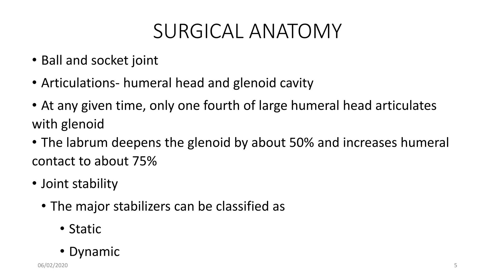
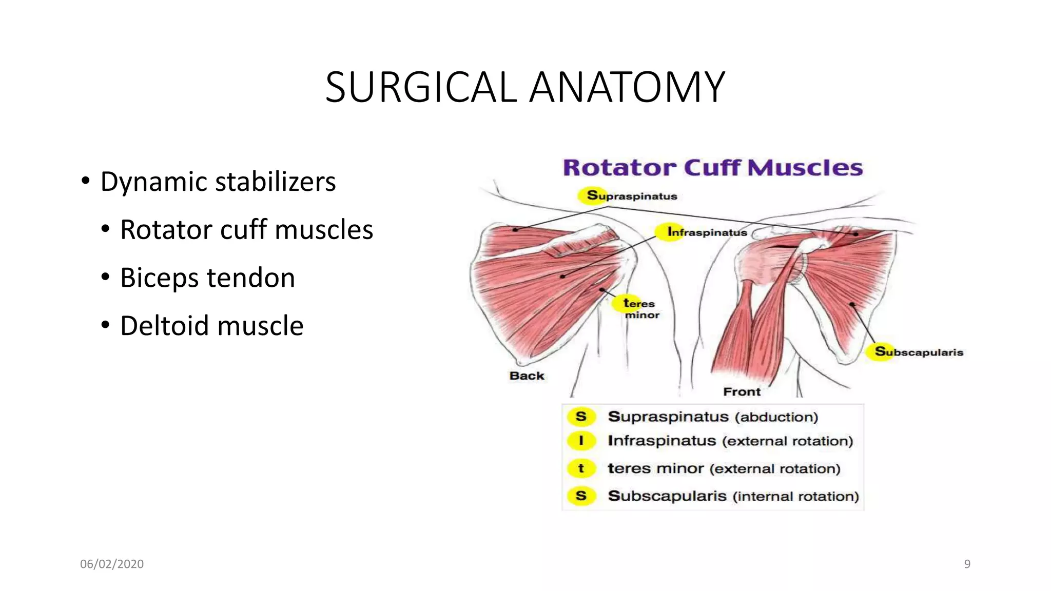
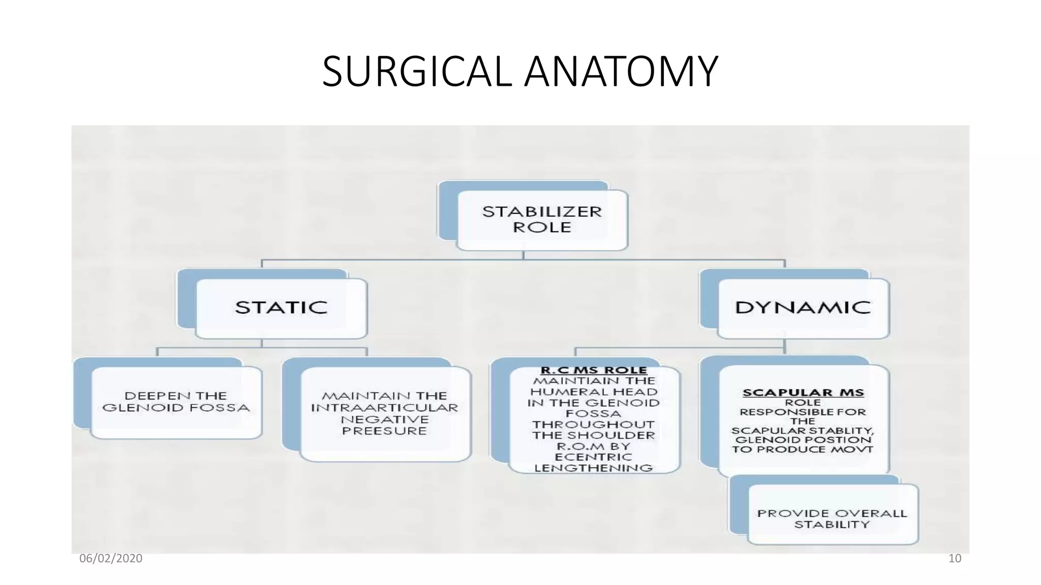
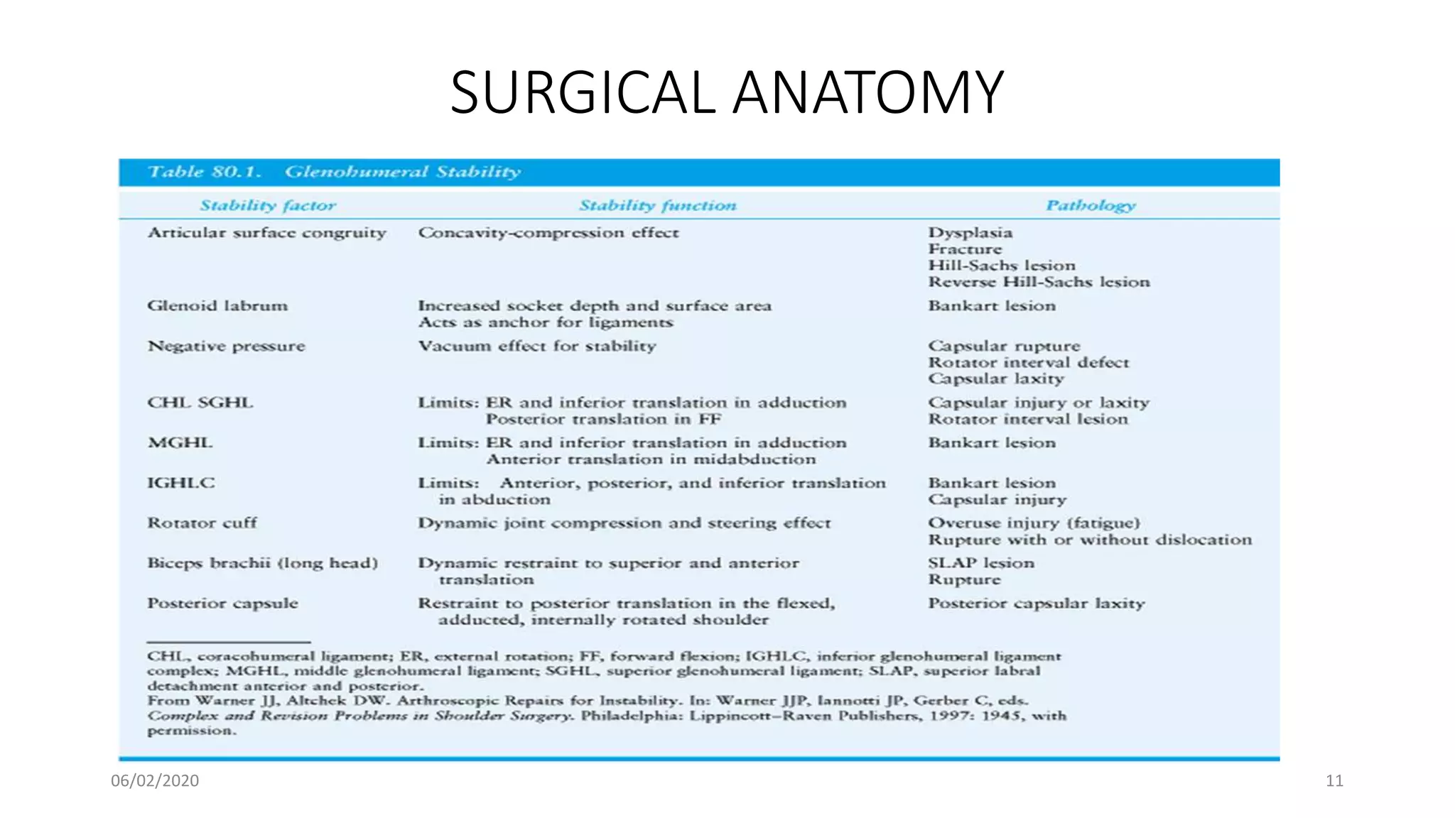
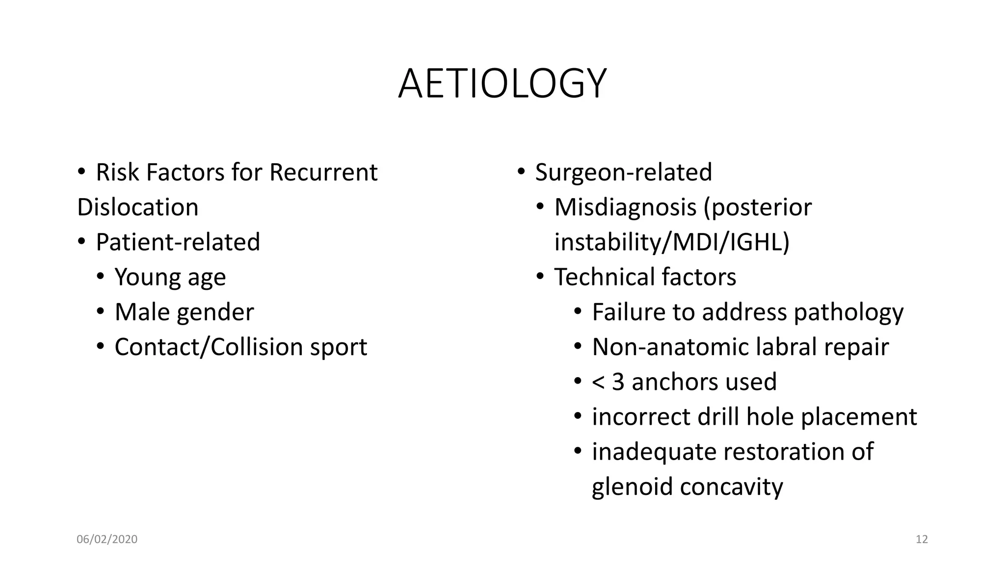
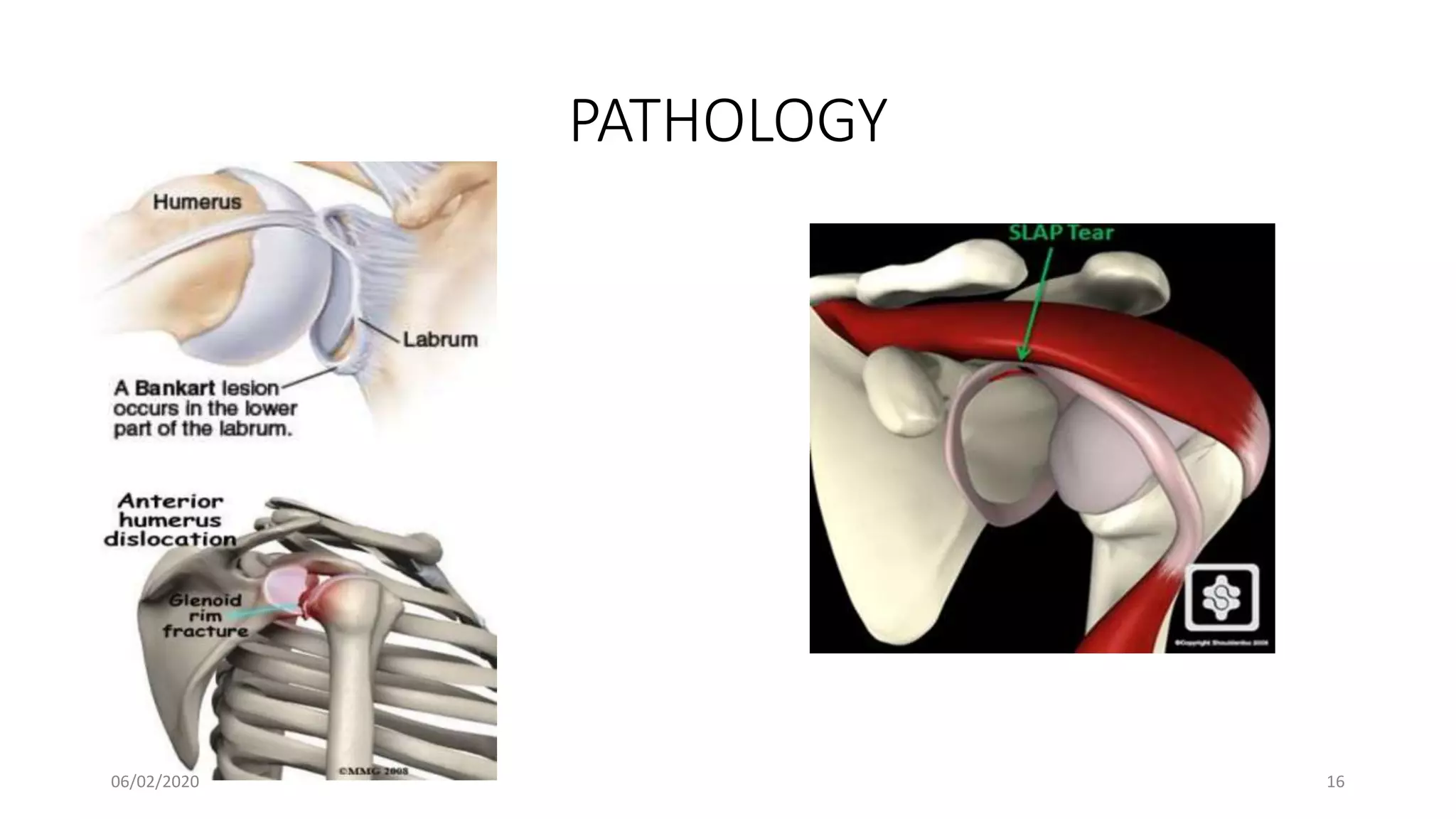
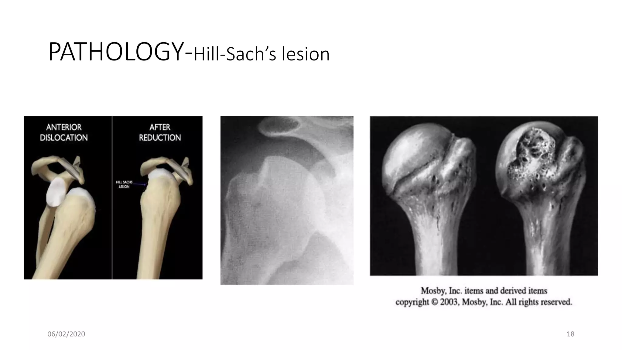
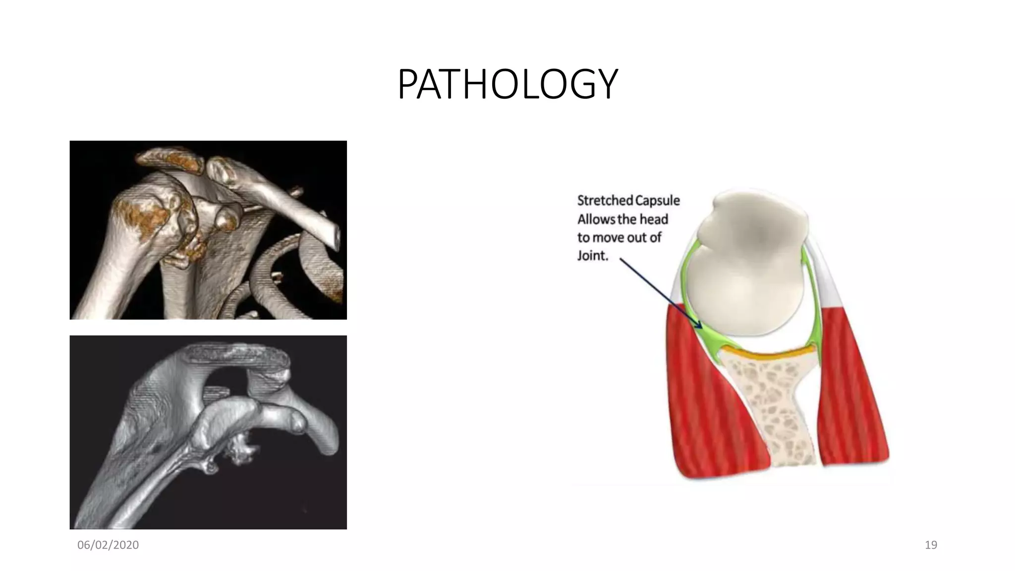
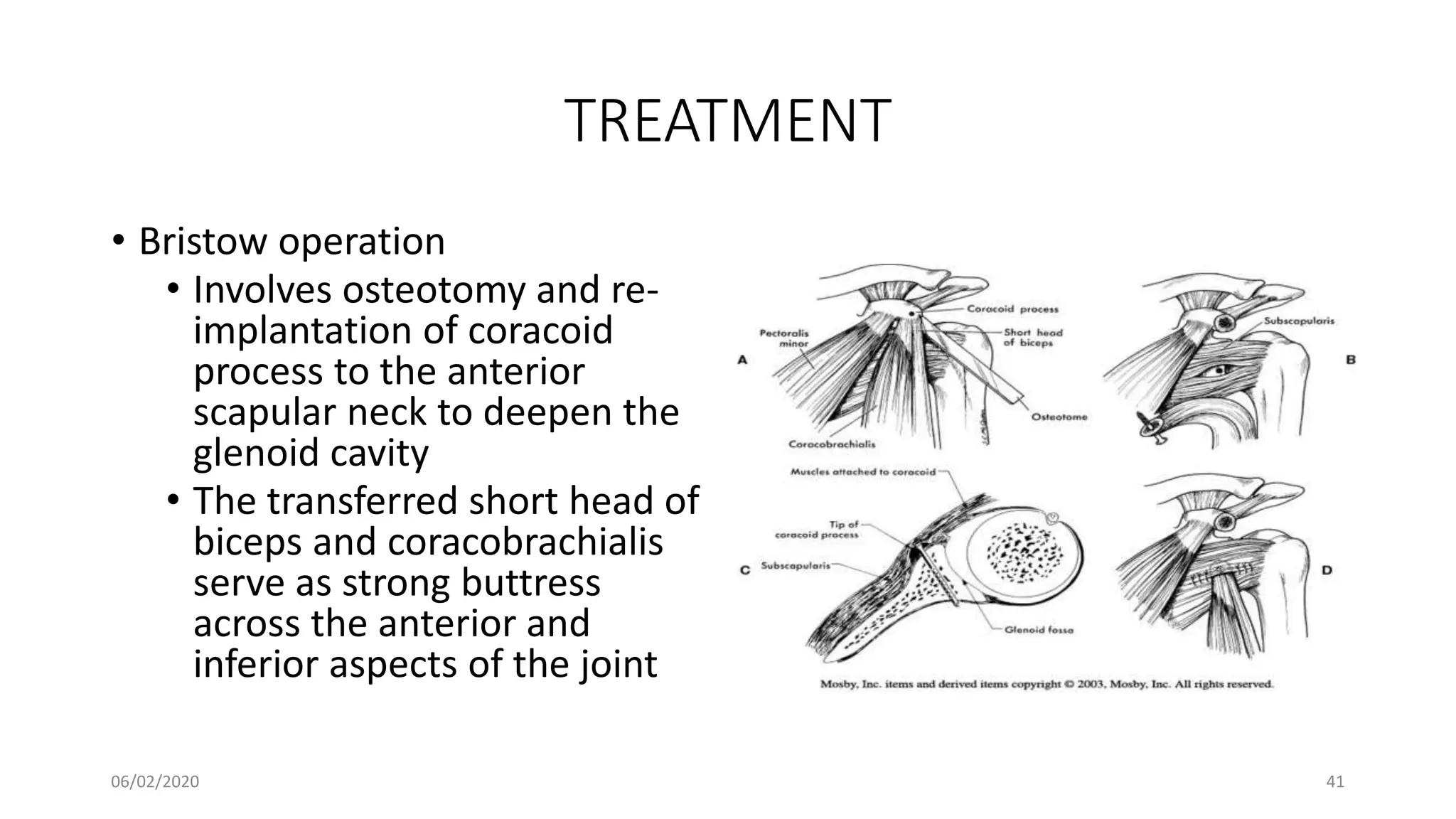
This document discusses the pathology and management of recurrent shoulder dislocations. It begins with an introduction covering the epidemiology and risk factors for recurrence. It then covers the surgical anatomy of the shoulder joint and stabilizing structures. The main pathological findings associated with recurrence include Bankart lesions, Hill-Sachs lesions, and ligamentous laxity. Treatment involves both surgical and non-surgical options depending on the individual case. The most common surgical procedures are Bankart repair and Latarjet procedure. Post-operative rehabilitation is important for recovery. Recurrence rates can be reduced with proper identification and treatment of the underlying causes.
Deformity: It’s the position of a limb/Joint, from which it cannot be brought back to its normal anatomical position.
Described as abnormalities of :
Length
Angulation
Rotation
Translation
Combination
Deformity: It’s the position of a limb/Joint, from which it cannot be brought back to its normal anatomical position.
Described as abnormalities of :
Length
Angulation
Rotation
Translation
Combination
Can read freely here
https://sethiortho.blogspot.com/
Challenges and Solutions in
Management of Distal Humerus Fractures
Epidemiology
Anatomy
Classification
Controversies and Recent studies
Approach
Implants selection
Plate configuration
Ulnar nerve transposition
Role of total elbow arthroplasty in DHF
Role of hemiarthroplasty in DHF
Metaphyseal comminution –
Anatomic complexity of the distal humerus
Positioning of the plates
TBW –
Skin closure
Osteoporotic nature of the bone –
Less BMD/Thin metaphysis
Screw Pullout strength is low
DHF account for 2% of all adult fractures
The common pattern of fracture
Intraarticular and involves both columns
Bimodal distribution
Peak incidence in young male and in older female patients
Young male – High-velocity injury
Older female - Osteoporosis
The distal humerus is flattened and expanded bony structure
It is composed of lateral and medial columns with the trochlea situated between these columns.
The location of the trochlea is central rather than medial
Formed by Medial SCR + M/Epicondyle
The distal end has 450 angulation with humeral shaft
M/ Epicondyle gives attachment for MCL & Common Flexor Origin
The MCL originates from the undersurface of the medial epicondyle where it is vulnerable to excessive dissection
Ulnar nerve
Formed by Lateral SCR and L/Epicondyle and Capitulum
Distal end has 200 with humeral shaft
L/ epicondyle gives attachment for LCL & common extensor origin
Its posterior surface is non articular and can be used as a site for a plate fixation
The lateral column curves anteriorly
Placement of a straight plate on the posterolateral surface of the humerus risks straightening of distal humerus.
The medial column including the medial epicondyle is in line with the humeral shaft.
It forms the center of the triangle
It has 30 - 80 – external rotation & 250 anterior divergent with the shaft
It forms a 40 - 80 degree valgus direction
X-ray -
Anterior-posterior view
lateral View
Traction View – This can help to define articular fragments and aid in pre-operative classification of the fracture.
NCCT – Elbow
Articular surfaces
Position of the fracture fragments
useful for identifying impacted fracture fragments that make reduction challenging
Olecranon Osteotomy Approach – 52-57%
Triceps sparing VS Olecranon osteotomy approach
The lateral column was often the first to fail as a result of excessive varus forces acting on the elbow during normal activities of daily living. Small anterior-posterior diameter
Smaller diameter of the humerus, permitting only one or two short screws for fixation.
Interruption of blood supply to the lateral column
blood supply to the lateral column is also derived from posterior segmental vessels. Sagittal plane plating has less risk of injuring these structures, which may improve the chances of union
Acl Reconstruction Surgery In Delhi Dr. Shekhar Srivastav 09971192233DelhiArthroscopy
ACL Reconstruction Surgery in Delhi by Dr. Shekhar Srivastav - Dr. Shekhar Srivastav is an Orthopedic Surgeon attached to Sant Parmanand Hospital, Delhi with special interest in Knee & Shoulder surgery. After obtaining his M.S. Orthopedics degree he has undergone training in various centers in India and Abroad which has helped him in understanding the Orthopedics problems and their Management. He did his AO/ ASIF fellowship at University Hospital, Salzburg, Austria in 2006 and recieved training in Arthroscopy & Sports Medicine at TUM, Munich (Germany) & Rush Orthopedics Centre, Chicago( USA). He has an experience of more than fifteen years of diagnosing and treating Orthopedics & Trauma patients.
Check Out Details at http://www.delhiarthroscopy.com
In the elderly osteoporotic fractures although the principles are the same but some special considerations in management of the soft tissues and the bony injuries are considered.
Objectives:
-Recognize the anatomy of the proximal tibia
-Describe initial evaluation and management
-Identify common fracture patterns
-Apply treatment principles and strategies for Partial articular fractures and Complete articular fractures
-Discuss rehabilitation and complications
-Learn Management in selected tibial plateau case scenarios
Fracture Neck of the femur with a case presentation and theory background
reference:
Apley's System of Orthopaedics and Fractures
Oxford Handbook of Orthopaedics and Trauma
At the end of this lecture you will be able to:-
Describe the anatomy of the ligaments stabilising the wrist, DRUJ and the MCP joints
Assess confidently the stability of these joints and identify the anatomy of the lesions
Identify and provide a management plan for patients with ligament injuries and their post-op rehabilitation.
Muktapishti is a traditional Ayurvedic preparation made from Shoditha Mukta (Purified Pearl), is believed to help regulate thyroid function and reduce symptoms of hyperthyroidism due to its cooling and balancing properties. Clinical evidence on its efficacy remains limited, necessitating further research to validate its therapeutic benefits.
263778731218 Abortion Clinic /Pills In Harare ,sisternakatoto
263778731218 Abortion Clinic /Pills In Harare ,ABORTION WOMEN’S CLINIC +27730423979 IN women clinic we believe that every woman should be able to make choices in her pregnancy. Our job is to provide compassionate care, safety,affordable and confidential services. That’s why we have won the trust from all generations of women all over the world. we use non surgical method(Abortion pills) to terminate…Dr.LISA +27730423979women Clinic is committed to providing the highest quality of obstetrical and gynecological care to women of all ages. Our dedicated staff aim to treat each patient and her health concerns with compassion and respect.Our dedicated group ABORTION WOMEN’S CLINIC +27730423979 IN women clinic we believe that every woman should be able to make choices in her pregnancy. Our job is to provide compassionate care, safety,affordable and confidential services. That’s why we have won the trust from all generations of women all over the world. we use non surgical method(Abortion pills) to terminate…Dr.LISA +27730423979women Clinic is committed to providing the highest quality of obstetrical and gynecological care to women of all ages. Our dedicated staff aim to treat each patient and her health concerns with compassion and respect.Our dedicated group of receptionists, nurses, and physicians have worked together as a teamof receptionists, nurses, and physicians have worked together as a team wwww.lisywomensclinic.co.za/
Title: Sense of Smell
Presenter: Dr. Faiza, Assistant Professor of Physiology
Qualifications:
MBBS (Best Graduate, AIMC Lahore)
FCPS Physiology
ICMT, CHPE, DHPE (STMU)
MPH (GC University, Faisalabad)
MBA (Virtual University of Pakistan)
Learning Objectives:
Describe the primary categories of smells and the concept of odor blindness.
Explain the structure and location of the olfactory membrane and mucosa, including the types and roles of cells involved in olfaction.
Describe the pathway and mechanisms of olfactory signal transmission from the olfactory receptors to the brain.
Illustrate the biochemical cascade triggered by odorant binding to olfactory receptors, including the role of G-proteins and second messengers in generating an action potential.
Identify different types of olfactory disorders such as anosmia, hyposmia, hyperosmia, and dysosmia, including their potential causes.
Key Topics:
Olfactory Genes:
3% of the human genome accounts for olfactory genes.
400 genes for odorant receptors.
Olfactory Membrane:
Located in the superior part of the nasal cavity.
Medially: Folds downward along the superior septum.
Laterally: Folds over the superior turbinate and upper surface of the middle turbinate.
Total surface area: 5-10 square centimeters.
Olfactory Mucosa:
Olfactory Cells: Bipolar nerve cells derived from the CNS (100 million), with 4-25 olfactory cilia per cell.
Sustentacular Cells: Produce mucus and maintain ionic and molecular environment.
Basal Cells: Replace worn-out olfactory cells with an average lifespan of 1-2 months.
Bowman’s Gland: Secretes mucus.
Stimulation of Olfactory Cells:
Odorant dissolves in mucus and attaches to receptors on olfactory cilia.
Involves a cascade effect through G-proteins and second messengers, leading to depolarization and action potential generation in the olfactory nerve.
Quality of a Good Odorant:
Small (3-20 Carbon atoms), volatile, water-soluble, and lipid-soluble.
Facilitated by odorant-binding proteins in mucus.
Membrane Potential and Action Potential:
Resting membrane potential: -55mV.
Action potential frequency in the olfactory nerve increases with odorant strength.
Adaptation Towards the Sense of Smell:
Rapid adaptation within the first second, with further slow adaptation.
Psychological adaptation greater than receptor adaptation, involving feedback inhibition from the central nervous system.
Primary Sensations of Smell:
Camphoraceous, Musky, Floral, Pepperminty, Ethereal, Pungent, Putrid.
Odor Detection Threshold:
Examples: Hydrogen sulfide (0.0005 ppm), Methyl-mercaptan (0.002 ppm).
Some toxic substances are odorless at lethal concentrations.
Characteristics of Smell:
Odor blindness for single substances due to lack of appropriate receptor protein.
Behavioral and emotional influences of smell.
Transmission of Olfactory Signals:
From olfactory cells to glomeruli in the olfactory bulb, involving lateral inhibition.
Primitive, less old, and new olfactory systems with different path
micro teaching on communication m.sc nursing.pdfAnurag Sharma
Microteaching is a unique model of practice teaching. It is a viable instrument for the. desired change in the teaching behavior or the behavior potential which, in specified types of real. classroom situations, tends to facilitate the achievement of specified types of objectives.
Title: Sense of Taste
Presenter: Dr. Faiza, Assistant Professor of Physiology
Qualifications:
MBBS (Best Graduate, AIMC Lahore)
FCPS Physiology
ICMT, CHPE, DHPE (STMU)
MPH (GC University, Faisalabad)
MBA (Virtual University of Pakistan)
Learning Objectives:
Describe the structure and function of taste buds.
Describe the relationship between the taste threshold and taste index of common substances.
Explain the chemical basis and signal transduction of taste perception for each type of primary taste sensation.
Recognize different abnormalities of taste perception and their causes.
Key Topics:
Significance of Taste Sensation:
Differentiation between pleasant and harmful food
Influence on behavior
Selection of food based on metabolic needs
Receptors of Taste:
Taste buds on the tongue
Influence of sense of smell, texture of food, and pain stimulation (e.g., by pepper)
Primary and Secondary Taste Sensations:
Primary taste sensations: Sweet, Sour, Salty, Bitter, Umami
Chemical basis and signal transduction mechanisms for each taste
Taste Threshold and Index:
Taste threshold values for Sweet (sucrose), Salty (NaCl), Sour (HCl), and Bitter (Quinine)
Taste index relationship: Inversely proportional to taste threshold
Taste Blindness:
Inability to taste certain substances, particularly thiourea compounds
Example: Phenylthiocarbamide
Structure and Function of Taste Buds:
Composition: Epithelial cells, Sustentacular/Supporting cells, Taste cells, Basal cells
Features: Taste pores, Taste hairs/microvilli, and Taste nerve fibers
Location of Taste Buds:
Found in papillae of the tongue (Fungiform, Circumvallate, Foliate)
Also present on the palate, tonsillar pillars, epiglottis, and proximal esophagus
Mechanism of Taste Stimulation:
Interaction of taste substances with receptors on microvilli
Signal transduction pathways for Umami, Sweet, Bitter, Sour, and Salty tastes
Taste Sensitivity and Adaptation:
Decrease in sensitivity with age
Rapid adaptation of taste sensation
Role of Saliva in Taste:
Dissolution of tastants to reach receptors
Washing away the stimulus
Taste Preferences and Aversions:
Mechanisms behind taste preference and aversion
Influence of receptors and neural pathways
Impact of Sensory Nerve Damage:
Degeneration of taste buds if the sensory nerve fiber is cut
Abnormalities of Taste Detection:
Conditions: Ageusia, Hypogeusia, Dysgeusia (parageusia)
Causes: Nerve damage, neurological disorders, infections, poor oral hygiene, adverse drug effects, deficiencies, aging, tobacco use, altered neurotransmitter levels
Neurotransmitters and Taste Threshold:
Effects of serotonin (5-HT) and norepinephrine (NE) on taste sensitivity
Supertasters:
25% of the population with heightened sensitivity to taste, especially bitterness
Increased number of fungiform papillae
NVBDCP.pptx Nation vector borne disease control programSapna Thakur
NVBDCP was launched in 2003-2004 . Vector-Borne Disease: Disease that results from an infection transmitted to humans and other animals by blood-feeding arthropods, such as mosquitoes, ticks, and fleas. Examples of vector-borne diseases include Dengue fever, West Nile Virus, Lyme disease, and malaria.
Rasamanikya is a excellent preparation in the field of Rasashastra, it is used in various Kushtha Roga, Shwasa, Vicharchika, Bhagandara, Vatarakta, and Phiranga Roga. In this article Preparation& Comparative analytical profile for both Formulationon i.e Rasamanikya prepared by Kushmanda swarasa & Churnodhaka Shodita Haratala. The study aims to provide insights into the comparative efficacy and analytical aspects of these formulations for enhanced therapeutic outcomes.
Ozempic: Preoperative Management of Patients on GLP-1 Receptor Agonists Saeid Safari
Preoperative Management of Patients on GLP-1 Receptor Agonists like Ozempic and Semiglutide
ASA GUIDELINE
NYSORA Guideline
2 Case Reports of Gastric Ultrasound
Top 10 Best Ayurvedic Kidney Stone Syrups in India
Pathology and management of recurrent shoulder dislocation
1. DISCUSS THE PATHOLOGY AND
MANAGEMENT OF RECURRENT
SHOULDER DISLOCATION
PRESENTER- SANUSI A.A
SUPERVISOR:-
DR T. SOUGH
CONSULTANT ORTHOPAEDICS AND TRAUMA SURGEON
UNIVERSITY OF ABUJA TEACHING HOSPITAL
06/02/2020 1
3. INTRODUCTION
• Dislocation of the shoulder is a common orthopaedic injury
• It is usually the result of trauma, although "atraumatic“ and even
voluntary dislocation of the shoulder may occur
• One of the most unstable and frequently dislocated joints in the body
• Greatest range of motion at expense of stability
• Recurrent shoulder dislocation is 2 or more repeated involuntary
dislocations in the same shoulder
• Recurrent shoulder dislocations are more commonly a problem seen
in young adults
06/02/2020 3
4. INTRODUCTION
• Epidemiology
• Incidence of shoulder dislocation
• 2% in general population
• About 50% of all dislocations
• Recurrence
• About 90% in patients <20 years
• 60% in patients 20-40 years
• 10% in patients > 40 years
06/02/2020 4
5. SURGICAL ANATOMY
• Ball and socket joint
• Articulations- humeral head and glenoid cavity
• At any given time, only one fourth of large humeral head articulates
with glenoid
• The labrum deepens the glenoid by about 50% and increases humeral
contact to about 75%
• Joint stability
• The major stabilizers can be classified as
• Static
• Dynamic
06/02/2020 5
7. SURGICAL ANATOMY
• Static stabilizers
• Superior glenohumeral ligament: Most important restraint at zero
degree of abduction
• Middle glenohumeral ligament: Most important restraint at lower
and middle range of abduction
• Inferior glenohumeral ligaments: Most important restraint at
more than 45 degrees of abduction
06/02/2020 7
14. PATHOLOGY
• No single deformity is responsible
for recurrent dislocation of shoulder
• Triad of essential lesions
• Labral injury
• Bankart lesion
• Impression fracture
• Hill Sach’s lesion
• Avulsion of glenohumeral
ligaments usually off the glenoid
• Others
• Capsular
laxity/stretching/tearing of
capsule
• Rotator cuff tear
06/02/2020 14
15. PATHOLOGY
• Bankart’s lesion
• Most commonly observed pathologic lesion
• Tear of the fibrocartilaginous labrum from almost the anterior half
of the rim of the glenoid cavity
• Also tear of the capsule and periosteum from the anterior surface
of the neck of the scapula
• Sometimes bony Bankart
• Loss of the labrum can reduce this stabilizing effect by 20%
06/02/2020 15
17. PATHOLOGY
• Hill Sach’s lesion
• A defect in the posterolateral aspect of the humeral head
• Instability results when the defect engages the glenoid rim
• Humeral head defects of 35% to 40% were shown to decrease
joint stability
• Secondary deficiencies from repeated dislocations
• Erosion of the anterior glenoid rim
• Stretching of the anterior capsule and subscapularis tendon, and
• Fraying and degeneration of the glenoid labrum
06/02/2020 17
24. CLINICAL FEATURES
• History
• Age
• Trauma
• The amount of initial trauma, if any, should be determined
• Hx of 1st dislocation
• Subsequent dislocations/subluxations
• Contact Sports, swimming, throwing or overhead activities
• Dislocation during sleep
06/02/2020 24
25. CLINICAL FEATURES
• Associated pains
• Position of dislocation
• The ease of relocation
• The signs and symptoms of any nerve injury should be elicited
• The physical limitations caused by the recurrent dislocation should be
documented
• The mental set of the patient must be evaluated
• History of underlying pathology
06/02/2020 25
26. CLINICAL FEATURES
• Physical examination
• Both shoulder should be thoroughly examined
• Look
• Atrophy or asymmetry, scapula winging, ecchymosis
• Previous scar
• Feel
• Differential warmth/Tenderness
• Palpable mass/bony defect
06/02/2020 26
27. CLINICAL FEATURES
• Move
• ROM (both active and passive)
• Evaluate for strength of the dynamic stabilizers
• Laxity and instability tests
• Sulcus sign
• Drawer test
• Apprehension test
• Load and shift test
• Jobe’s relocation test
Sulcus
sign
06/02/2020 27
32. TREATMENT
• Decision making
• Key questions
• What is the problem? Soft tissue, bony or both
• If bony, where is the problem? Glenoid, humeral or both
• Also consider the mental state of the patient
06/02/2020 32
33. TREATMENT
• Treatment for recurrent anterior dislocation
• Non operative treatment
• Indications
• Long interval of recurrence
• Recurrent instability without prior treatment
• Recurrent atraumatic instability
• Chronic dislocation with good function and no pain
• Patient medically unstable for surgery
• Unstable epilepsy
06/02/2020 33
34. TREATMENT
• Non operative treatment
• Mainstay of non operative treatment is physical therapy
• It involves muscle strengthening exercises especially for
atraumatic multidirectional recurrent shoulder dislocation
• Avoidance of provocative activities and some specific voluntary
maneuvers.
• Pre and post-operative physiotherapy
• Psychiatric evaluation for voluntary habitual dislocators
06/02/2020 34
35. TREATMENT
• Operative treatments
• Factors favouring surgical treatment
• Young age
• High activity level
• Frequent dislocation
• There is no single best procedure
06/02/2020 35
36. TREATMENT
• Ideally, the procedure for
recurrent dislocation/instability
should include the following
factors:
• Low recurrence rate
• Low complication rate
• Low reoperation rate
• Does no harm (arthritis)
• Maintains motion
• Applicable in most cases
• Allows observation of the joint
• Corrects the pathologic
condition, and
• Is not too difficult
06/02/2020 36
38. TREATMENT
• Bankart repair
• Most commonly performed procedure
• Anterior labral defect identified, mobilized and re attached to
original anatomic site with suture anchor
• Can be done open or arthroscopic
• Modifications available
• Capsular repair also recommended
06/02/2020 38
40. TREATMENT
• Putti-Platt operation
• Subscapularis and capsule
incised vertically
• Lateral leaf sutured to the
labrum & medial leaf
imbricated
• Subscapularis is advanced
laterally
• Gross limitation of ext.
rotation
06/02/2020 40
41. TREATMENT
• Bristow operation
• Involves osteotomy and re-
implantation of coracoid
process to the anterior
scapular neck to deepen the
glenoid cavity
• The transferred short head of
biceps and coracobrachialis
serve as strong buttress
across the anterior and
inferior aspects of the joint
06/02/2020 41
42. TREATMENT
• Procedures for recurrent posterior dislocation
• Open or arthroscopic posterior labral repair
• Open or arthroscopic capsular shift and rotator interval closure
• Capsular shift reconstruction and posterior glenoid opening wedge
osteotomy
06/02/2020 42
43. REHABILITATION
• Phase I (Post op to 3 weeks)
• Rest and immobilization
• Pain control with nonsteroidal anti-inflammatory drugs and ice
applied to the shoulder
• Phase II (3 to 6 weeks)
• Isometric strengthening, Isotonic strengthening
• Begin exercises with shoulder in adducted, forward- flexed
position, progressing to abducted position
06/02/2020 43
44. REHABILITATION
• Phase III ( 6 weeks to 3 months)
• Endurance building along with strengthening exercises
• Goal: the patient reaches 90% strength in the injured shoulder
compared with the uninjured shoulder
• Phase IV (3 months to 6 months)
• Increase activity to sport- or job-specific activities
06/02/2020 44
46. PROGNOSIS
• Good if the aetiological factor is properly identified and properly
treated
• Traumatic structural type is better than other types
• Inferior instability has very good prognosis if well attended to
• Poor prognosis for non-structural types esp. for individuals seeking
compensation from work places or other sources
06/02/2020 46
47. CONCLUSIONS
• Recurrent shoulder dislocation requires a meticulous and systematic
management plan
• Good understanding of the pathological anatomy will enhance better
diagnosis and adequate treatment for a good outcome
• The trend now is towards anatomic repair of aetiologic structural
lesions
• Prognosis is good if proper diagnosis is made and adequate treatment
and rehabilitation given
06/02/2020 47
48. REFERENCES
• Andrew Cole; The Shoulder and Pectoral Girdle, in Apley and
Solomon’s System of Orthopaedics and Trauma, 10th ed. 2018; 13:
367-372
• Barry B. Phillips; Recurrent Dislocations, in Campbell’s Operative
Orthopaedics, 13th ed. 2017; 47:2364-2390
• CDR Mathew T. Provencher et al; Recurrent Shoulder Instability:
Current Concepts for Evaluation and Management of Glenoid Bone
loss, in The Journal of Bone and Joint Surgery; 2010;92:133-151
06/02/2020 48
49. REFERENCES
• Chris Sinopidi; Chronic Instability of Shoulder, in Textbook of
Orthopaedics and Trauma, 2nd ed. Vol 3, 2008;266:2560-2568
• J. Crawford Adams; Recurrent Dislocation of the Shoulder, in the
Journal of Bone and Joint Surgery, vol 30B, No.1, February 1948; 26-
38
• James V. Nepola, MD; Recurrent Shoulder Dislocation, in The Iowa
Orthopaedics Journal, vol 13:97-106
06/02/2020 49
50. REFERENCES
• John Ebenezer; Injuries of the Shoulder Joint, in Textbook of
Orthopaedics, 5th ed. 2010; 12:121-130
• Williams H. Rossy et al; Current Trend in the management of
Recurrent Anterior Shoulder Instability, in Bulletinof the Hospital for
Joint Diseases; 2014; 72(3)210-6
06/02/2020 50