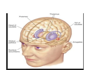
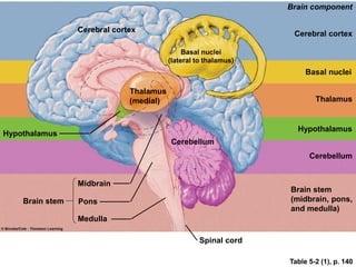
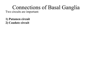
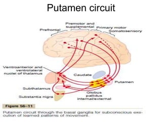
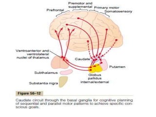
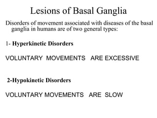
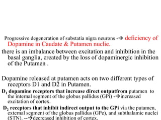
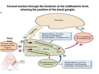
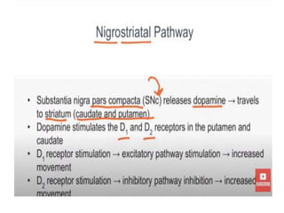
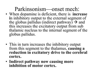
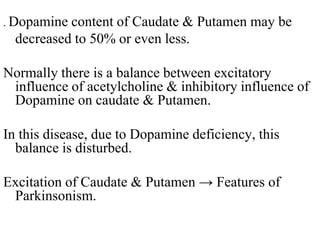
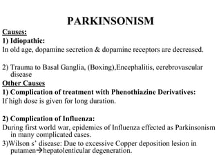
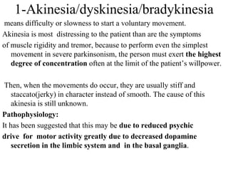
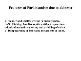
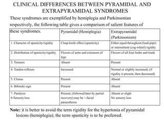
Parkinsonism
Dr Saboohi Saeed
Learning Obejctives
• After attending this lecture the student will be
able to
• 1-Define Parkinsonism
• 2-List the features of this disease
• 3-Explain the pathophysiology of its sign and
symptoms
• 4-Understands the management lines
• 5- Differentiate between Parkinsonism and
Hemiplegia
Table 5-2 (1), p. 140
Hypothalamus
Brain stem
Cerebral cortex
Thalamus
(medial)
Basal nuclei
(lateral to thalamus)
Cerebellum
Spinal cord
Midbrain
Pons
Medulla
Brain component
Cerebral cortex
Basal nuclei
Thalamus
Hypothalamus
Cerebellum
Brain stem
(midbrain, pons,
and medulla)
Connections of Basal Ganglia
Two circuits are important:
1) Putamen circuit
2) Caudate circuit
Putamen circuit
FUNCTIONS OF SPECIFIC
NEUROTRANSMITTER SUBSTANCES IN
THE BASAL GANGLIAL SYSTEM
(1) acetylcholine pathways from the cortex to the caudate nucleus and
putamen
; (2) gamma-aminobutyric acid (GABA) pathways from the caudate
nucleus and putamen to the globus pallidus and substantia nigra;
(3) dopamine pathwaysfrom the substantia nigra to the caudate nucleus
and putamen
(4) multiple general pathways from the brain stem that secrete
norepinephrine, serotonin, enkephalin, and several others.
(5) there are multiple glutamate pathways that provide most of the
excitatory signals that balance out the large numbers of inhibitory
signals transmitted especially by the dopamine, GABA, and serotonin
inhibitory transmitters.
(6) GABA neurons in the feedback loops from the cortex
through the basal ganglia and then back to the cortex make
virtually all these loops negative feedback loops, thus
lending stability to the motor control systems.
(7)Dopamine also functions as an inhibitory neurotransmitter
in most parts of the brain, so it also functions as a stabilizer
Lesions of Basal Ganglia
Disorders of movement associated with diseases of the basal
ganglia in humans are of two general types:
1- Hyperkinetic Disorders
VOLUNTARY MOVEMENTS ARE EXCESSIVE
2-Hypokinetic Disorders
VOLUNTARY MOVEMENTS ARE SLOW
Lesions of Basal Ganglia-Putamen circuit
Hyperkinetic disorders
• 1-Multiple small lesions in the putamen lead to flicking movements
in the hands, face, and other parts of the body, called chorea.
• 2-lesions in the globus pallidus frequently lead to spontaneous and
often continuous writhing movements of a hand, an arm, the neck, or
the face—movements called athetosis.
• 3-A lesion in the subthalamus often leads to sudden flailing
movements of an entire limb to half body -a condition called
hemiballismus.
Lesions of Basal Ganglia-Putamen circuit
Hypokinetic disorders
• Lesions of the substantia nigra lead to the
common and extremely severe disease of
rigidity, akinesia, and tremors known as
Parkinson’s disease.
Parkinsonism
• It is a disease of motor system caused by damage of
basal ganglia Substantia Nigra(Pars compacta)
resulting in deficiency of dopamine characterized by
Akinesia, rigidity,tremors , postural and gait
changes.
• First discovered by James Parkinson
Progressive degeneration of substatia nigra neurons - deficiency of
Dopamine in Caudate & Putamen nuclie.
there is an imbalance between excitation and inhibition in the
basal ganglia, created by the loss of dopaminergic inhibition
of the Putamen .
Dopamine released at putamen acts on two different types of
receptors D1 and D2 in Putamen.
D1 dopamine receptors that increase direct outputfrom putamen to
the internal segment of the globus pallidus (GPi) -increased
excitation of cortex.
D2 receptors that inhibit indirect output to the GPi via the putamen,
external segment of the globus pallidus (GPe), and subthalamic nuclei
(STN). --decreased inhibition of cortex.
Coronal section through the forebrain at the midthalamic level,
showing the position of the basal ganglia.
D1
D2
Dopa
mine
D2
Parkinsonism—onset mech:
• When dopamine is deficient, there is increase
in inhibitory output to the external segment of
the globus pallidus (indirect pathway)  and
this increases the excitatory output from sub
thalamic nucleus to the internal segment of the
globus pallidus.
• This in turn increases the inhibitory output
from this segment to the thalamus, causing a
reduction in excitatory drive to the cerebral
cortex.
• Indirect pathway now causing more
inhibition of motor cortex.
. Dopamine content of Caudate & Putamen may be
decreased to 50% or even less.
Normally there is a balance between excitatory
influence of acetylcholine & inhibitory influence of
Dopamine on caudate & Putamen.
In this disease, due to Dopamine deficiency, this
balance is disturbed.
Excitation of Caudate & Putamen → Features of
Parkinsonism.
PARKINSONISM
Causes:
1) Idiopathic:
In old age, dopamine secretion & dopamine receptors are decreased.
2) Trauma to Basal Ganglia, (Boxing),Encephalitis, cerebrovascular
disease
Other Causes
1) Complication of treatment with Phenothiazine Derivatives:
If high dose is given for long duration.
2) Complication of Influenza:
During first world war, epidemics of Influenza effected as Parkinsonism
in many complicated cases.
3)Wilson s’ disease: Due to excessive Copper deposition lesion in
putamenhepatolenticular degeneration.
PARKINSONISM-causes
4-Maganese intoxication:damages SN.
5-Encephalitis:if lesion in GP and SN.(post
encephalitis parkinsonism).
6-Cerebrovascular disease.
7-MPTP intoxication:Methyl-phenyl-tetrahydro-
pyridine is used in experimental animal to produce
parkinsonism in experimental animal.
Features of Parkinsonism
.
1) Dyskinesia and Akinesia
2) Rigidity and stooped (flexed) posture
3) Resting Tremors
4) Festinant gait
1-Akinesia/dyskinesia/bradykinesia
means difficulty or slowness to start a voluntary movement.
Akinesia is most distressing to the patient than are the symptoms
of muscle rigidity and tremor, because to perform even the simplest
movement in severe parkinsonism, the person must exert the highest
degree of concentration often at the limit of the patient’s willpower.
Then, when the movements do occur, they are usually stiff and
staccato(jerky) in character instead of smooth. The cause of this
akinesia is still unknown.
Pathophysiology:
It has been suggested that this may be due to reduced psychic
drive for motor activity greatly due to decreased dopamine
secretion in the limbic system and in the basal ganglia.
Features of Parkinsonism due to akinesia
a- Smaller and smaller writing-micrographia.
b-No blinking, face like reptiles without expression.
c-Lack of normal swallowing and dribbling of saliva.
d- Disappearance of associated movements of limbs.
.
2-Rigidity
A- Cog-wheel rigidity:
When one limb is passively flexed, there is intermittent resistance to the
flexion.Occurs due to enhanced gamma efferent activity.
B- Lead Pipe rigidity
When the limb is passively flexed, there is a feeling of bending of lead
pipe, that is called lead pipe rigidity.
It is due to increased motor neuron discharge to both agonist and the
antagonist. Because of rigidity, the back is flexed, arms are flexed &
adducted, knees are bent.In severe cases, there is so marked rigidity
that the patient moves like a statue. Stooped posture.
Pathophysiology: destruction of the dopaminergic neurons in the
substantia nigra allow the caudate nucleus and putamen to become
overly active and cause continuous output of excitatory signals to the
corticospinal motor control system. These signals excite many or all
of the muscles of the body,thus leading to rigidity in patients with
Parkinson’s disease.
3) Tremors:
present at rest, static, nonintentional, at a fixed rate of 3 to 6 cycles
per second, These are regular, rhythmic, alternate contraction and
relaxation of agonist & antagonist.
Tremors involve fingers, hands and may involve tongue & lips.
Pill-rolling type of tremors are present. Tremors are present at rest. So
there are static tremors. These are absent during sleep.
Tremors becomes worse during emotional states and when patient is
conscious.
Pathophysiology:
Some of the feedback circuits might easily oscillate because
of high feedback gains after loss of their inhibition,
leading to the tremor of Parkinson’s disease.
4) Gait of the patient:
Patient takes short steps and unable to stop
movement. Festinant gait. There is also
decreased associated movement, such as
swinging of arms during walking
Other Features of Parkinsonism
5-There is loss of facial expression. So there is a ‘mask’ like
face.
6- Babinski’s sign is not present
7. No loss of superficial reflexes like superficial abdominal
reflexes are present.
8. Tendon jerks are difficult to be elicited because of rigidity
otherwise they are present.
9. No sensory loss .
Treatment of Parkinson's Disease
• 1) ) LEVO Dopa is given which passes through blood brain barrier
and it converts into dopamine in brain. Dopamine due to its
chemical structure cannot be given because it doesn’t pass through
the blood brain barrier. So it causes marked improvement in the
condition of patient. restores the normal balance between inhibition
and excitation in the caudate nucleus and putamen. dopamine has a
chemical structure that will not allow it to pass through the blood
brain barrier.
• 2-Mono amine oxidase inhibitors: Treatment with l-
Deprenylinhibits monoamine oxidase, which is responsible for
destruction of most of the dopamine after it has been secreted.
• 3-Treatment with Transplanted Fetal Dopamine
Cells:transplanataion of fetal dopamine cells in the caudate and
putamen is carried out. cells do not live for more than a few months.
• 4- Anticholinergic drugs:
Are given to inhibit over activity of acetylcholine.
• 5- Treatment by Destroying Part of the Feedback Circuitry
in the Basal Ganglia. Electro-coagulation of thalamic
nuclei-Ventro-anterior & Ventro-lateral thalamic nuclei:
can give benefit and are helpful.
CLINICAL DIFFERENCES BETWEEN PYRAMIDAL AND
EXTRAPYRAMIDAL SYNDROMES
These syndromes are exemplified by hemiplegia and Parkinsonian
respectively, the following table gives a comparison of salient features of
these syndromes.
Note: it is better to avoid the term rigidity for the hypertonia of pyramidal
lesions (hemiplegia); the term spasticity is to be preferred.
Pyramidal (Hemiplegia) Extrapyramidal
(Parkinsonism)
1. Character of spasticity/rigidity Clasp-knife effect (spasticity) Either equal throughout (lead-pipe)
or intermittent (cog-wheel) rigidity
2. Distribution of spasticity/rigidity Flexors of arms and extensors of
legs
Flexors of all four limbs and trunk
3. Tremors Absent Present
4. Tendon reflexes Increased Normal or slightly increased; (if
rigidity is present, then decreased)
5. Clonus Present Absent
6. Bibinski sign Present Absent
7. Paralysis
8-Sensory loss
Present; (followed later by partial
recovery) may be +,facial
paraesthesia
Absent or slight
No sensory loss
• https://www.youtube.com/watch?v=UtacL
Pnoa28

Parkinsonism.ppt

  • 1.
  • 2.
    Learning Obejctives • Afterattending this lecture the student will be able to • 1-Define Parkinsonism • 2-List the features of this disease • 3-Explain the pathophysiology of its sign and symptoms • 4-Understands the management lines • 5- Differentiate between Parkinsonism and Hemiplegia
  • 4.
    Table 5-2 (1),p. 140 Hypothalamus Brain stem Cerebral cortex Thalamus (medial) Basal nuclei (lateral to thalamus) Cerebellum Spinal cord Midbrain Pons Medulla Brain component Cerebral cortex Basal nuclei Thalamus Hypothalamus Cerebellum Brain stem (midbrain, pons, and medulla)
  • 5.
    Connections of BasalGanglia Two circuits are important: 1) Putamen circuit 2) Caudate circuit
  • 6.
  • 8.
    FUNCTIONS OF SPECIFIC NEUROTRANSMITTERSUBSTANCES IN THE BASAL GANGLIAL SYSTEM (1) acetylcholine pathways from the cortex to the caudate nucleus and putamen ; (2) gamma-aminobutyric acid (GABA) pathways from the caudate nucleus and putamen to the globus pallidus and substantia nigra; (3) dopamine pathwaysfrom the substantia nigra to the caudate nucleus and putamen (4) multiple general pathways from the brain stem that secrete norepinephrine, serotonin, enkephalin, and several others. (5) there are multiple glutamate pathways that provide most of the excitatory signals that balance out the large numbers of inhibitory signals transmitted especially by the dopamine, GABA, and serotonin inhibitory transmitters.
  • 9.
    (6) GABA neuronsin the feedback loops from the cortex through the basal ganglia and then back to the cortex make virtually all these loops negative feedback loops, thus lending stability to the motor control systems. (7)Dopamine also functions as an inhibitory neurotransmitter in most parts of the brain, so it also functions as a stabilizer
  • 14.
    Lesions of BasalGanglia Disorders of movement associated with diseases of the basal ganglia in humans are of two general types: 1- Hyperkinetic Disorders VOLUNTARY MOVEMENTS ARE EXCESSIVE 2-Hypokinetic Disorders VOLUNTARY MOVEMENTS ARE SLOW
  • 15.
    Lesions of BasalGanglia-Putamen circuit Hyperkinetic disorders • 1-Multiple small lesions in the putamen lead to flicking movements in the hands, face, and other parts of the body, called chorea. • 2-lesions in the globus pallidus frequently lead to spontaneous and often continuous writhing movements of a hand, an arm, the neck, or the face—movements called athetosis. • 3-A lesion in the subthalamus often leads to sudden flailing movements of an entire limb to half body -a condition called hemiballismus.
  • 16.
    Lesions of BasalGanglia-Putamen circuit Hypokinetic disorders • Lesions of the substantia nigra lead to the common and extremely severe disease of rigidity, akinesia, and tremors known as Parkinson’s disease.
  • 17.
    Parkinsonism • It isa disease of motor system caused by damage of basal ganglia Substantia Nigra(Pars compacta) resulting in deficiency of dopamine characterized by Akinesia, rigidity,tremors , postural and gait changes. • First discovered by James Parkinson
  • 18.
    Progressive degeneration ofsubstatia nigra neurons - deficiency of Dopamine in Caudate & Putamen nuclie. there is an imbalance between excitation and inhibition in the basal ganglia, created by the loss of dopaminergic inhibition of the Putamen . Dopamine released at putamen acts on two different types of receptors D1 and D2 in Putamen. D1 dopamine receptors that increase direct outputfrom putamen to the internal segment of the globus pallidus (GPi) -increased excitation of cortex. D2 receptors that inhibit indirect output to the GPi via the putamen, external segment of the globus pallidus (GPe), and subthalamic nuclei (STN). --decreased inhibition of cortex.
  • 19.
    Coronal section throughthe forebrain at the midthalamic level, showing the position of the basal ganglia. D1 D2 Dopa mine D2
  • 21.
    Parkinsonism—onset mech: • Whendopamine is deficient, there is increase in inhibitory output to the external segment of the globus pallidus (indirect pathway)  and this increases the excitatory output from sub thalamic nucleus to the internal segment of the globus pallidus. • This in turn increases the inhibitory output from this segment to the thalamus, causing a reduction in excitatory drive to the cerebral cortex. • Indirect pathway now causing more inhibition of motor cortex.
  • 22.
    . Dopamine contentof Caudate & Putamen may be decreased to 50% or even less. Normally there is a balance between excitatory influence of acetylcholine & inhibitory influence of Dopamine on caudate & Putamen. In this disease, due to Dopamine deficiency, this balance is disturbed. Excitation of Caudate & Putamen → Features of Parkinsonism.
  • 23.
    PARKINSONISM Causes: 1) Idiopathic: In oldage, dopamine secretion & dopamine receptors are decreased. 2) Trauma to Basal Ganglia, (Boxing),Encephalitis, cerebrovascular disease Other Causes 1) Complication of treatment with Phenothiazine Derivatives: If high dose is given for long duration. 2) Complication of Influenza: During first world war, epidemics of Influenza effected as Parkinsonism in many complicated cases. 3)Wilson s’ disease: Due to excessive Copper deposition lesion in putamenhepatolenticular degeneration.
  • 24.
    PARKINSONISM-causes 4-Maganese intoxication:damages SN. 5-Encephalitis:iflesion in GP and SN.(post encephalitis parkinsonism). 6-Cerebrovascular disease. 7-MPTP intoxication:Methyl-phenyl-tetrahydro- pyridine is used in experimental animal to produce parkinsonism in experimental animal.
  • 25.
    Features of Parkinsonism . 1)Dyskinesia and Akinesia 2) Rigidity and stooped (flexed) posture 3) Resting Tremors 4) Festinant gait
  • 26.
    1-Akinesia/dyskinesia/bradykinesia means difficulty orslowness to start a voluntary movement. Akinesia is most distressing to the patient than are the symptoms of muscle rigidity and tremor, because to perform even the simplest movement in severe parkinsonism, the person must exert the highest degree of concentration often at the limit of the patient’s willpower. Then, when the movements do occur, they are usually stiff and staccato(jerky) in character instead of smooth. The cause of this akinesia is still unknown. Pathophysiology: It has been suggested that this may be due to reduced psychic drive for motor activity greatly due to decreased dopamine secretion in the limbic system and in the basal ganglia.
  • 27.
    Features of Parkinsonismdue to akinesia a- Smaller and smaller writing-micrographia. b-No blinking, face like reptiles without expression. c-Lack of normal swallowing and dribbling of saliva. d- Disappearance of associated movements of limbs. .
  • 28.
    2-Rigidity A- Cog-wheel rigidity: Whenone limb is passively flexed, there is intermittent resistance to the flexion.Occurs due to enhanced gamma efferent activity. B- Lead Pipe rigidity When the limb is passively flexed, there is a feeling of bending of lead pipe, that is called lead pipe rigidity. It is due to increased motor neuron discharge to both agonist and the antagonist. Because of rigidity, the back is flexed, arms are flexed & adducted, knees are bent.In severe cases, there is so marked rigidity that the patient moves like a statue. Stooped posture. Pathophysiology: destruction of the dopaminergic neurons in the substantia nigra allow the caudate nucleus and putamen to become overly active and cause continuous output of excitatory signals to the corticospinal motor control system. These signals excite many or all of the muscles of the body,thus leading to rigidity in patients with Parkinson’s disease.
  • 33.
    3) Tremors: present atrest, static, nonintentional, at a fixed rate of 3 to 6 cycles per second, These are regular, rhythmic, alternate contraction and relaxation of agonist & antagonist. Tremors involve fingers, hands and may involve tongue & lips. Pill-rolling type of tremors are present. Tremors are present at rest. So there are static tremors. These are absent during sleep. Tremors becomes worse during emotional states and when patient is conscious. Pathophysiology: Some of the feedback circuits might easily oscillate because of high feedback gains after loss of their inhibition, leading to the tremor of Parkinson’s disease. 4) Gait of the patient:
  • 34.
    4) Gait ofthe patient: Patient takes short steps and unable to stop movement. Festinant gait. There is also decreased associated movement, such as swinging of arms during walking
  • 35.
    Other Features ofParkinsonism 5-There is loss of facial expression. So there is a ‘mask’ like face. 6- Babinski’s sign is not present 7. No loss of superficial reflexes like superficial abdominal reflexes are present. 8. Tendon jerks are difficult to be elicited because of rigidity otherwise they are present. 9. No sensory loss .
  • 36.
    Treatment of Parkinson'sDisease • 1) ) LEVO Dopa is given which passes through blood brain barrier and it converts into dopamine in brain. Dopamine due to its chemical structure cannot be given because it doesn’t pass through the blood brain barrier. So it causes marked improvement in the condition of patient. restores the normal balance between inhibition and excitation in the caudate nucleus and putamen. dopamine has a chemical structure that will not allow it to pass through the blood brain barrier. • 2-Mono amine oxidase inhibitors: Treatment with l- Deprenylinhibits monoamine oxidase, which is responsible for destruction of most of the dopamine after it has been secreted. • 3-Treatment with Transplanted Fetal Dopamine Cells:transplanataion of fetal dopamine cells in the caudate and putamen is carried out. cells do not live for more than a few months.
  • 37.
    • 4- Anticholinergicdrugs: Are given to inhibit over activity of acetylcholine. • 5- Treatment by Destroying Part of the Feedback Circuitry in the Basal Ganglia. Electro-coagulation of thalamic nuclei-Ventro-anterior & Ventro-lateral thalamic nuclei: can give benefit and are helpful.
  • 38.
    CLINICAL DIFFERENCES BETWEENPYRAMIDAL AND EXTRAPYRAMIDAL SYNDROMES These syndromes are exemplified by hemiplegia and Parkinsonian respectively, the following table gives a comparison of salient features of these syndromes. Note: it is better to avoid the term rigidity for the hypertonia of pyramidal lesions (hemiplegia); the term spasticity is to be preferred. Pyramidal (Hemiplegia) Extrapyramidal (Parkinsonism) 1. Character of spasticity/rigidity Clasp-knife effect (spasticity) Either equal throughout (lead-pipe) or intermittent (cog-wheel) rigidity 2. Distribution of spasticity/rigidity Flexors of arms and extensors of legs Flexors of all four limbs and trunk 3. Tremors Absent Present 4. Tendon reflexes Increased Normal or slightly increased; (if rigidity is present, then decreased) 5. Clonus Present Absent 6. Bibinski sign Present Absent 7. Paralysis 8-Sensory loss Present; (followed later by partial recovery) may be +,facial paraesthesia Absent or slight No sensory loss
  • 39.