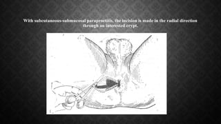
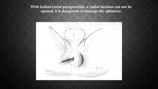
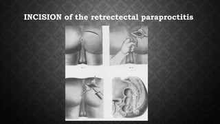
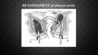
Paraproctitis
paraproctitis
•
(Pararectal abscess) - acute or chronic bacterial
inflammation of the peritoneal tissue
etiology
• Paraproctitis is one of the most frequent proctologic diseases
(20-40% of all diseases of the rectum). Paraproctitis in
frequency is the 4th place after hemorrhoids, anal fissures
and colitis. Men suffer from paraproctitis more often than
women.This ratio ranges from 1.5: 1 to 4.7: 1. Paraproctitis is
a disease of adults: there are rare descriptions of fistulas of
the rectum in children.
Etiology (flora)
• E. coli;
• Anaerobes;
• Staphylococcus aureus;
• White staphylococcus aureus;
• streptococcus;
• Enterococcus;
• Clostridial anaerobes: Cl.perfringens, Cl.Hystoliticum, Cl.oedematiens,
Cl.septicum
Pathogenesis. Ways of introduction of
infection into pararectal tissue
Columns of Morgagni
(columnae Morgani)
• Infection can penetrate through the anal glands, damaged rectum
mucosa, hematogenous and lymphogenous pathway, from
neighboring organs affected by the inflammatory process. A
number of researchers consider paraproctitis a fairly frequent
complication of ulcerative colitis and Crohn's disease. Purulent
processes in the cells around the rectum are observed in diseases
of the prostate gland, urethra, paraurethral, Cooper glands,​​
female sexual organs (perimetritis, bartholinitis), sometimes with
osteomyelitis of pelvic bones, spinal tuberculosis.
Typical ways of spreading the inflammatory
process
• The entrance gates are anal crypts, which open the ducts of the anal
glands, or (much less often) the damaged mucosa of the anal canal.
• In paraproctitis, fistulous holes are described on the scrotum, thigh,
anterior abdominal wall. In addition, it is possible that abscess can
break through into the lumen of the intestine at the level of the
ampullar part of it and even into the abdominal cavity.
Classification of paraproctitis.
The etiological principle distinguishes:
Nonspecific paraproctitis,
Specific paraproctitis,
Post traumatic paraproctitis.
By the activity of the inflammatory process:
Acute paraproctitis,
Recurrent paraproctitis,
Chronic paraproctitis (fistulas of the rectum).
Localization of abscesses
- Subcutaneous paraproctitis;
- Submucosal paraproctitis;
- Ischial-rectal paraproctitis;
- Behind-rectal paraproctitis;
- Pelvic-rectal paraproctitis (pelvio-rectal).
52%
4%
42%
2%
частота различных форм
подкожный подслизистый -седалищно прямокишечный -тазово прямокишечный
• 1-subcutaneous;
• 2 submucosal;
• 3-ischial-rectal;
• 4-pelvis and rectum;
• 5- behind-rectal.
Retrorectal paraproctitis
clinics
• PECULIARITIES OF CLINIC AND PARAPROCTITIS ARE DETERMINED BY
LOCALIZATION OF THE FOCUS IN THE CELLULAR SPACES OF THE PELVIS
• At the beginning of the disease there is a short period with malaise, weakness and headache.
There is an increase in temperature above 37.5 ° C with chill
Subcutaneous paraproctitis
• Submucosal abscess is located under the mucosa of the rectum.
Symptoms in this form of location are similar to subcutaneous
paraproctitis, however, the pain syndrome and skin changes are
less pronounced
• With ischiorectal abscess, a purulent focus is located above the muscle
that raises the anus. Because of the deeper location of the abscess, the
local symptoms are more vague: blunt pulsating pains in the small
pelvis and rectum, intensifying with defecation. Changes from the skin
in the form of redness, swelling, swelling, asymmetry of the buttocks
occur later on the 5th-6th day from the onset of pain.The general state
of health is severe: the temperature can rise to 38 ° C, intoxication is
expressed
• The most severe is pelvio-rectal abscess.This is a rare form of acute
paraproctitis, when the purulent focus is located above the muscles
that form the pelvic floor, a thin layer of the peritoneum separates it
from the abdominal cavity. At the beginning of the disease, severe
fever, chills, joint pain predominate. Local symptoms: pain in the pelvis
and in the lower abdomen. After 10-12 days of pain intensifies, there is
a delay of stool and urine
Chronic paraproctitis
• Manifested festering fistula.The mouth of the fistulous passage can be located near
the anal opening of the rectum or at a distance from it on the buttocks.The pain is
usually not expressed. From the mouth of the fistula pus often diffuses feces. During
the development of chronic paraproctitis, the opening of the fistula can be closed,
pus stagnation occurs, abscesses develop, new tissue defects appear, pus
breakthrough and outflow into the rectum and outward, necrosis and other tissue
changes that significantly complicate the fistula.Thus, there are complex fistulous
systems with branching fistula, cavity depots and a lot of holes.
Factors of chronic course
• The internal fistula opening (covered with the epithelial lining of the mouth of an infected
one or more anal glands opening in the Morgan crypt) is a source of permanent or periodic
infection of the pararectal tissue.
• Presence of a formed fistulous course, single or branched, located superficially or deep in
the pelvic tissues.The healing is impeded by the epithelialization of the fistula wall, which
prevents their collapse and makes the lumen open for reinfection.The internal surface of
the fistulous course is lined with granulation tissue or glandular epithelium.
• Activation of dormant surgical infection. Infection can stay in the tissues of the body more or
less for a long time without causing a disease (latent phase), but then under the influence of
various factors manifests itself in the form of acute paraproctitis (active phase). In
paraproctitis, the focus of a dormant infection can have a morphological substrate.They are
either scars at the site of the open abscess, or the remainder of the fistulous course, or a
ligated, undisclosed stitch
Diagnostics
• The basis of diagnosis are typical complaints and pains in the area of the rectum and​​
anus.When making a diagnosis, you need a digital rectal examination, sometimes
performed under anesthesia in the operating room because of pain.
• From laboratory methods, it is necessary to study the blood and urine, blood for
glucose, and in case of doubt - ultrasound examination of the perianal region,
including using a rectal sensor. In the presence of fistulous strokes, fistulography is
shown, the introduction of a contrast medium into the fistula with the execution of a
series of X-ray photographs.
treatment
• In the very initial stage of the disease, with a small infiltrate in the
perianal area, conservative treatment methods are used: sedentary
warm baths with potassium permanganate solution, lumbar procaine
blockade, warmers, UHF therapy, etc. All thermal procedures are
combined with antibiotic therapy.
Operative treatment
• Surgical treatment of acute paraproctitis includes early emergency
surgical intervention by opening the abscess with removal of pus and
necrotic tissues, examination of the abscess cavity with a finger,
separation of the bridges and drainage of the cavity.
The choice of method of anesthesia
-Infiltration anesthesia
(Vishnevsky AS, Ryzhikh AN);
- cerebrospinal anesthesia
           (Yudin SS);
-operational anesthesia
(Yakovlev NA, Makhaev NM);
-General anesthesia.
The patient's position on the operating table
POSITION ON DEPAGE
Radical operations in acute paraproctitis can be
grouped as follows
• 1. opening and draining of the abscess, excision of the affected crypt and dissection
of the purulent course into the lumen of the intestine;
• 2. opening and draining of the abscess, excision of the affected crypt and
sphincterotomy;
• 3. dissection and drainage of the abscess, excision of the affected crypt, ligation;
• 4. dissection and drainage of the abscess, delayed excision of the affected crypt and
movement of the gut flap to interrupt the infection pathway from the lumen of the
rectum
• Apply radial, semilunar, crosswise incisions, the most convenient of
them - semilunar and radial.They provide a gaping wound and an
outflow of purulent exudate, as well as less traumatic. Strict bed rest is
necessary.
• When putrefied necrotic paraproctitis during the operation, complete
excision of dead fiber is performed within healthy tissues, and two or
three additional cuts of the skin and subcutaneous tissue for draining
are performed.
The incisions used in
acute paraproctitis:
1 - perianal abscess; 2
- behind-mousse; 3 -
Ischiorectal.
• Surgical approaches used for ischiorectal (1) and pelviorectal (2) abscesses.
DISSECTION with subcutaneous
paraproctitis
With subcutaneous-submucosal paraproctitis, the incision is made in the radial direction
through an interested crypt.
With ischial-rectal paraproctitis, a radial incision can not be
opened, it is dangerous to damage the sphincter.
INCISION of the retrectectal paraproctitis
REASSESSMENT of abscess cavity
Complications of paraproctitis
Purulent fusion of the rectum wall with the spread of intestinal contents into the
pararectal tissue.Widespread access to pararectal tissue for the contents of the
intestine can lead to even more severe consequences as follows:
• Formation of purulent fistula between the rectum and the vagina • Purulent melting of
the urethra, the transition of the purulent process to the scrotum followed by gangrene •
Breakdown of pus into the free Abdominal cavity and retroperitoneal tissue with the
development of peritonitis and retroperitoneal phlegmon, which lead to sepsis and
death.? Necrosis of the skin in the area of dissemination of the abscess​​
Complications of acute
paraproctitis
Prevention of recurrence of the disease
• To achieve a lasting effect, in addition to the radical surgery performed, it is
necessary to adhere to a number of rules.
• • Fighting with constipation. It is necessary to achieve a regular soft stool. •
The diet should contain plant foods rich in fiber (fresh fruit vegetables,
especially beets, cabbage), and fermented milk products (kefir, curdled milk,
cottage cheese).You should avoid a dough product. • In diabetes, the normal
blood sugar level should be maintained, otherwise purulent diseases can not
be avoided • Maintaining normal body weight • Observance of personal
hygiene rules - regular washing after stools • Treatment of chronic
hemorrhoids And anal fissures.
Paraproctitis

Paraproctitis

  • 1.
  • 2.
    paraproctitis • (Pararectal abscess) -acute or chronic bacterial inflammation of the peritoneal tissue
  • 3.
    etiology • Paraproctitis isone of the most frequent proctologic diseases (20-40% of all diseases of the rectum). Paraproctitis in frequency is the 4th place after hemorrhoids, anal fissures and colitis. Men suffer from paraproctitis more often than women.This ratio ranges from 1.5: 1 to 4.7: 1. Paraproctitis is a disease of adults: there are rare descriptions of fistulas of the rectum in children.
  • 4.
    Etiology (flora) • E.coli; • Anaerobes; • Staphylococcus aureus; • White staphylococcus aureus; • streptococcus; • Enterococcus; • Clostridial anaerobes: Cl.perfringens, Cl.Hystoliticum, Cl.oedematiens, Cl.septicum
  • 5.
    Pathogenesis. Ways ofintroduction of infection into pararectal tissue
  • 6.
  • 7.
    • Infection canpenetrate through the anal glands, damaged rectum mucosa, hematogenous and lymphogenous pathway, from neighboring organs affected by the inflammatory process. A number of researchers consider paraproctitis a fairly frequent complication of ulcerative colitis and Crohn's disease. Purulent processes in the cells around the rectum are observed in diseases of the prostate gland, urethra, paraurethral, Cooper glands,​​ female sexual organs (perimetritis, bartholinitis), sometimes with osteomyelitis of pelvic bones, spinal tuberculosis.
  • 8.
    Typical ways ofspreading the inflammatory process • The entrance gates are anal crypts, which open the ducts of the anal glands, or (much less often) the damaged mucosa of the anal canal. • In paraproctitis, fistulous holes are described on the scrotum, thigh, anterior abdominal wall. In addition, it is possible that abscess can break through into the lumen of the intestine at the level of the ampullar part of it and even into the abdominal cavity.
  • 9.
    Classification of paraproctitis. Theetiological principle distinguishes: Nonspecific paraproctitis, Specific paraproctitis, Post traumatic paraproctitis. By the activity of the inflammatory process: Acute paraproctitis, Recurrent paraproctitis, Chronic paraproctitis (fistulas of the rectum).
  • 10.
    Localization of abscesses -Subcutaneous paraproctitis; - Submucosal paraproctitis; - Ischial-rectal paraproctitis; - Behind-rectal paraproctitis; - Pelvic-rectal paraproctitis (pelvio-rectal).
  • 11.
    52% 4% 42% 2% частота различных форм подкожныйподслизистый -седалищно прямокишечный -тазово прямокишечный
  • 12.
    • 1-subcutaneous; • 2submucosal; • 3-ischial-rectal; • 4-pelvis and rectum; • 5- behind-rectal.
  • 13.
  • 14.
    clinics • PECULIARITIES OFCLINIC AND PARAPROCTITIS ARE DETERMINED BY LOCALIZATION OF THE FOCUS IN THE CELLULAR SPACES OF THE PELVIS • At the beginning of the disease there is a short period with malaise, weakness and headache. There is an increase in temperature above 37.5 ° C with chill
  • 15.
  • 16.
    • Submucosal abscessis located under the mucosa of the rectum. Symptoms in this form of location are similar to subcutaneous paraproctitis, however, the pain syndrome and skin changes are less pronounced
  • 17.
    • With ischiorectalabscess, a purulent focus is located above the muscle that raises the anus. Because of the deeper location of the abscess, the local symptoms are more vague: blunt pulsating pains in the small pelvis and rectum, intensifying with defecation. Changes from the skin in the form of redness, swelling, swelling, asymmetry of the buttocks occur later on the 5th-6th day from the onset of pain.The general state of health is severe: the temperature can rise to 38 ° C, intoxication is expressed
  • 18.
    • The mostsevere is pelvio-rectal abscess.This is a rare form of acute paraproctitis, when the purulent focus is located above the muscles that form the pelvic floor, a thin layer of the peritoneum separates it from the abdominal cavity. At the beginning of the disease, severe fever, chills, joint pain predominate. Local symptoms: pain in the pelvis and in the lower abdomen. After 10-12 days of pain intensifies, there is a delay of stool and urine
  • 19.
    Chronic paraproctitis • Manifestedfestering fistula.The mouth of the fistulous passage can be located near the anal opening of the rectum or at a distance from it on the buttocks.The pain is usually not expressed. From the mouth of the fistula pus often diffuses feces. During the development of chronic paraproctitis, the opening of the fistula can be closed, pus stagnation occurs, abscesses develop, new tissue defects appear, pus breakthrough and outflow into the rectum and outward, necrosis and other tissue changes that significantly complicate the fistula.Thus, there are complex fistulous systems with branching fistula, cavity depots and a lot of holes.
  • 21.
    Factors of chroniccourse • The internal fistula opening (covered with the epithelial lining of the mouth of an infected one or more anal glands opening in the Morgan crypt) is a source of permanent or periodic infection of the pararectal tissue. • Presence of a formed fistulous course, single or branched, located superficially or deep in the pelvic tissues.The healing is impeded by the epithelialization of the fistula wall, which prevents their collapse and makes the lumen open for reinfection.The internal surface of the fistulous course is lined with granulation tissue or glandular epithelium. • Activation of dormant surgical infection. Infection can stay in the tissues of the body more or less for a long time without causing a disease (latent phase), but then under the influence of various factors manifests itself in the form of acute paraproctitis (active phase). In paraproctitis, the focus of a dormant infection can have a morphological substrate.They are either scars at the site of the open abscess, or the remainder of the fistulous course, or a ligated, undisclosed stitch
  • 22.
    Diagnostics • The basisof diagnosis are typical complaints and pains in the area of the rectum and​​ anus.When making a diagnosis, you need a digital rectal examination, sometimes performed under anesthesia in the operating room because of pain. • From laboratory methods, it is necessary to study the blood and urine, blood for glucose, and in case of doubt - ultrasound examination of the perianal region, including using a rectal sensor. In the presence of fistulous strokes, fistulography is shown, the introduction of a contrast medium into the fistula with the execution of a series of X-ray photographs.
  • 23.
    treatment • In thevery initial stage of the disease, with a small infiltrate in the perianal area, conservative treatment methods are used: sedentary warm baths with potassium permanganate solution, lumbar procaine blockade, warmers, UHF therapy, etc. All thermal procedures are combined with antibiotic therapy.
  • 24.
    Operative treatment • Surgicaltreatment of acute paraproctitis includes early emergency surgical intervention by opening the abscess with removal of pus and necrotic tissues, examination of the abscess cavity with a finger, separation of the bridges and drainage of the cavity.
  • 25.
    The choice ofmethod of anesthesia -Infiltration anesthesia (Vishnevsky AS, Ryzhikh AN); - cerebrospinal anesthesia            (Yudin SS); -operational anesthesia (Yakovlev NA, Makhaev NM); -General anesthesia.
  • 26.
    The patient's positionon the operating table
  • 27.
  • 28.
    Radical operations inacute paraproctitis can be grouped as follows • 1. opening and draining of the abscess, excision of the affected crypt and dissection of the purulent course into the lumen of the intestine; • 2. opening and draining of the abscess, excision of the affected crypt and sphincterotomy; • 3. dissection and drainage of the abscess, excision of the affected crypt, ligation; • 4. dissection and drainage of the abscess, delayed excision of the affected crypt and movement of the gut flap to interrupt the infection pathway from the lumen of the rectum
  • 29.
    • Apply radial,semilunar, crosswise incisions, the most convenient of them - semilunar and radial.They provide a gaping wound and an outflow of purulent exudate, as well as less traumatic. Strict bed rest is necessary. • When putrefied necrotic paraproctitis during the operation, complete excision of dead fiber is performed within healthy tissues, and two or three additional cuts of the skin and subcutaneous tissue for draining are performed.
  • 30.
    The incisions usedin acute paraproctitis: 1 - perianal abscess; 2 - behind-mousse; 3 - Ischiorectal.
  • 31.
    • Surgical approachesused for ischiorectal (1) and pelviorectal (2) abscesses.
  • 32.
  • 33.
    With subcutaneous-submucosal paraproctitis,the incision is made in the radial direction through an interested crypt.
  • 34.
    With ischial-rectal paraproctitis,a radial incision can not be opened, it is dangerous to damage the sphincter.
  • 35.
    INCISION of theretrectectal paraproctitis
  • 36.
  • 37.
    Complications of paraproctitis Purulentfusion of the rectum wall with the spread of intestinal contents into the pararectal tissue.Widespread access to pararectal tissue for the contents of the intestine can lead to even more severe consequences as follows: • Formation of purulent fistula between the rectum and the vagina • Purulent melting of the urethra, the transition of the purulent process to the scrotum followed by gangrene • Breakdown of pus into the free Abdominal cavity and retroperitoneal tissue with the development of peritonitis and retroperitoneal phlegmon, which lead to sepsis and death.? Necrosis of the skin in the area of dissemination of the abscess​​
  • 38.
  • 39.
    Prevention of recurrenceof the disease • To achieve a lasting effect, in addition to the radical surgery performed, it is necessary to adhere to a number of rules. • • Fighting with constipation. It is necessary to achieve a regular soft stool. • The diet should contain plant foods rich in fiber (fresh fruit vegetables, especially beets, cabbage), and fermented milk products (kefir, curdled milk, cottage cheese).You should avoid a dough product. • In diabetes, the normal blood sugar level should be maintained, otherwise purulent diseases can not be avoided • Maintaining normal body weight • Observance of personal hygiene rules - regular washing after stools • Treatment of chronic hemorrhoids And anal fissures.

Editor's Notes

  • #5 Парапроктит вызывается смешанной микрофлорой, но преобладающее значение имеет E. coli. В этиологии парапроктита ведущее значение имеет внедрение инфекции в параректальную клетчатку Предрасполагающими факторами возникновения гнойного процесса в параректальной клетчатке являются: ослабление местного и гуморального иммунитета при истощении, алкоголизме, вследствие острой или хронической инфекции (ангина, грипп, сепсис); сосудистые изменения при сахарном диабете, атеросклерозе; функциональные нарушения (запор, понос); наличие геморроя, трещин заднего прохода, криптита и др. <number>
  • #7 На 2 см выше анального отверстия слизистая оболочка прямой кишки образует ряд вертикально расположенных параллельных возвышений. Это так называемые столбики Морганьи. Число их различно (от 6 до 14), они имеют форму продольных валиков, поднимающихся над уровнем слизистой на 2—4 мм. Столбики Морганьи образованы складкой слизистой оболочки. Между каждыми двумя столбиками получается углубление в виде желобка, который заканчивается слепым кармашком (криптой). Крипты играют большую роль. В них нередко задерживаются инородные тела или частицы кала, которые могут вызвать воспаления и привести к развитию парапроктита. <number>
  • #8 <number>
  • #9 Распространение инфекции при парапроктите идет со стороны прямой кишки. Вначале воспаляется какая-либо крипта, где и формируется внутреннее отверстие свища. Затем инфекция проникает по протокам анальных желез в их разветвления, локализующиеся в подслизистом или межмышечном слоях. При проникновении инфекции через поврежденную слизистую оболочку кишки дальнейшее ее распространение возможно по межтканевым щелям, а также лимфогенным и гематогенным путем. В связи с рыхлостью подслизистого слоя кишки гнойники этой локализации легко и быстро распространяются вверх, отслаивая слизистую оболочку. Затеки гноя, распространяясь по ходу сосудистых ветвей, проникают в межмышечные пространства между циркулярным и продольным слоями мышц кишечной стенки. Распространяясь по разветвлениям анальных желез и разрушая их, гной проникает в параректальные клетчаточные пространства (подслизистое, подкожное, подвздошно-прямокишечное, тазово-прямокишечное и позадипрямокишечное). В зависимости от степени вирулентности флоры, реактивности организма процесс может быть отграниченным в какой-либо одной области или распространенным по клетчатке соседних анатомических областей. При распространении гнойного процесса имеют значение и условия оттока. Если гнойник хорошо дренируется по широкому сообщению с кишкой или имеет выход на кожу, можно ожидать минимальных поражений клетчаточных пространств таза. Если таких условий нет, вокруг прямой кишки может образоваться множество гнойных ходов с затеками в различные клетчаточные пространства. Возможны прорывы гнойника на кожу в различных участках (наружные свищевые отверстия). <number>
  • #15 Клиническая картина парапроктита существенно варьирует в зависимости от расположения гнойного очага. Заболевание, как правило, начинается остро. Вслед за коротким продромальным периодом с недомоганием, слабостью, головной болью появляется нарастающая боль в прямой кишке, промежности или в тазу, сопровождающаяся повышением температуры тела и ознобом. Степень выраженности симптомов острого парапроктита зависит от локализации воспалительного процесса, распространенности его, характера возбудителя, реактивности организма. При локализации гнойника в подкожной клетчатке клинические проявления выражены более ярко и определенно: болезненный инфильтрат в области заднего прохода, гиперемия кожи, повышение температуры тела вынуждают, как правило, обратиться к врачу в первые дни после начала заболевания <number>
  • #16 При подкожном парапроктите, когда гнойник располагается около анального отверстия под кожей, симптомы наиболее яркие: болезненная опухоль в области заднего прохода, с покраснением кожи над ней. Боли постепенно нарастают, приобретая интенсивный пульсирующий характер, мешая спать, сидеть, дефекация становится крайне болезненной, над опухолью появляется размягчение. Эта форма парапроктита встречается наиболее часто. <number>
  • #17 . <number>
  • #18 <number>
  • #19 Читать отсюда Наиболее тяжело протекает пельвиоректальный парапроктит, при котором гнойник располагается глубоко в тазу. В первые дни болезни преобладают общие симптомы воспаления: лихорадка, ознобы, головная боль, боль в суставах, в тазу, внизу живота. Часто пациент обращается к хирургу, урологу, женщины — к гинекологу. Нередко их лечат по поводу острого респираторного заболевания, гриппа. Продолжительность этого периода иногда достигает 10-12 дней. В дальнейшем отмечаются усиление боли в тазу и прямой кишке, задержка стула, мочи и выраженная интоксикация. Если происходит гнойное расплавление стенки прямой кишки, гной прорывается в просвет прямой кишки, у женщин может быть прорыв гноя из пельвиоректального пространства во влагалище. Осложненное течение пельвиоректального парапроктита с опорожнением гнойника в кишку или влагалище часто смазывает картину заболевания, так как уменьшается боль и снижается температура. В этих случаях выделения гноя и крови из кишки и влагалища могут навести на мысль о дизентерии, гинекологических заболеваниях и т. д. Такие симптомы, как задержка стула, тенезмы, дизурические расстройства, боль внизу живота, особенно часто бывают при локализации гнойника в пельвиоректальном пространстве, но могут быть при любом виде парапроктита <number>
  • #20 <number>
  • #21 На рисунке показано насколько многочисленны и разнообразны могут быть системы свищевых ходов при хроническом парапроктите <number>
  • #22 Современные исследователи рассматривают острый и хронический парапроктит как стадии одного заболевания. В прежние годы широко обсуждался вопрос о том, почему после вскрытия острого парапроктита так часто формируется свищ прямой кишки (хронический парапроктит). <number>
  • #23 Достаточно проста диагностика подкожного парапроктита. Гнойник, локализующийся в подкожной клетчатке перианальной зоны, проявляется быстро и ярко, но флюктуации вначале может и не быть — это поздний симптом. Несмотря на то что диагноз кажется несомненным уже при наружном осмотре и пальпации, необходимо выполнять пальцевое исследование прямой кишки и анального канала. При этом нужно не только установить связь гнойника с кишкой, найти пораженную крипту, но и помнить, что гной в подкожной клетчатке может появиться вследствие прорыва его из других клетчаточных пространств, чаще всего из ишиоректального. Введение пальца в кишку следует делать осторожно по стенке анального канала, противоположной расположению гнойника. При подкожном парапроктите верхняя граница абсцесса определяется ниже аноректальной линии. Выше этой зоны стенка кишки эластична. Таким образом, диагноз острого подкожного парапроктита можно поставить на основании клинической картины, наружного осмотра, пальпации и пальцевого исследования прямой кишки. Ано-, ректороманоскопия, сфинктерометрия и другие исследования обычно не проводятся в связи с тем, что инструментальные исследования при остром парапроктите очень болезненны. Ишиоректальный парапроктит может давать изменения, видимые глазом, в поздней стадии, когда появляются выраженная асимметрия ягодиц, сглаживание перианальных складок. Поэтому, если больной обратился по поводу познабливания, ухудшения самочувствия, сна и тупой постоянной боли в прямой кишке и тазу, усиливающейся при дефекации, но без видимых на глаз изменений в области заднего прохода, следует выполнить пальцевое исследование прямой кишки. Уже в самые ранние сроки заболевания можно выявить уплощение стенки кишки выше анального канала, сглаженность складок слизистой оболочки на стороне поражения. К концу 1-й недели заболевания инфильтрат выбухает в просвет прямой кишки, местно отмечается повышение температуры. Воспалительный инфильтрат может распространиться на предстательную железу и уретру, в этом случае пальпация их вызывает болезненный позыв к мочеиспусканию. Характерные признаки острого ишиоректального парапроктита — наличие инфильтрата в анальном канале на уровне и выше аноректальной линии (при этом пальцем удается достичь верхнюю границу этого инфильтрата), усиление боли при толчкообразном исследовании со стороны промежности. Если диагноз ясен, инструментальные методы исследования так же, как и при подкожном парапроктите, не применяются. Подслизистый парапроктит диагностируется при пальцевом исследовании прямой кишки. Обычно выбухание гнойника в просвет кишки бывает выраженным, гной может распространяться вниз в подкожное клетчаточное пространство или идти вверх, отслаивая слизистую нижнеампулярного отдела прямой кишки. Подслизистый парапроктит часто вскрывается самостоятельно в просвет кишки, и если дренаж достаточный, может наступить выздоровление. Тазово-прямокишечный (пельвиоректальный) зачастую диагностируется поздно. Воспалительный процесс локализуется глубоко в тазу. Верхняя граница пельвиоректального пространства — тазовая брюшина, нижняя граница — мышцы, поднимающие задний проход. От кожи промежности до пельвиоректальной клетчатки два клетчаточных пространства — подкожное и ишиоректальное, поэтому наружный осмотр промежности обычно не позволяет диагностировать пельвиоректальный парапроктит. Признаки пельвиоректального парапроктита, видимые при наружном осмотре пациента, появляются только в том случае, если гнойный процесс распространяется на ишиоректальную область и на подкожную клетчатку, т. е. в поздней стадии. Уже в начальной стадии пельвиоректального парапроктита при пальцевом исследовании можно определить болезненность одной из стенок средне- или верхнеампулярного отдела прямой кишки, можно обнаружить тестоватость кишечной стенки или инфильтрат за ее пределами. В более поздней стадии стенка кишки утолщается, отмечается сдавление ее извне, а еще позднее — выбухание в просвет кишки эластичного опухолевидного образования, иногда определяется флюктуация. Следует отметить, что при пельвиоректальном парапроктите верхний полюс гнойника пальцем обычно не достигается даже при положении больного на корточках. Пельвиоректальный гнойник, как правило, можно распознать, применив только пальцевое исследование прямой кишки. Зачастую необходимости в применении инструментальных методов диагностики нет. Но если диагноз неясен, нужно использовать ректороманоскопию и ультрасонографию. Эндоскопическая картина при тазово-прямокишечном парапроктите имеет особенности: слизистая оболочка ампулярного отдела кишки над областью инфильтрации гиперемирована, сосудистый рисунок усилен, сетчатый. В поздней стадии, когда инфильтрат выбухает в просвет кишки, слизистая оболочка над ним гладкая, ярко-красного цвета, контактно кровоточит. Если произошел прорыв гноя в просвет кишки, при надавливании тубусом ректоскопа на стенку кишки в месте инфильтрации выделяется гной. Увидеть отверстие в кишке при этом удается не всегда. Ультрасонография открыла большие возможности в диагностике парапроктита. Исследование позволяет установить локализацию, размер гнойника, характер изменений в окружающих тканях, применение ректального датчика помогает в топической диагностике гнойного хода и пораженной крипты. И все-таки основными в диагностике острого пельвиоректального парапроктита на сегодняшний день остаются оценка клинической картины и данные пальцевого исследования прямой кишки. Разновидностью пельвиоректального парапроктита считается позадипрямокишечный, или ретроректальный. Особенностью клинической картины при локализации воспаления в ретроректальном пространстве является выраженный болевой синдром с самого начала заболевания. И при этой форме парапроктита важнейшим диагностическим методом является пальцевое исследование прямой кишки. Гнойник располагается позади прямой кишки выше заднепроходно-копчиковой связки, исследование обычно очень болезненно. Нередко при этой форме парапроктита четко определяется пораженная задняя крипта. Межмышечный парапроктит возникает при распространении гноя в пространство между внутренним и наружным сфинктером. Диагностируется он не так часто. Дело в том, что при наружном осмотре видимых изменений не бывает, процесс локализуется в стенке анального канала. Локализацию и границы инфильтрата можно определить с помощью пальпации большим пальцем со стороны перианальной кожи и указательным пальцем, введенным в кишку. Плотный и очень болезненный инфильтрат указывает на наличие межмышечного парапроктита. Зачастую в такой ситуации ставится диагноз инфильтративного парапроктита и начинается консервативная терапия. Как правило, через сутки гной распространяется на другие клетчаточные пространства и больной оперируется уже по поводу абсцесса.. Итак, мы обсудили вопросы диагностики гнойного процесса в параректальных клетчаточных пространствах: в подкожном, подслизистом, ишиоректальном, пельвиоректальном и ретроректальном, межмышечном. Установили, что зачастую правильно проведенное пальцевое исследование позволяет своевременно поставить диагноз. Лишь в отдельных случаях, особенно при запущенных и осложненных воспалительных процессах, приходится прибегать к инструментальным методам исследования. Уже упоминалось о том, что гнойник — это только часть патологического процесса при остром парапроктите. Есть еще пораженная крипта, куда открываются протоки воспаленных анальных желез, и есть гнойный ход, по которому гной распространился на клетчаточные пространства. Иногда уже во время первого осмотра при пальцевом исследовании прямой кишки удается пропальпировать пораженную крипту. Но более достоверные сведения получают в операционной, когда можно не только пропальпировать область всех крипт, но и, введя красящий раствор (метиленовый синий с добавлением перекиси водорода) в полость абсцесса, увидеть окрашивание заинтересованной крипты. Гнойный ход тоже чаще выявляется уже при операции. Если процесс острый, нет рубцов, свидетельствующих о предшествующем воспалении, пропальпировать даже поверхностный ход удается не всегда. Чаще всего гнойный ход проходит через подкожную порцию наружного сфинктера, и можно иногда через перианальную кожу пропальпировать тяж, идущий от подкожного абсцесса к прямой кишке. Очень редко гнойный ход располагается строго кнутри от наружного жома. Если же гнойный ход проходит через поверхностную, а тем более глубокую, порцию анального сфинктера, пропальпировать его, как правило, невозможно. При остром парапроктите ход еще не окружен рубцовой тканью, диаметр его невелик — 1—2 мм, зондом удается пройти по нему, если ход располагается достаточно поверхностно. При высоком расположении хода зондом можно сделать ложный ход. После проведенного первичного осмотра мы можем сформулировать диагноз острого парапроктита с указанием локализации гнойника в клетчаточном пространстве, ориентировочно в ряде случаев предположить локализацию пораженной крипты и гнойного хода по отношению к наружному сфинктеру: интрасфинктерный, транссфинктерный и экстрасфинктерный. Интрасфинктерный гнойный ход бывает очень редко, чаще всего мы имеем дело с транссфинктерным ходом, он может пронизывать сфинктер на разных уровнях. По клинической картине мы можем судить о возбудителе парапроктита. Как правило, процесс вызывается ассоциацией микроорганизмов и налицо классические симптомы воспаления: боль, отек, гиперемия, гной. Сведения, получаемые нами при первом осмотре пациента, дополняются в дальнейшем при проведении операции. Но здесь могут быть затруднения. Так, если локализацию гнойника и распространенность воспаления по параректальным клетчаточным пространствам действительно можно установить описанными выше методами, то локализацию гнойного хода определить бывает очень трудно. Использование допплеро-, контрастной абсцессографии не дало желаемого эффекта. Наиболее перспективным для обнаружения гнойного хода зарекомендовало себя ультразвуковое исследование с использованием ректального датчика. <number>
  • #26 Сначала естественно надо как-то обезболить пациента. Здесь представлены применяемые методы <number>
  • #29 Основные задачи радикальной операции — обязательное вскрытие гнойника, дренирование его, поиск и нахождение пораженной крипты и гнойного хода, ликвидация крипты и хода. Если ликвидировать связь с кишкой, можно рассчитывать на полное выздоровление пациента. В условиях неспециализированных стационаров чаще всего выполняются только вскрытие и дренирование гнойника, радикальные операции, в основном, выполняются в колопроктологических отделениях. Стремление во что бы то ни стало сразу выполнить радикальное вмешательство без диагностических навыков и достаточных знаний анатомии анального жома и параректальных клетчаточных пространств может привести к тому, что вместе с гнойным ходом будет иссечена часть наружного сфинктера и возникнет его недостаточность <number>
  • #37 <number>
  • #38 Хронические свищи покрываются внутри кишечным эпителем и самостоятельно зажить не могут. Длительно существующие (в среднем коло 10 лет )свищи могут озлокачествляться. <number>