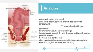
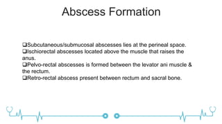
Paraproctitis
GROUP- 414
BY- Arora Vrushit
Baxi Niyanta
Bhanderi Bhakti
Joshi Deep
Zala Chandni
Definition
• It is a purulent inflammation of the paraproctium
(cellular tissues around the rectum and anus).
• i.e, collection of pus in anorectal area.
• ETIOLOGY: E.coli; Staphylococcus aureus;
Streptococcus; Clostridial anaerobes; Cl. Hystoliticum.
Anatomy
Anus, rectum and anal canal
Anal canal has muscles: (i) internal anal sphincter
(involuntary)
(ii) external anal sphincter
(voluntary)
Levator ant muscular pelvic diaphragm
Support pelvic vessels & control urinary and faecal muscles
peritoneum lines.
Pectinate line/ dentate line
Above pectinate line endoderm origin below pectinate is
ectoderm origin. ( sensitive as their skin).
Classification
Paraproctitis
Subcutaneous
Submucosa
Ischiorectal
Pelvorectal
Retro Rectal
Abscess Formation
Subcutaneous/submucosal abscesses lies at the perineal space.
Ischiorectal abscesses located above the muscle that raises the
anus.
Pelvo-rectal abscesses is formed between the levator ani muscle &
the rectum.
Retro-rectal abscess present between rectum and sacral bone.
Indication: inflammation of
tissue around the rectum and
anus.
Surgical approach: Radical
Instruments: scalpel, Kocher
clamp, rectal speculum,
grooved probe.
Procedure: drainage of
abscess elimination of its
internal opening, semilunar
incision, pus is drained,
grooved probe is delivered
from the wound through the
opening of rectum.
Operation
Procedure
Complications
• Spreading of inflammation to the cellular tissue
of the pelvis, purulent fusion of the wall of the
rectum higher than the level of anorectal line.
• Break of pus into the abdominal cavity and
retroperitoneal space.
• Burst of pus from the abscess cavity through the
perineal skin.
• Rupture of abscess in lumen of rectum or
vagina.
THANK YOU

Paraproctitis 41401.ppt

  • 1.
    Paraproctitis GROUP- 414 BY- AroraVrushit Baxi Niyanta Bhanderi Bhakti Joshi Deep Zala Chandni
  • 2.
    Definition • It isa purulent inflammation of the paraproctium (cellular tissues around the rectum and anus). • i.e, collection of pus in anorectal area. • ETIOLOGY: E.coli; Staphylococcus aureus; Streptococcus; Clostridial anaerobes; Cl. Hystoliticum.
  • 3.
    Anatomy Anus, rectum andanal canal Anal canal has muscles: (i) internal anal sphincter (involuntary) (ii) external anal sphincter (voluntary) Levator ant muscular pelvic diaphragm Support pelvic vessels & control urinary and faecal muscles peritoneum lines. Pectinate line/ dentate line Above pectinate line endoderm origin below pectinate is ectoderm origin. ( sensitive as their skin).
  • 4.
  • 5.
    Abscess Formation Subcutaneous/submucosal abscesseslies at the perineal space. Ischiorectal abscesses located above the muscle that raises the anus. Pelvo-rectal abscesses is formed between the levator ani muscle & the rectum. Retro-rectal abscess present between rectum and sacral bone.
  • 6.
    Indication: inflammation of tissuearound the rectum and anus. Surgical approach: Radical Instruments: scalpel, Kocher clamp, rectal speculum, grooved probe. Procedure: drainage of abscess elimination of its internal opening, semilunar incision, pus is drained, grooved probe is delivered from the wound through the opening of rectum. Operation Procedure
  • 7.
    Complications • Spreading ofinflammation to the cellular tissue of the pelvis, purulent fusion of the wall of the rectum higher than the level of anorectal line. • Break of pus into the abdominal cavity and retroperitoneal space. • Burst of pus from the abscess cavity through the perineal skin. • Rupture of abscess in lumen of rectum or vagina.
  • 8.