Downloaded 2,040 times

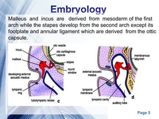


![Page 17
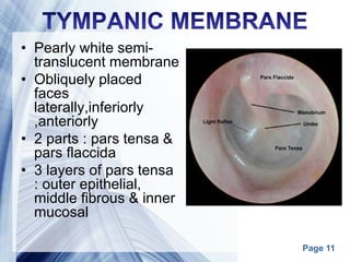
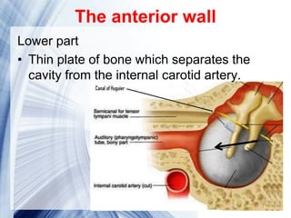
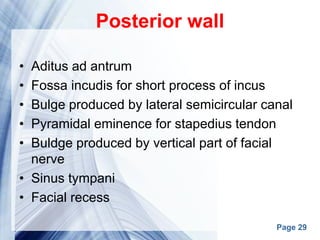
The floor
• Thin plate of bone which seperates
tympanic cavity from the jugular bulb.
• Near the medial border of the floor is a
small aperture, through which the
tympanic branch from the
glossopharyngeal nerve [IX] enters the
middle ear](https://image.slidesharecdn.com/middleearanatomymamoon-170317174955/85/Middle-ear-anatomy-17-320.jpg)

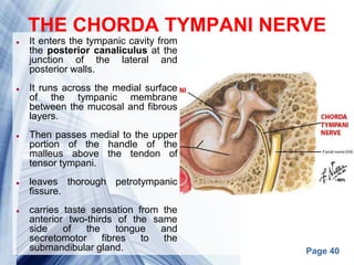
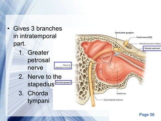
![Page 38
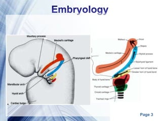
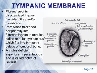
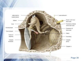
MUSCLE ORIGIN INSERTION NERVE
SUPPLY
ACTION
Tensor
typmani
Cartilaginous
part of ET, its
own bony
canal
Upper part of
handle of
malleus
Branch from
mandibular
nerve [V3]
tensing
tympanic
membrane to
reduce the
force of
vibrations in
response to
loud noises
Muscles of the middle ear](https://image.slidesharecdn.com/middleearanatomymamoon-170317174955/85/Middle-ear-anatomy-38-320.jpg)
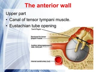
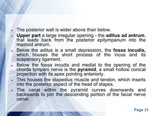
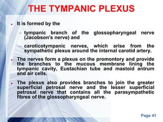
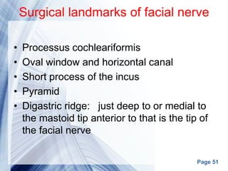
![Page 39
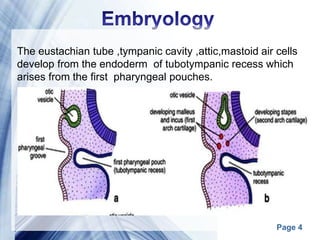
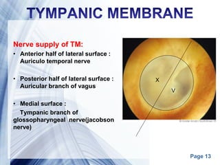
Stapedius pyramidal
eminence
Neck of
stapes
Branch of
facial nerve
[VII]
pulls the
stapes
posteriorly
and prevents
excessive
oscillation in
loud noises](https://image.slidesharecdn.com/middleearanatomymamoon-170317174955/85/Middle-ear-anatomy-39-320.jpg)
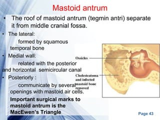


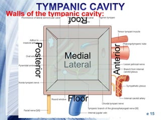
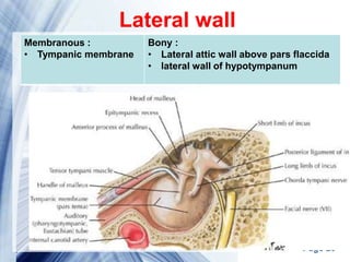
This document provides an anatomical overview of the structures of the middle ear and mastoid region. It describes the development, features, and contents of the eustachian tube, tympanic cavity, mastoid air cells, and related structures. Key structures discussed include the ossicles, muscles, nerves, blood supply, and the walls, openings and recesses of the middle ear cavity. Comparisons are made between adult and infant anatomy.


![CTEV [ clubfoot] DR ARUN LAL ,DR MOHAMED ASHRAF travancore medical college k...](https://cdn.slidesharecdn.com/ss_thumbnails/ctevclubfootdrarunlaldrmohamedashraftravancoremedicalcollegekollamkeralaindia-260208063247-18fc466c-thumbnail.jpg?width=640&height=640&fit=bounds)


