Analysis and Management of Tripod Fractures: Our Experience
Ijsron1201396
1. International Journal of Science and Research (IJSR), India Online ISSN: 2319-7064
Volume 2 Issue 5, April 2013
www.ijsr.net
A Case Report on Benign Sinonasal Paraganglioma
Smrity Rupa Borah Dutta1
, Sachender Pal Singh2
, Aakanksha Rathor3
1
Assistant Professor; OtrhinolOtorhinolaryngology,
Silchar Medical College & Hospital, Silchar
2
PGT; Otorhinolaryngology,
Silchar Medical College & Hospital, Silchar
3
PGT; Otorhinolaryngology,
Silchar Medical College & Hospital, Silchar
Abstract: We report a case of sinonasal paraganglioma presenting with episodes of epistaxis. A 55 year old male presented with a
nasal mass. It is an uncommon site of presentation and in an uncommon age group. A high grade of suspicion is required to diagnose
sino nasal paraganglioma. However, CT Scan and histopathology helps in early diagnosis and treatment. Surgical excision done with
cranialization of frontal sinus with fascia lata graft, followed up for 1 year without any evidence of disease recurrence.
Keywords: Sinonasal; Paraganglioma; Fascia Lata.
1. Introduction
Rarely paraganglioma have been described in areas like the
sinonasal tract where there is apparently no paraganglionic
tissue and very few cases of definite paraganglioma arising
primarily in the nose or paranasal sinuses have been reported,
presenting with nasal obstruction, profuse epistaxis and facial
swelling. Complete excision of the paraganglioma is normally
curative. We report a case of nasal paraganglioma and discuss
the diagnosis and therapy. Malignant transformation of
benign paraganglioma is rare and transformation of
paragangliomas to other types of malignancies is even rarer
2. Case Report
A 55 year old man presented in emergency hours in the
department of otorhinlaryngology, silchar medical college &
hospital, silchar with history of massive bleeding from nose
for last 3 days and a swelling at the root of nose for last 4
months. There had been several episodes of mild intermittent
nasal bleeding . There was no complain of nasal obstruction
but hyposmia. Immediately anterior nasal pack was given and
1 unit of whole blood was transfused.
Figure 1: Preoperative photo of sinonasal paraganglioma.
2.1. On gross examination, a smooth, firm, non tender,
nonpulsatile, diffuse swelling about 4X5 cm size in its
greatest dimensions extending from root of nose over its
dorsum. The overlying skin was normal with no local
rise of temperature.
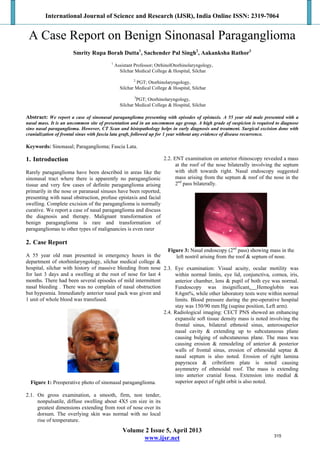
2.2. ENT examination on anterior rhinoscopy revealed a mass
at the roof of the nose bilaterally involving the septum
with shift towards right. Nasal endoscopy suggested
mass arising from the septum & roof of the nose in the
2nd
pass bilaterally.
Figure 3: Nasal endoscopy (2nd
pass) showing mass in the
left nostril arising from the roof & septum of nose.
2.3. Eye examination: Visual acuity, ocular motility was
within normal limits, eye lid, conjunctiva, cornea, iris,
anterior chamber, lens & pupil of both eye was normal.
Fundoscopy was insignificant. Hemoglobin was
8.6gm%, while other laboratory tests were within normal
limits. Blood pressure during the pre-operative hospital
stay was 150/90 mm Hg (supine position, Left arm).
2.4. Radiological imaging: CECT PNS showed an enhancing
expansile soft tissue density mass is noted involving the
frontal sinus, bilateral ethmoid sinus, anterosuperior
nasal cavity & extending up to subcutaneous plane
causing bulging of subcutaneous plane. The mass was
causing erosion & remodeling of anterior & posterior
walls of frontal sinus, erosion of ethmoidal septae &
nasal septum is also noted. Erosion of right lamina
papyracea & cribriform plate is noted causing
asymmetry of ethmoidal roof. The mass is extending
into anterior cranial fossa. Extension into medial &
superior aspect of right orbit is also noted.
315
2. International Journal of Science and Research (IJSR), India Online ISSN: 2319-7064
Volume 2 Issue 5, April 2013
www.ijsr.net
Figure 4.1: CT findings: An enhancing expansile soft tissue
density mass is noted involving the frontal sinuses, bilateral
anterior ethmoid sinuses, anterosuperior nasal cavity and
extending upto subcutaneous plane.
Figure 4.2: Coronal postcontrast image showing extension
into anterior cranial fossa.
Figure 5.1: MR shows well defined lobulated lesion in
fronto-nasal-ethmoidal region which is isointense to brain in
T1W and mildly hyperintense on T2W image.
Figure 5.2: Sagittal MR image shows extraaxial extension of
lesion into anterior cranial fossa .
Figure 5.3: FLAIR image show the lesion to be hyperintense
with extension into superior aspect of orbit.
3.Operative Details
Under all aseptic & antiseptic conditions general anaesthesia
was given. Lumbar drainage catheter was put in l3-l4 space in
order to prevent post operative rise of intracranial tension.
Bicoronal flap was raised & tumour was assessed & removed
along with surrounding mucosa. Posterior wall of frontal
sinus was also removed & tumor was found to be restricted in
extradural space. Dura was intact. Estimated blood loss
intraoperatively was around 1500ml intraoperatively patient
was transfused with 3 units of whole blood & 2000ml of
crystalloids.
Cranialization of frontal sinus was done with lattismus dorsi
flap. No intraoperative or postoperative complications were
found. Lumbar drainage catheter removed after 48 hours
when nasal pack was removed after confirming no elevations
in the intracranial pressure over 2 days & no CSF leakage.
Figure 6.1: Intra operative photo showing removal of tumor.
Figure 6.2: Intraoperative photo after the complete removal
of tumor.
Figure 6.3: Intraoperative photo showing fascial lata graft.
316
3. International Journal of Science and Research (IJSR), India Online ISSN: 2319-7064
Volume 2 Issue 5, April 2013
www.ijsr.net
4.Histopathology Report
A histopathological finding shows it to be a paraganglioma.
Figure 7.1: Histopathological slide (4X magnification)
showing capsule & zellabalen pattern
Figure 7.2: Histopathological slide (40X magnification)
showing zellabalen pattern
5. Discussion
Paraganglionic chemoreceptor cells of neural crest origin give
rise to benign, slow growing but locally invasive tumour
known as paraganglioma [1].
Almost half of these occur in the temporal bone, arising from
either the cochlear promontory (i.e. typanicum) or the jugular
blub (i.e. jugulare), nearly 1/3rd
in the carotid body, nearly
1/8th
in the region of the high cervical vagus and the rest at
various sites of the head and neck[2]. The most common site
of occurrence being adrenal glands. In the head and neck area,
common sites of occurrence are the carotid body, orbit,
larynx, and the nasopharynx, but paragangliomas are rare in
the nasal cavity and paranasal sinus.
In the nasal cavity, the middle turbinate, lateral nasal wall and
superior nasal vault are the most common sites. In paranasal
sinuses, the ethmoid sinus is the most common site of
occurrence [4, 5, 6]. Nearly 10 percent of paragangliomas are
malignant [7].
In approximately 10 percent of patients, tumours are
multifocal and up to 5 percent of tumours secrete
catacholamines. [3]
Common symptoms include recurrent epistaxis, nasal
obstruction and frontal headache. The clinical presentation
depends on the localisation of the tumour. In this case, the
patient presented with epistaxis, facial swelling & hyposmia.
Hyposmia is probably due to mechanical obstruction by the
lesion.
They may also be associated with some syndromes such as
multiple endocrine neoplasia type 2b (MEN IIB), von Hippel–
Lindau disease, neurofibromatosis types I [8]. In this case, we
did not have any syndromic involvement. It is estimated that
about 10–50% of paraganglioma cases are familial (autosomal
dominant) [1]. The genes for the familial paraganglioma have
been recently identified at the 11q23 locus [9]. Also, 4–19%
of all head and neck paragangliomas have been reported to be
malignant [10].
The presence of metastasis is the only definite criteria for
malignancy as there are no reliable histopathological criteria
to distinguish between benign and malignant paragangliomas
[1,11] and since the lesions are almost impossible to remove
completely, postoperative radiotherapy is then mandatory [3].
In our case, we could not demonstrate any evidence of
metastasis to the regional nodes or distant organs. He has
been followed up for 12 months, and no additional symptoms
or signs indicating recurrence have been identified.
6. Conclusion
Rarely, are the paragangliomas of the sinonasal region
reported in the literature. Benign paraganglioma, may
occasionally,both clinically and radiologically resemble
malignant sinonasal tumour so a high grade of clinical
suspicion is required to diagnose such a rare & curable tumor.
It may be provisionally diagnosed in any patient with nasal
mass associated with severe epistaxis. To conclude,
histopathology is the spine for definitive diagnosis.
References
[1] Pellitteri PK, Rinaldo A, Myssiorek D, Gary JC, Bradley
PJ, Devaney KO et al (2004) Paragangliomas of the
head and neck. Oral Oncol 40:563–575.
[2] Zak FG, Lawson W. The paraganglionic
chemoreceptorsystem. Physiology, pathology and
clinical medicine. New York: Springer Verlag. 1982.
[3] Scott Brown’s Otorhinolaryngology, Head & Neck
Surgery; 7th
edition 2008.
[4] Sharma HS, Madhavan M, Othman NH, Muhamad M,
Abdullah JM. Malignant paraganglioma of
frontoethmoidal region. Auris Nasus larynx
1999;26:487–93.
[5] Welkoborsky HJ, Gosepath J, Jacob R, Mann WJ,
Amedee RG. Biologic characteristics of paragangliomas
of the nasal cavity and paranasal sinuses. Am J Rhinol
2000;14:419–26.
[6] Mevio E, bignami M, Luinetti O, Villani L. Nasal
paraganglioma: a case report. Acta Oto-rhino-
laryngologica Belg 2001; 55:247–9.
[7] Conley JJ. The carotid body tumour: A review of 23
cases. Archieves of Otolaryngology. 1965;81 : 187-93.
[8] Bijlenga P, Dulguerov P, Richter M, de Tribolet N
(2004) Nasopharynx paraganglioma with extension in
the clivus. Acta Neurochir (Wien) 146:1355–1359.
[9] Baysal BE, Van Schothorst EM, Farr JE, Grashof P,
Myssiorek D, Rubinstein WS et al (1999) Repositioning
the hereditary paraganglioma critical region on
chromosome band 11q23. Hum Genet 104:219–225.
[10] Kuhn JA, Aronoff BL (1989) Nasal and nasopharyngeal
paraganglioma. J Surg Oncol 40:38–45.
[11] Deb P, Sharma MC, Gaikwad S, Gupta A, Mehta VS,
Sarkar C (2005) Cerebellopontine angle
paraganglioma—report of a case and review of the
317
4. International Journal of Science and Research (IJSR), India Online ISSN: 2319-7064
Volume 2 Issue 5, April 2013
www.ijsr.net
literature. J Neuro-oncol 74:65–69Subsidiary, H.
Etemad and L. S, Sulude (eds.), Croom‐Helm,
London, 1986. (book chapter style)
[12] K. Deb, S. Agrawal, A. Pratab, T. Meyarivan, “A Fast
Elitist Non-dominated Sorting Genetic Algorithms for
Multiobjective Optimization: NSGA II,” KanGAL
report 200001, Indian Institute of Technology, Kanpur,
India, 2000. (technical report style)
[13] J. Geralds, "Sega Ends Production of Dreamcast,"
vnunet.com, para. 2, Jan. 31, 2001. [Online]. Available:
http://nl1.vnunet.com/news/1116995. [Accessed: Sept.
12, 2004]. (General Internet site)
Author Profile
Dr. Smrity Rupa Borah Dutta completed her
M.B.B.S and M.S degrees from Assam Medical
College, Dibrugarh, Assam in the years 2000 and
2004 respectively. Presently she is the Assistant
Professor in the Department of Otorhinolaryngology,
Silchar Medical College, Assam, India.
318