SlideShare a Scribd company logo
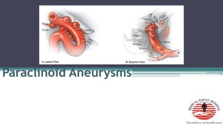
Paraclinoid Aneurysms
• Introduction
• Anatomy
• Classification
• Indications for Treatment
• Pre-op evaluation
• Management techniques
Paraclinoid aneurysms
• Nutik
• Arise from the ICA distal to the cavernous
sinus but proximal to the PCoM
• 5 – 10% of all intracranial aneurysms arise
from this segment of the ICA
• Female preponderance - 9:1(Shimizu et al; AJNR 2016)
• High incidence of being multiple
• Rupture rate is lower than supra-clinoid
aneurysms
• Half of pts with C6 segment aneurysms have
additional intracranial aneurysms elsewhere
Seven segments
• Cervical – C1
• Petrous – C2
• Lacerum – C3
• Cavernous – C4
• Clinoid – C5
• Ophthalmic – C6
• Communicating/Terminal segment – C7
C5 and C6 segments
• Clinoid segment
▫ Optic strut
▫ ACP
▫ Superiorly – DR
▫ Inferiorly – COM
▫ Interdural
• Ophthalmic segment
▫ Sub-arachnoid space
▫ Ophthalmic artery
▫ Sup Hypophyseal artery
Anterior Clinoid process
• Cline – Greek – bed
• Lesser sphenoid wing
• Superior orbital fissure
• Optic canal
• Cavernous sinus
• Carotid sulcus
• Infra-clinoid carotid groove
Dural reflections off ACP
• Falciform ligament
▫ Covers posterior optic canal
• Dural ring
▫ Superior dural surface fuses with ICA adventitia
▫ Distal dural ring
▫ Diaphragma sellae
• Carotid-oculomotor membrane(COM)
▫ Periosteal dura
▫ Marks exit of ICA from C.S
Distal Dural ring
• Slopes down
▫ Anterior to posterior
▫ Lateral to medial
• Medial to ICA - subarachnoid recess –
Carotid cave
Carotid cave
• Kobayashi(1989)
• Sup – Medial surface of DR
• Lat – Medial wall of ICA
• Medially – Carotid groove
• Cave communicates with the
clinoidal venous plexus
• Subarachnoid space
• 3 patterns
▫ Slit (34%)
▫ Pocket(24%)
▫ Mesh(10%)
• Carotid collar attaches to
ICA only at Upper dural ring
• Space between collar and ICA
– Clinoid venous plexus
• Lower ring – Perneczky’s
ring
• Middle clinoid process
• Osseous bridge
• Carotico-clinoidal
foramen
Neural relationship
• Optic Nerve
▫ Optic canal
▫ Optic strut
▫ Falciform ligament
Arterial bends - 2
• 1st
▫ Sharp posterior turn
▫ Superior vector stress on the anterior and dorsal
wall
• 2nd
▫ Medial to lateral curve
▫ Stress on the medial aspect
Branch points
• Ophthalmic artery
▫ Dorso-medial ICA surface
▫ Just above DR
▫ Infero-lateral to Optic nerve
▫ Optic canal
• Superior hypophyseal
artery
▫ Largest perforator from
Ophthalmic segment
▫ Runs medially
▫ Optic chiasm
▫ Optic tracts
Barami classification(2003)
• Type Ia – Arising from the ophthalmic segment of the ICA and
related to the ophthalmic artery
• Type Ib – arising from the superior ophthalmic segment but
without relationship with ICA branches
•
• Type II – from ventral ophthalmic segment of ICA without ICA
branches relation
• Type IIIa – from medial ophthalmic segment of the ICA and
related to the SHA
• Type IIIb – from the medial clinoid segment below the DR
• Type IV –Large aneurysms involving the clinoid and the
ophthalmic segment of the ICA
• Ia – Most common - 43%
• Clinoidal segment
▫ Antero-lateral variant
▫ Medial variant
• Ophthalmic segment
▫ Ophthalmic artery aneurysms
▫ Sup Hypophyseal artery aneurysms
▫ Dorsal variant
• Carotid cave aneurysms
• Transitional aneurysms
Anterolateral variant
• From anterolateral surface of C5
• Superior projecting towards ACP
• Erode optic strut – visual loss
Medial variant
• From medial surface of clinoidal segment
• Enlarges towards sella and sphenoid sinus
• Expands beneath the diaphragma sellae into the pituitary fossa
• Hypopituitarism
• Visual loss
• Rupture - Simulate apoplexy
Ophthalmic artery aneurysms
• From posterior bend of ICA just distal to origin
of Ophthalmic artery and DR
• Project dorsally or dorsomedially
• Can elevate the Optic nerve against the falciform
ligament
• Monocular inferior nasal field defect
• Entire nasal field
• Superior temporal field loss in C/L eye
Large ophthalmic artery aneurysms
• Thickened or calcified walls
• Antero-inferior portion
Superior hypophyseal artery
aneurysms
• Medial surface of ICA
• Just distal to the DR
• Surrounded by Superior hypophyseal artery perforators
• Can project into the carotid cave – unlikely to rupture
when small
• When it fills the cave and extends into the
suprasellar region – Risk of hemorrhage rises
• Thickening or calcification along the anterior
medial aspect near the origin
• Giant aneurysms – compress chiasm like
pituitary tumors
Dorsal variant aneurysms
• Dorsal aspect of ICA
• Distal to ophthalmic artery origin
• Blisters
• Hemorrhage when small and expand as fusiform lesions
externally
• Very fragile
Carotid cave aneurysms
• Arises proximal to ophthalmic artery
• Below distal dural ring
• Points ventromedially into the cave
Clinical presentation
• Sudden severe headache
• Decreased visual acuity/field
defects
• Changes in colour vision
• Diplopia
• Panhypopituitarism
• Pituitary apoplexy
• Epistaxis
• Facial numbness
Indication for treatment
• Small <1cm asymptomatic
clinoid segment aneurysms –
conservative
• Small symptomatic lesions
(visual deficits, focal
unrelenting headaches) –
Rx
• Lesion whose protective ACP
roof has been removed - Rx
• >1cm – often extend into
subarachnoid space –
Increased risk of rupture – Rx
even if asymptomatic
• Ophthalmic segment aneurysms – low risk of
rupture. Expn – dorsal variant
• All symptomatic aneurysms > 1cm – Rx
• Large or giant lesions – Flow diverters
Pre-operative evaluation
• Cranial nerve deficits
▫ Visual acuity and fields
▫ EOM
▫ Facial sensations
• Endocrine status
• DSA
• CT angiography – DR can be
identified. Atheroma +
calcifications
• Anterolat v/s Ophthalmic in
CT
▫ Erosion of ACP
▫ Proximal origin
▫ Dorsolat v/s dorsomedial
▫ Angiographic waist
Clinoidal medial Sup Hypophyseal
• Projects medially
• Originates below diaphragma
sellae
• Superior surface is flattened
by the diaphragma
• Narrow neck(b/w DR and
COM)
• Projects medially
• Originates above diaphragma
sellae
• Not flattened
• Wide neck
• EVD or Lumbar drain
• Positioned supine with a shoulder roll ipsilateral
to the lesion
• Head is elevated above the heart
• Rotated 45 to 60 degrees away from the side of
the aneurysm
• Cervical carotid exposed
• Skin incision
• Pterional craniotomy with an orbital osteotomy
• Lesser wing is removed extra-durally down to
the base of ACP
Anterior clinoidectomy
• Extradural – Dolenc approach
• Intradural approach
• Intradural advantage – visualisation of
aneurysm throughout the procedure
Extradural ACP removal -EDAC
• Avoid extradural ACP removal if a clinoidal segment
anterolateral variant is suspected
• Extend medial dissection
• Diamond drill
• Dural attachments released
Intradural ACP removal
• Basal removal
• Dura opened in a curvilinear fashion
• Sylvian fissure split
• Aneurysm, ICA and optic apparatus visualised
• Falciform ligament sectioned
• Better visualization of optic apparatus and
aneurysm
• Optic canal – unroofed
• Optic strut drilled down
• Optic nerve sheath - sectioned
• DR sectioned circumferetially
• ICA can me mobilized
Techniques
• Temporary proximal cervical carotid ligation
• Trapping of aneurysm
• Intra-aneurysmal suction decompression
• Fenestrated clips – calcified lesion
• ICG – perforator patency
• Intraoperative angiography
Clinoidal segment
• Anterolateral variant – gently curved or side
angled clip parallel to the anterolateral surface
of ICA
• Medial variant
Ophthalmic segment
• Calcification along anterior aspect
• Sup Hypophyseal - Right angled fenestrated clip
• Dorsal wall aneurysms
▫ Blister variety – Clipping after trapping
▫ Sling of fascia or PTFE – wrap and clip
Complications
• Delayed stenosis or thrombosis
• Post-operative visual deterioration – 8.7%
(Nanda and Javalkar et al – Neurosurgery 2011)
• Heat from drilling
• Manipulation of Optic nerve
• Perforators
• Most patients have improved visual function after
surgery(De Oliveira et al – Neurosurg Focus 2009)
• 3rd, 4th, 6th nerve palsies
• Avoid excessive cranial nerve manipulation or
cavernous sinus packing
• CSF rhinorrhea – ACP drilling – Yo –Yo
technique
• Detachable coils
• Stenting and flow diverters
• Balloon assisted coiling
• Adjunct to surgery – Suction decompression +
Intraop angiography
Complications
• Coil compaction – refilling of aneurysm
• Blockage of parent vessel
• Progression of aneurysm thrombosis to parent
vessel
• Embolization of aneurysm thrombus
PED v/s Coiling v/s Clipping(JNS 2018 – Michael A
silva et al)
• PED has highest rate of visual improvement –
93%
• Complication rates 9% - PED
• Complete occlusion – Clipping(97) > PED(89) >
Coiling(78)
• CN palsy – 29% clipping > 13% PED > 0%
coiling
• SHA – coiling
• Ophthalmic – clipping
• Large aneurysms – PED and coiling
Bibliography
• Youmans
• Ramamurthi
• 7 Aneurysms

More Related Content

What's hot

Sch.36 surgical management of sphenoid wing meningioma
Sch.36 surgical management of sphenoid wing meningiomaSch.36 surgical management of sphenoid wing meningioma
Sch.36 surgical management of sphenoid wing meningioma
Neurosurgery Vajira
 
SAH (Subarachnoid Haemorrhage)
SAH (Subarachnoid Haemorrhage)SAH (Subarachnoid Haemorrhage)
SAH (Subarachnoid Haemorrhage)
Jigar Padalia
 
Intracranial pressure - waveforms and monitoring
Intracranial pressure - waveforms and monitoringIntracranial pressure - waveforms and monitoring
Intracranial pressure - waveforms and monitoring
joemdas
 

What's hot (20)

Caroticocavernous fistula CCF
Caroticocavernous fistula CCFCaroticocavernous fistula CCF
Caroticocavernous fistula CCF
 
Stroke EVT- A Discussion
Stroke EVT- A DiscussionStroke EVT- A Discussion
Stroke EVT- A Discussion
 
Clipping or Coiling for MCA Aneurysm
Clipping or Coiling for MCA AneurysmClipping or Coiling for MCA Aneurysm
Clipping or Coiling for MCA Aneurysm
 
Stroke syndromes
Stroke syndromesStroke syndromes
Stroke syndromes
 
Carotid cavernous fistula
Carotid cavernous fistulaCarotid cavernous fistula
Carotid cavernous fistula
 
Vascular anatomy of brain
Vascular anatomy of brain Vascular anatomy of brain
Vascular anatomy of brain
 
10 triangles 360°
10 triangles 360°10 triangles 360°
10 triangles 360°
 
Sch.36 surgical management of sphenoid wing meningioma
Sch.36 surgical management of sphenoid wing meningiomaSch.36 surgical management of sphenoid wing meningioma
Sch.36 surgical management of sphenoid wing meningioma
 
009 youmans cerebral edema
009 youmans cerebral edema009 youmans cerebral edema
009 youmans cerebral edema
 
Subaxial spine
Subaxial spineSubaxial spine
Subaxial spine
 
Orbital apex syndrome
Orbital apex syndromeOrbital apex syndrome
Orbital apex syndrome
 
Cavernous sinus
Cavernous sinusCavernous sinus
Cavernous sinus
 
Caroticocavernous fistula HAKU
Caroticocavernous fistula HAKUCaroticocavernous fistula HAKU
Caroticocavernous fistula HAKU
 
Petroclival Meningioma
Petroclival MeningiomaPetroclival Meningioma
Petroclival Meningioma
 
Chronic subdural hematoma
Chronic subdural hematomaChronic subdural hematoma
Chronic subdural hematoma
 
SAH (Subarachnoid Haemorrhage)
SAH (Subarachnoid Haemorrhage)SAH (Subarachnoid Haemorrhage)
SAH (Subarachnoid Haemorrhage)
 
Right AICA PICA stroke
Right AICA PICA strokeRight AICA PICA stroke
Right AICA PICA stroke
 
Intracranial pressure - waveforms and monitoring
Intracranial pressure - waveforms and monitoringIntracranial pressure - waveforms and monitoring
Intracranial pressure - waveforms and monitoring
 
Posterior circulation - Applied Anatomy
Posterior circulation - Applied AnatomyPosterior circulation - Applied Anatomy
Posterior circulation - Applied Anatomy
 
Fourth ventricular tumors
Fourth ventricular tumorsFourth ventricular tumors
Fourth ventricular tumors
 

Similar to Paraclinoid aneurysms

Orbital imaging (X-RAY,CT SCAN,AND MRI)
Orbital imaging (X-RAY,CT SCAN,AND MRI)Orbital imaging (X-RAY,CT SCAN,AND MRI)
Orbital imaging (X-RAY,CT SCAN,AND MRI)
Prashant Patel
 

Similar to Paraclinoid aneurysms (20)

367 Intracranial internal carotid artery aneurysm
367 Intracranial internal carotid artery aneurysm367 Intracranial internal carotid artery aneurysm
367 Intracranial internal carotid artery aneurysm
 
371 Microsurgery of VA PICA VBJ aneurysm
371 Microsurgery of VA PICA VBJ aneurysm371 Microsurgery of VA PICA VBJ aneurysm
371 Microsurgery of VA PICA VBJ aneurysm
 
Sch 33 surgical approach to falcine meningioma
Sch 33 surgical approach to falcine meningiomaSch 33 surgical approach to falcine meningioma
Sch 33 surgical approach to falcine meningioma
 
brain anatomy and vascular supply.pptx
brain anatomy and vascular supply.pptxbrain anatomy and vascular supply.pptx
brain anatomy and vascular supply.pptx
 
CP angle.pptx
CP angle.pptxCP angle.pptx
CP angle.pptx
 
Orbital imaging (X-RAY,CT SCAN,AND MRI)
Orbital imaging (X-RAY,CT SCAN,AND MRI)Orbital imaging (X-RAY,CT SCAN,AND MRI)
Orbital imaging (X-RAY,CT SCAN,AND MRI)
 
Ncct and cect brain and orbit
Ncct and cect brain and orbitNcct and cect brain and orbit
Ncct and cect brain and orbit
 
Complications of cataract surgery by Dr. Iddi.pptx
Complications of cataract surgery by Dr. Iddi.pptxComplications of cataract surgery by Dr. Iddi.pptx
Complications of cataract surgery by Dr. Iddi.pptx
 
The Imaging of the Orbit
The Imaging of the OrbitThe Imaging of the Orbit
The Imaging of the Orbit
 
Ptosis
PtosisPtosis
Ptosis
 
Approach to a patient with ectropion, entropion, symblepharon.pptx
Approach to a patient with ectropion, entropion, symblepharon.pptxApproach to a patient with ectropion, entropion, symblepharon.pptx
Approach to a patient with ectropion, entropion, symblepharon.pptx
 
Parasagittal meningioma
Parasagittal meningioma Parasagittal meningioma
Parasagittal meningioma
 
CTCA - CT Coronary Angiography Plaque Characterization
CTCA - CT Coronary Angiography Plaque CharacterizationCTCA - CT Coronary Angiography Plaque Characterization
CTCA - CT Coronary Angiography Plaque Characterization
 
REFRACTIVE SURGERY PRESENTATION.pptx
REFRACTIVE SURGERY PRESENTATION.pptxREFRACTIVE SURGERY PRESENTATION.pptx
REFRACTIVE SURGERY PRESENTATION.pptx
 
Carotid in neck , eca, ima &amp;
Carotid in neck , eca, ima &amp;Carotid in neck , eca, ima &amp;
Carotid in neck , eca, ima &amp;
 
178 arachnoid cysts
178 arachnoid cysts178 arachnoid cysts
178 arachnoid cysts
 
Neck dissection
Neck dissectionNeck dissection
Neck dissection
 
335 Surgical management of traumatic brain injury
335 Surgical management of traumatic brain injury335 Surgical management of traumatic brain injury
335 Surgical management of traumatic brain injury
 
ANGLE ANATOMY
ANGLE ANATOMYANGLE ANATOMY
ANGLE ANATOMY
 
Endoscopic skull base surgery level iii
Endoscopic skull base surgery level iiiEndoscopic skull base surgery level iii
Endoscopic skull base surgery level iii
 

Recently uploaded

Mastering Wealth: A Path to Financial Freedom
Mastering Wealth: A Path to Financial FreedomMastering Wealth: A Path to Financial Freedom
Mastering Wealth: A Path to Financial Freedom
FatimaMary4
 

Recently uploaded (20)

1130525--家醫計畫2.0糖尿病照護研討會-社團法人高雄市醫師公會.pdf
1130525--家醫計畫2.0糖尿病照護研討會-社團法人高雄市醫師公會.pdf1130525--家醫計畫2.0糖尿病照護研討會-社團法人高雄市醫師公會.pdf
1130525--家醫計畫2.0糖尿病照護研討會-社團法人高雄市醫師公會.pdf
 
Compare home pulse pressure components collected directly from home
Compare home pulse pressure components collected directly from homeCompare home pulse pressure components collected directly from home
Compare home pulse pressure components collected directly from home
 
Aptopadesha Pramana / Pariksha: The Verbal Testimony
Aptopadesha Pramana / Pariksha: The Verbal TestimonyAptopadesha Pramana / Pariksha: The Verbal Testimony
Aptopadesha Pramana / Pariksha: The Verbal Testimony
 
The POPPY STUDY (Preconception to post-partum cardiovascular function in prim...
The POPPY STUDY (Preconception to post-partum cardiovascular function in prim...The POPPY STUDY (Preconception to post-partum cardiovascular function in prim...
The POPPY STUDY (Preconception to post-partum cardiovascular function in prim...
 
Young at heart: Cardiovascular health stations to empower healthy lifestyle b...
Young at heart: Cardiovascular health stations to empower healthy lifestyle b...Young at heart: Cardiovascular health stations to empower healthy lifestyle b...
Young at heart: Cardiovascular health stations to empower healthy lifestyle b...
 
DECIPHERING COMMON ECG FINDINGS IN ED.pptx
DECIPHERING COMMON ECG FINDINGS IN ED.pptxDECIPHERING COMMON ECG FINDINGS IN ED.pptx
DECIPHERING COMMON ECG FINDINGS IN ED.pptx
 
Scientificity and feasibility study of non-invasive central arterial pressure...
Scientificity and feasibility study of non-invasive central arterial pressure...Scientificity and feasibility study of non-invasive central arterial pressure...
Scientificity and feasibility study of non-invasive central arterial pressure...
 
TEST BANK For Williams' Essentials of Nutrition and Diet Therapy, 13th Editio...
TEST BANK For Williams' Essentials of Nutrition and Diet Therapy, 13th Editio...TEST BANK For Williams' Essentials of Nutrition and Diet Therapy, 13th Editio...
TEST BANK For Williams' Essentials of Nutrition and Diet Therapy, 13th Editio...
 
Temporal, Infratemporal & Pterygopalatine BY Dr.RIG.pptx
Temporal, Infratemporal & Pterygopalatine BY Dr.RIG.pptxTemporal, Infratemporal & Pterygopalatine BY Dr.RIG.pptx
Temporal, Infratemporal & Pterygopalatine BY Dr.RIG.pptx
 
Retinal consideration in cataract surgery
Retinal consideration in cataract surgeryRetinal consideration in cataract surgery
Retinal consideration in cataract surgery
 
Gauri Gawande(9) Constipation Final.pptx
Gauri Gawande(9) Constipation Final.pptxGauri Gawande(9) Constipation Final.pptx
Gauri Gawande(9) Constipation Final.pptx
 
Evaluation of antidepressant activity of clitoris ternatea in animals
Evaluation of antidepressant activity of clitoris ternatea in animalsEvaluation of antidepressant activity of clitoris ternatea in animals
Evaluation of antidepressant activity of clitoris ternatea in animals
 
Presentació "Advancing Emergency Medicine Education through Virtual Reality"
Presentació "Advancing Emergency Medicine Education through Virtual Reality"Presentació "Advancing Emergency Medicine Education through Virtual Reality"
Presentació "Advancing Emergency Medicine Education through Virtual Reality"
 
Relationship between vascular system disfunction, neurofluid flow and Alzheim...
Relationship between vascular system disfunction, neurofluid flow and Alzheim...Relationship between vascular system disfunction, neurofluid flow and Alzheim...
Relationship between vascular system disfunction, neurofluid flow and Alzheim...
 
PT MANAGEMENT OF URINARY INCONTINENCE.pptx
PT MANAGEMENT OF URINARY INCONTINENCE.pptxPT MANAGEMENT OF URINARY INCONTINENCE.pptx
PT MANAGEMENT OF URINARY INCONTINENCE.pptx
 
Mastering Wealth: A Path to Financial Freedom
Mastering Wealth: A Path to Financial FreedomMastering Wealth: A Path to Financial Freedom
Mastering Wealth: A Path to Financial Freedom
 
ANATOMY OF THE LOWER URINARY TRACT AND MALE [Autosaved] [Autosaved].pptx
ANATOMY OF THE LOWER URINARY TRACT AND MALE [Autosaved] [Autosaved].pptxANATOMY OF THE LOWER URINARY TRACT AND MALE [Autosaved] [Autosaved].pptx
ANATOMY OF THE LOWER URINARY TRACT AND MALE [Autosaved] [Autosaved].pptx
 
Antiulcer drugs Advance Pharmacology .pptx
Antiulcer drugs Advance Pharmacology .pptxAntiulcer drugs Advance Pharmacology .pptx
Antiulcer drugs Advance Pharmacology .pptx
 
Fundamental of Radiobiology -SABBU.pptx
Fundamental of Radiobiology  -SABBU.pptxFundamental of Radiobiology  -SABBU.pptx
Fundamental of Radiobiology -SABBU.pptx
 
How to Give Better Lectures: Some Tips for Doctors
How to Give Better Lectures: Some Tips for DoctorsHow to Give Better Lectures: Some Tips for Doctors
How to Give Better Lectures: Some Tips for Doctors
 

Paraclinoid aneurysms

  • 2. • Introduction • Anatomy • Classification • Indications for Treatment • Pre-op evaluation • Management techniques
  • 3. Paraclinoid aneurysms • Nutik • Arise from the ICA distal to the cavernous sinus but proximal to the PCoM • 5 – 10% of all intracranial aneurysms arise from this segment of the ICA
  • 4. • Female preponderance - 9:1(Shimizu et al; AJNR 2016) • High incidence of being multiple • Rupture rate is lower than supra-clinoid aneurysms • Half of pts with C6 segment aneurysms have additional intracranial aneurysms elsewhere
  • 5.
  • 6. Seven segments • Cervical – C1 • Petrous – C2 • Lacerum – C3 • Cavernous – C4 • Clinoid – C5 • Ophthalmic – C6 • Communicating/Terminal segment – C7
  • 7.
  • 8.
  • 9. C5 and C6 segments • Clinoid segment ▫ Optic strut ▫ ACP ▫ Superiorly – DR ▫ Inferiorly – COM ▫ Interdural • Ophthalmic segment ▫ Sub-arachnoid space ▫ Ophthalmic artery ▫ Sup Hypophyseal artery
  • 10.
  • 11.
  • 12. Anterior Clinoid process • Cline – Greek – bed • Lesser sphenoid wing • Superior orbital fissure • Optic canal • Cavernous sinus
  • 13.
  • 14.
  • 15.
  • 16. • Carotid sulcus • Infra-clinoid carotid groove
  • 17. Dural reflections off ACP • Falciform ligament ▫ Covers posterior optic canal • Dural ring ▫ Superior dural surface fuses with ICA adventitia ▫ Distal dural ring ▫ Diaphragma sellae • Carotid-oculomotor membrane(COM) ▫ Periosteal dura ▫ Marks exit of ICA from C.S
  • 18.
  • 19.
  • 20.
  • 21. Distal Dural ring • Slopes down ▫ Anterior to posterior ▫ Lateral to medial • Medial to ICA - subarachnoid recess – Carotid cave
  • 22.
  • 23. Carotid cave • Kobayashi(1989) • Sup – Medial surface of DR • Lat – Medial wall of ICA • Medially – Carotid groove • Cave communicates with the clinoidal venous plexus • Subarachnoid space • 3 patterns ▫ Slit (34%) ▫ Pocket(24%) ▫ Mesh(10%)
  • 24. • Carotid collar attaches to ICA only at Upper dural ring • Space between collar and ICA – Clinoid venous plexus • Lower ring – Perneczky’s ring
  • 25. • Middle clinoid process • Osseous bridge • Carotico-clinoidal foramen
  • 26. Neural relationship • Optic Nerve ▫ Optic canal ▫ Optic strut ▫ Falciform ligament
  • 27.
  • 28. Arterial bends - 2 • 1st ▫ Sharp posterior turn ▫ Superior vector stress on the anterior and dorsal wall • 2nd ▫ Medial to lateral curve ▫ Stress on the medial aspect
  • 29.
  • 30. Branch points • Ophthalmic artery ▫ Dorso-medial ICA surface ▫ Just above DR ▫ Infero-lateral to Optic nerve ▫ Optic canal
  • 31.
  • 32. • Superior hypophyseal artery ▫ Largest perforator from Ophthalmic segment ▫ Runs medially ▫ Optic chiasm ▫ Optic tracts
  • 33.
  • 34. Barami classification(2003) • Type Ia – Arising from the ophthalmic segment of the ICA and related to the ophthalmic artery • Type Ib – arising from the superior ophthalmic segment but without relationship with ICA branches • • Type II – from ventral ophthalmic segment of ICA without ICA branches relation
  • 35.
  • 36. • Type IIIa – from medial ophthalmic segment of the ICA and related to the SHA • Type IIIb – from the medial clinoid segment below the DR • Type IV –Large aneurysms involving the clinoid and the ophthalmic segment of the ICA • Ia – Most common - 43%
  • 37. • Clinoidal segment ▫ Antero-lateral variant ▫ Medial variant • Ophthalmic segment ▫ Ophthalmic artery aneurysms ▫ Sup Hypophyseal artery aneurysms ▫ Dorsal variant • Carotid cave aneurysms • Transitional aneurysms
  • 38.
  • 39. Anterolateral variant • From anterolateral surface of C5 • Superior projecting towards ACP • Erode optic strut – visual loss
  • 40. Medial variant • From medial surface of clinoidal segment • Enlarges towards sella and sphenoid sinus • Expands beneath the diaphragma sellae into the pituitary fossa • Hypopituitarism • Visual loss • Rupture - Simulate apoplexy
  • 41.
  • 42.
  • 43.
  • 44.
  • 45.
  • 46. Ophthalmic artery aneurysms • From posterior bend of ICA just distal to origin of Ophthalmic artery and DR • Project dorsally or dorsomedially • Can elevate the Optic nerve against the falciform ligament
  • 47. • Monocular inferior nasal field defect • Entire nasal field • Superior temporal field loss in C/L eye
  • 48. Large ophthalmic artery aneurysms • Thickened or calcified walls • Antero-inferior portion
  • 49. Superior hypophyseal artery aneurysms • Medial surface of ICA • Just distal to the DR • Surrounded by Superior hypophyseal artery perforators • Can project into the carotid cave – unlikely to rupture when small
  • 50.
  • 51. • When it fills the cave and extends into the suprasellar region – Risk of hemorrhage rises • Thickening or calcification along the anterior medial aspect near the origin • Giant aneurysms – compress chiasm like pituitary tumors
  • 52. Dorsal variant aneurysms • Dorsal aspect of ICA • Distal to ophthalmic artery origin • Blisters • Hemorrhage when small and expand as fusiform lesions externally • Very fragile
  • 53.
  • 54. Carotid cave aneurysms • Arises proximal to ophthalmic artery • Below distal dural ring • Points ventromedially into the cave
  • 55. Clinical presentation • Sudden severe headache • Decreased visual acuity/field defects • Changes in colour vision • Diplopia • Panhypopituitarism • Pituitary apoplexy • Epistaxis • Facial numbness
  • 56. Indication for treatment • Small <1cm asymptomatic clinoid segment aneurysms – conservative • Small symptomatic lesions (visual deficits, focal unrelenting headaches) – Rx • Lesion whose protective ACP roof has been removed - Rx • >1cm – often extend into subarachnoid space – Increased risk of rupture – Rx even if asymptomatic
  • 57. • Ophthalmic segment aneurysms – low risk of rupture. Expn – dorsal variant • All symptomatic aneurysms > 1cm – Rx • Large or giant lesions – Flow diverters
  • 58. Pre-operative evaluation • Cranial nerve deficits ▫ Visual acuity and fields ▫ EOM ▫ Facial sensations • Endocrine status • DSA • CT angiography – DR can be identified. Atheroma + calcifications • Anterolat v/s Ophthalmic in CT ▫ Erosion of ACP ▫ Proximal origin ▫ Dorsolat v/s dorsomedial ▫ Angiographic waist
  • 59. Clinoidal medial Sup Hypophyseal • Projects medially • Originates below diaphragma sellae • Superior surface is flattened by the diaphragma • Narrow neck(b/w DR and COM) • Projects medially • Originates above diaphragma sellae • Not flattened • Wide neck
  • 60.
  • 61. • EVD or Lumbar drain • Positioned supine with a shoulder roll ipsilateral to the lesion • Head is elevated above the heart • Rotated 45 to 60 degrees away from the side of the aneurysm
  • 62. • Cervical carotid exposed • Skin incision • Pterional craniotomy with an orbital osteotomy • Lesser wing is removed extra-durally down to the base of ACP
  • 63.
  • 64. Anterior clinoidectomy • Extradural – Dolenc approach • Intradural approach • Intradural advantage – visualisation of aneurysm throughout the procedure
  • 65. Extradural ACP removal -EDAC • Avoid extradural ACP removal if a clinoidal segment anterolateral variant is suspected • Extend medial dissection • Diamond drill • Dural attachments released
  • 66.
  • 67.
  • 68. Intradural ACP removal • Basal removal • Dura opened in a curvilinear fashion • Sylvian fissure split • Aneurysm, ICA and optic apparatus visualised
  • 69.
  • 70. • Falciform ligament sectioned • Better visualization of optic apparatus and aneurysm • Optic canal – unroofed • Optic strut drilled down • Optic nerve sheath - sectioned
  • 71.
  • 72. • DR sectioned circumferetially • ICA can me mobilized
  • 73.
  • 74. Techniques • Temporary proximal cervical carotid ligation • Trapping of aneurysm • Intra-aneurysmal suction decompression • Fenestrated clips – calcified lesion • ICG – perforator patency • Intraoperative angiography
  • 75. Clinoidal segment • Anterolateral variant – gently curved or side angled clip parallel to the anterolateral surface of ICA • Medial variant
  • 76. Ophthalmic segment • Calcification along anterior aspect • Sup Hypophyseal - Right angled fenestrated clip • Dorsal wall aneurysms ▫ Blister variety – Clipping after trapping ▫ Sling of fascia or PTFE – wrap and clip
  • 77.
  • 78.
  • 79.
  • 80.
  • 81.
  • 82.
  • 83.
  • 84. Complications • Delayed stenosis or thrombosis • Post-operative visual deterioration – 8.7% (Nanda and Javalkar et al – Neurosurgery 2011) • Heat from drilling • Manipulation of Optic nerve • Perforators • Most patients have improved visual function after surgery(De Oliveira et al – Neurosurg Focus 2009)
  • 85. • 3rd, 4th, 6th nerve palsies • Avoid excessive cranial nerve manipulation or cavernous sinus packing • CSF rhinorrhea – ACP drilling – Yo –Yo technique
  • 86.
  • 87. • Detachable coils • Stenting and flow diverters • Balloon assisted coiling • Adjunct to surgery – Suction decompression + Intraop angiography
  • 88. Complications • Coil compaction – refilling of aneurysm • Blockage of parent vessel • Progression of aneurysm thrombosis to parent vessel • Embolization of aneurysm thrombus
  • 89.
  • 90. PED v/s Coiling v/s Clipping(JNS 2018 – Michael A silva et al) • PED has highest rate of visual improvement – 93% • Complication rates 9% - PED • Complete occlusion – Clipping(97) > PED(89) > Coiling(78)
  • 91. • CN palsy – 29% clipping > 13% PED > 0% coiling • SHA – coiling • Ophthalmic – clipping • Large aneurysms – PED and coiling