Downloaded 2,064 times
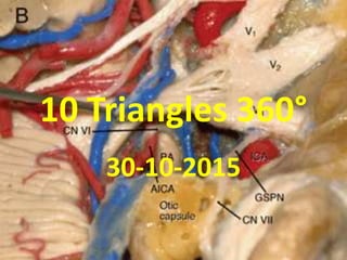


![Anterior clinoid drilling videos in FTOZ
[ neurosurgery skull base ]
1. https://www.youtube.com/watch?v=wO2cWHiOdO0
2. https://www.youtube.com/watch?v=4dkQY3zxJHU
3. https://www.youtube.com/watch?v=vd4_lPVIUvE
4. https://www.youtube.com/watch?v=_dvYB1InGMc
5. https://www.youtube.com/watch?v=83_VuKHXOmQ
6. https://www.youtube.com/watch?v=0KwBhTqNXA4
7. https://www.youtube.com/watch?v=pCURjQ83HzU
8. https://www.youtube.com/watch?v=DNIy0L3oFgY
9. https://www.youtube.com/watch?v=GT4eBB2x58Q
10. https://www.youtube.com/watch?v=OS4Mc0X8tlU
11. https://www.youtube.com/watch?v=_xq9e3p1cc4](https://image.slidesharecdn.com/10triangles360-150127035719-conversion-gate01/85/10-triangles-360-9-320.jpg)
![1. SOF present between two structs
2. OS [ optic struct separates optic canal from SOF ]](https://image.slidesharecdn.com/10triangles360-150127035719-conversion-gate01/85/10-triangles-360-13-320.jpg)
![1. SOF present between two structs
2. OS [ optic struct separates optic canal from SOF ]](https://image.slidesharecdn.com/10triangles360-150127035719-conversion-gate01/85/10-triangles-360-14-320.jpg)
![Anterior clinoid process [ ACP ] has 3 roots of attachements :
1. Anterior root – ACP attachment to sphenoid planum medial
to falciform ligament
2. posterior root = OS = L-OCR
3. 3rd root to lesser wing of sphenoid](https://image.slidesharecdn.com/10triangles360-150127035719-conversion-gate01/85/10-triangles-360-16-320.jpg)
![Optic strut [ OS ] =
L-OCR
[ Pneumatisation
of OS ] =
Posterior root of
Anterior clinoid
process [ ACP ]
OS = L-OCR =
posterior root of
ACP](https://image.slidesharecdn.com/10triangles360-150127035719-conversion-gate01/85/10-triangles-360-17-320.jpg)
![1. Surpa-optic pneumatisation starts from anterior root of ACP & goes to ACP
, infra-optic pneumatization starts in posterior root of ACP [ = OS = L-OCR ] &
may goes into ACP
2. In ACP drilling if there is pneumatization we will directly open into sphenoid
so we have to plug with fat after ACP drilling in neurosurgical skull base](https://image.slidesharecdn.com/10triangles360-150127035719-conversion-gate01/85/10-triangles-360-18-320.jpg)
![Surpa-optic pneumatisation starts from anterior root of ACP & goes to
ACP , infra-optic pneumatization starts in posterior root of ACP [ = OS
= L-OCR ] & may goes into ACP](https://image.slidesharecdn.com/10triangles360-150127035719-conversion-gate01/85/10-triangles-360-19-320.jpg)
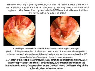
![The lower dural ring is given by the COM [ Carotid-oculomotor
membrane ] , that lines the inferior surface of the ACP. It can be visible, through a
transcranial route, only by removing the ACP. The lower dural ring is also called
Perneczky’s ring. Medially the COM blends with the dura that lines the carotid sulcus
(Yasuda et al. 2005 )
Endoscopic supraorbital view with a 30°
down-facing lens -The right portion of the
planum sphenoidale is seen from above.
Right side](https://image.slidesharecdn.com/10triangles360-150127035719-conversion-gate01/85/10-triangles-360-21-320.jpg)
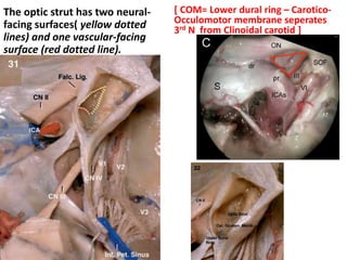
![The optic strut has two neural-
facing surfaces( yellow dotted
lines) and one vascular-facing
surface (red dotted line).
[ COM= Lower dural ring – Carotico-
Occulomotor membrane seperates
3rd N from Clinoidal carotid ]](https://image.slidesharecdn.com/10triangles360-150127035719-conversion-gate01/85/10-triangles-360-25-320.jpg)
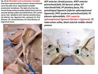
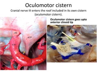
![Oculomotor triangle [ 3rd N. , 4th N. & Pcom ] is seen in Posterosuperior
compartment [ virtual compartment ] of cavernous sinus –better
understanding see cavernous sinus PPT
http://www.slideshare.net/muralichandnallamothu/cavernous-sinus-360](https://image.slidesharecdn.com/10triangles360-150127035719-conversion-gate01/85/10-triangles-360-30-320.jpg)
![Note the aperture for 3rd nerve & 4th nerve anterior & posterior to
posterior petro-clival fold [ PPCF ]](https://image.slidesharecdn.com/10triangles360-150127035719-conversion-gate01/85/10-triangles-360-32-320.jpg)
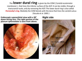
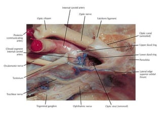
![The lower dural ring is given by the COM [ Carotid-oculomotor
membrane ] , that lines the inferior surface of the ACP. It can be visible, through a
transcranial route, only by removing the ACP. The lower dural ring is also called
Perneczky’s ring. Medially the COM blends with the dura that lines the carotid sulcus
(Yasuda et al. 2005 )
Endoscopic supraorbital view with a 30°
down-facing lens -The right portion of the
planum sphenoidale is seen from above.
Right side](https://image.slidesharecdn.com/10triangles360-150127035719-conversion-gate01/85/10-triangles-360-35-320.jpg)
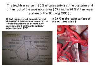
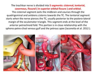
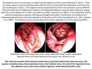
![The trochlear nerve in 80 % of cases enters at the posterior end
of the roof of the cavernous sinus ( CS ) and in 20 % at the lower
surface of the TC (Lang 1995 ) .
80 % of cases enters at the posterior end
of the roof of the cavernous sinus ( CS ) ---
---Note the aperture for 3rd nerve & 4th
nerve anterior & posterior to posterior
petro-clival fold [ PPCF ]
in 20 % at the lower surface of
the TC (Lang 1995 )](https://image.slidesharecdn.com/10triangles360-150127035719-conversion-gate01/85/10-triangles-360-36-320.jpg)
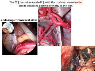
![The TC [ tentorium cerebelli ], with the trochlear nerve inside,
can be visualized passing inferiorly to the IIIcn.
endoscopic transclival view](https://image.slidesharecdn.com/10triangles360-150127035719-conversion-gate01/85/10-triangles-360-38-320.jpg)
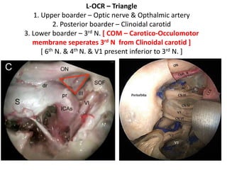
![L-OCR – Triangle
1. Upper boarder – Optic nerve & Opthalmic artery
2. Posterior boarder – Clinoidal carotid
3. Lower boarder – 3rd N. [ COM – Carotico-Occulomotor
membrane seperates 3rd N from Clinoidal carotid ]
[ 6th N. & 4th N. & V1 present inferior to 3rd N. ]](https://image.slidesharecdn.com/10triangles360-150127035719-conversion-gate01/85/10-triangles-360-40-320.jpg)
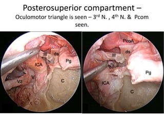
![Oculomotor triangle is seen [ 3rd N. , 4th N. & Pcom ] seen in
Posterosuperior compartment [ virtual compartment ] of cavernous sinus –
better understanding see cavernous sinus PPT
http://www.slideshare.net/muralichandnallamothu/cavernous-sinus-360](https://image.slidesharecdn.com/10triangles360-150127035719-conversion-gate01/85/10-triangles-360-41-320.jpg)
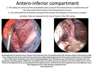
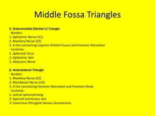
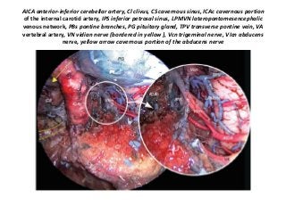
![Antero-inferior compartment [ virtual compartment ] of cavernous sinus
– for better understanding see cavernous sinus PPT
http://www.slideshare.net/muralichandnallamothu/cavernous-sinus-360
1. The abducens nerve and the sympathetic plexus around the intracavernous carotid artery are the only
nerves which have a real intracavernous course.
2. The anteroinferior and lateral compartments contain the abducens nerve and, as surgical corridors, they
are exposed to the risk of injury to the VIth nerve.
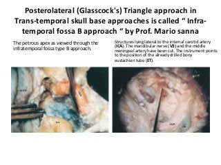
BS basisphenoid, CS cavernous sinus, CSd dura of the cavernous sinus, ET eustachian tube, ICAc cavernous portion of the internal carotid
artery, ICAh horizontal portion of the internal carotid artery, ICAp parapharyngeal portion of the internal carotid artery, ILT inferolateral
trunk, LVPM levator veli palatini muscle, MHT meningohypophyseal trunk, PAp petrous apex, PCFd posterior cranial fossa dura and
periosteum, PG pituitary gland, TVPM tensor veli palatini muscle, VN vidian nerve, IIIcn oculomotor nerve, IVcn trochlear nerve, V1 fi rst
branch of the trigeminal nerve, V2 second branch of the trigeminal nerve, V3 third branch of the trigeminal nerve, VIcn abducens nerve,
XIIcn hypoglossal nerve, white asterisks sympathetic fi bres
connecting the VIcn](https://image.slidesharecdn.com/10triangles360-150127035719-conversion-gate01/85/10-triangles-360-42-320.jpg)
![Anterior clinoid drilling videos in FTOZ
[ neurosurgery skull base ]
1. https://www.youtube.com/watch?v=wO2cWHiOdO0
2. https://www.youtube.com/watch?v=4dkQY3zxJHU
3. https://www.youtube.com/watch?v=vd4_lPVIUvE
4. https://www.youtube.com/watch?v=_dvYB1InGMc
5. https://www.youtube.com/watch?v=83_VuKHXOmQ
6. https://www.youtube.com/watch?v=0KwBhTqNXA4
7. https://www.youtube.com/watch?v=pCURjQ83HzU
8. https://www.youtube.com/watch?v=DNIy0L3oFgY
9. https://www.youtube.com/watch?v=GT4eBB2x58Q
10. https://www.youtube.com/watch?v=OS4Mc0X8tlU
11. https://www.youtube.com/watch?v=_xq9e3p1cc4](https://image.slidesharecdn.com/10triangles360-150127035719-conversion-gate01/85/10-triangles-360-43-320.jpg)


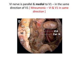
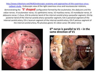
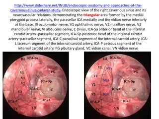
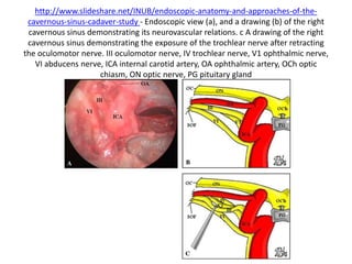
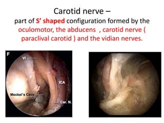
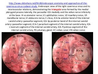
![http://www.slideshare.net/INUB/endoscopic-anatomy-and-approaches-of-the-cavernous-sinus-
cadaver-study - Endoscopic view of the right cavernous sinus and neurovascular relations,
demonstrating the ‘S’ shaped configuration formed by the oculomotor, the
abducens , carotid nerve ( paraclival carotid ) and the vidian nerves.
III oculomotor nerve, V1 ophthalmic nerve, V2 maxillary nerve, V3 mandibular nerve, VI abducens
nerve, C clivus, ICA-Sa anterior bend of the internal carotid artery–parasellar segment, ICA-Sp posterior
bend of the internal carotid artery–parasellar segment, ICA-C paraclival segment of the internal carotid
artery, ICA-L lacerum segment of the internal carotid artery, ICA-P petrous segment of the internal
carotid artery, PG pituitary gland, VC vidian canal, VN vidian nerve
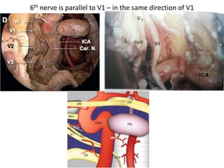
VI nerve is parallel & medial to V1 –
in the same direction of V1 [
Mneumonic – VI & V1 in same
direction ]](https://image.slidesharecdn.com/10triangles360-150127035719-conversion-gate01/85/10-triangles-360-53-320.jpg)

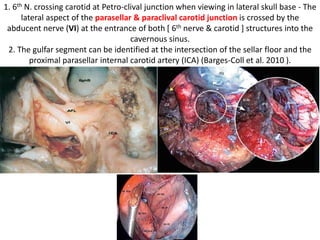
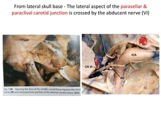
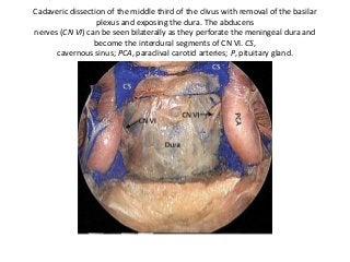
![1. 6th N. crossing carotid at Petro-clival junction when viewing in lateral skull base - The
lateral aspect of the parasellar & paraclival carotid junction is crossed by the
abducent nerve (VI) at the entrance of both [ 6th nerve & carotid ] structures into the
cavernous sinus.
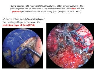
2. The gulfar segment can be identified at the intersection of the sellar floor and the
proximal parasellar internal carotid artery (ICA) (Barges-Coll et al. 2010 ).](https://image.slidesharecdn.com/10triangles360-150127035719-conversion-gate01/85/10-triangles-360-55-320.jpg)
![1. 6th N. crossing carotid at Petro-clival junction when viewing in lateral skull
base - The lateral aspect of the parasellar & paraclival carotid junction is
crossed by the abducent nerve (VI) at the entrance of both [ 6th nerve &
carotid ] structures into the cavernous sinus.
2. The gulfar segment can be identified at the intersection of the sellar floor
and the proximal parasellar internal carotid artery (ICA) (Barges-Coll et al.
2010 ).](https://image.slidesharecdn.com/10triangles360-150127035719-conversion-gate01/85/10-triangles-360-56-320.jpg)

![VI nerve is parallel & medial to V1 – in the same direction of V1 [ Mneumonic – VI &
V1 in same direction ]](https://image.slidesharecdn.com/10triangles360-150127035719-conversion-gate01/85/10-triangles-360-58-320.jpg)


![VI nerve is parallel & medial to V1 – in the same
direction of V1 [ Mneumonic – VI & V1 in same
direction ]](https://image.slidesharecdn.com/10triangles360-150127035719-conversion-gate01/85/10-triangles-360-71-320.jpg)
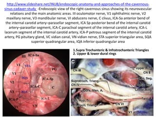
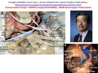
![Triangles of Middle cranial fossa – see Ant. Medial & Ant. Lateral triangles in both photos.
http://www.eneurosurgery.com/surgicaltrianglesofthecavernoussinus.html
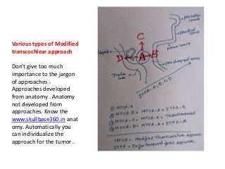
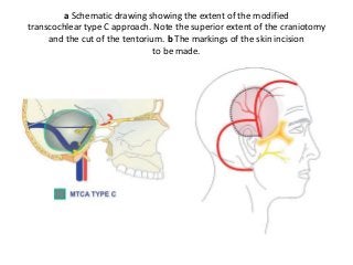
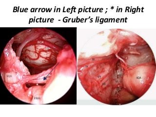
Postero-medial Triangle = KAWASE triangle [Prof.KAWASE , JAPAN Neurosurgeon -below photo]](https://image.slidesharecdn.com/10triangles360-150127035719-conversion-gate01/85/10-triangles-360-103-320.jpg)
![1. 6th N. crossing carotid at Petro-clival junction when viewing in lateral skull base - The
lateral aspect of the parasellar & paraclival carotid junction is crossed by the
abducent nerve (VI) at the entrance of both [ 6th nerve & carotid ] structures into the
cavernous sinus.
2. The gulfar segment can be identified at the intersection of the sellar floor and the
proximal parasellar internal carotid artery (ICA) (Barges-Coll et al. 2010 ).](https://image.slidesharecdn.com/10triangles360-150127035719-conversion-gate01/85/10-triangles-360-120-320.jpg)
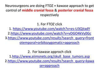
![1. 6th N. crossing carotid at Petro-clival junction when viewing in lateral skull
base - The lateral aspect of the parasellar & paraclival carotid junction is
crossed by the abducent nerve (VI) at the entrance of both [ 6th nerve &
carotid ] structures into the cavernous sinus.
2. The gulfar segment can be identified at the intersection of the sellar floor
and the proximal parasellar internal carotid artery (ICA) (Barges-Coll et al.
2010 ).](https://image.slidesharecdn.com/10triangles360-150127035719-conversion-gate01/85/10-triangles-360-121-320.jpg)
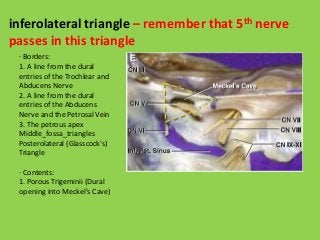
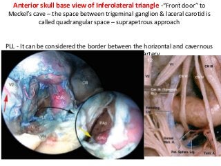


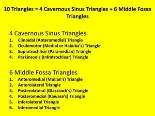
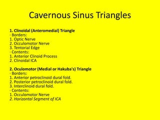
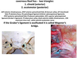
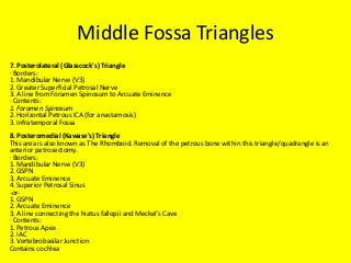
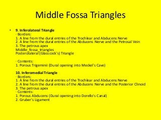
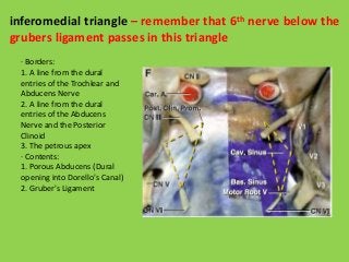
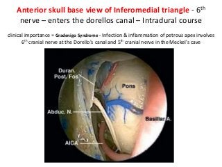
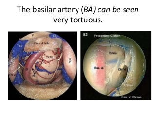
This document discusses the anatomy of the skull base triangles. It begins by naming the 10 triangles, which are divided into 4 cavernous sinus triangles and 6 middle fossa triangles. Each triangle is then defined by its borders and contents. Key structures discussed include the anterior clinoid process, carotid oculomotor membrane, cavernous segment of the internal carotid artery, and cranial nerves III, IV, V and VI. The relationships between these structures are illustrated in several diagrams. Videos are also provided that demonstrate anterior clinoid drilling techniques.


