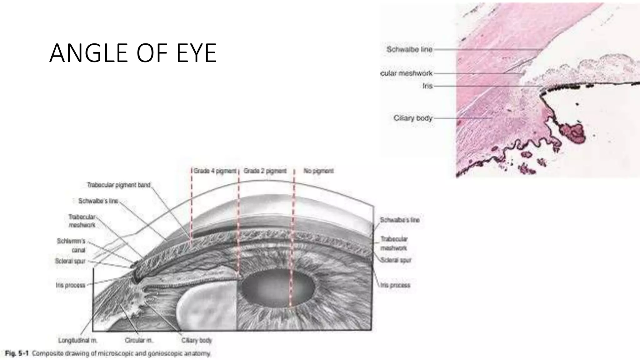
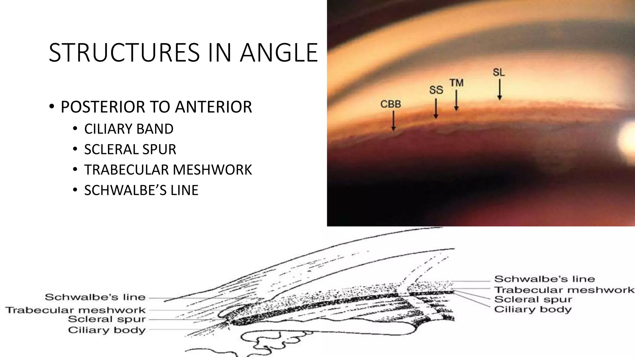
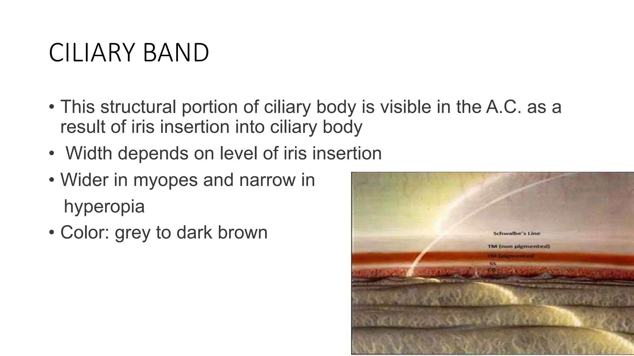
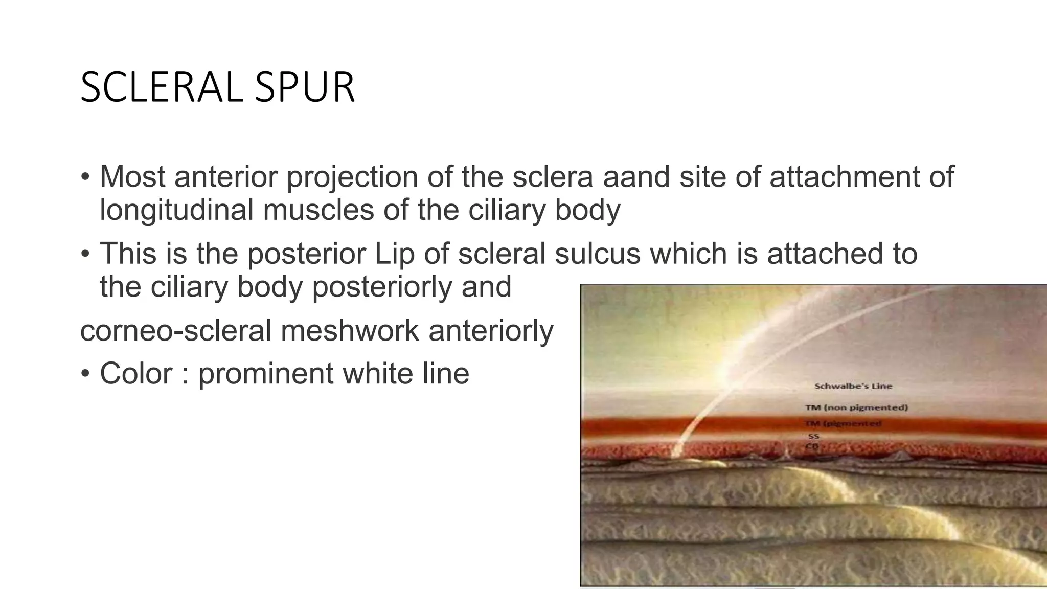
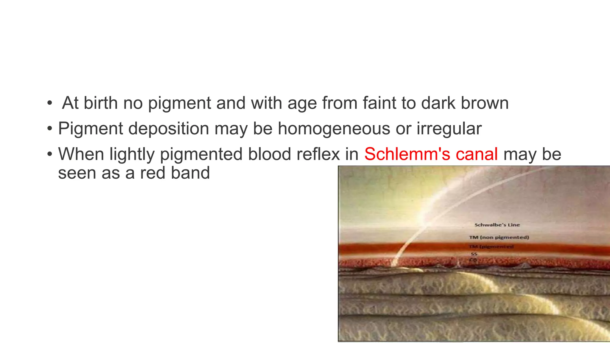
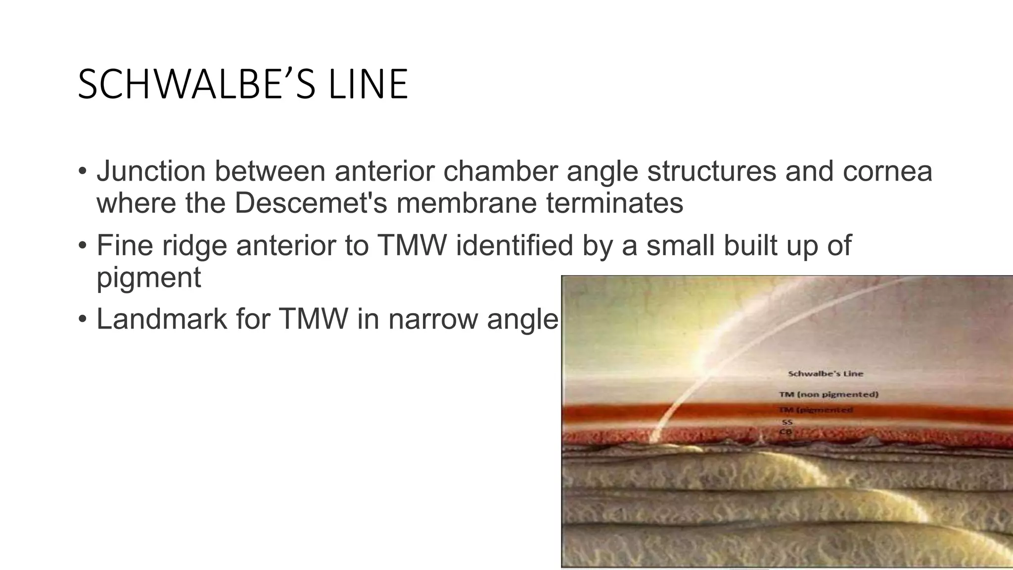
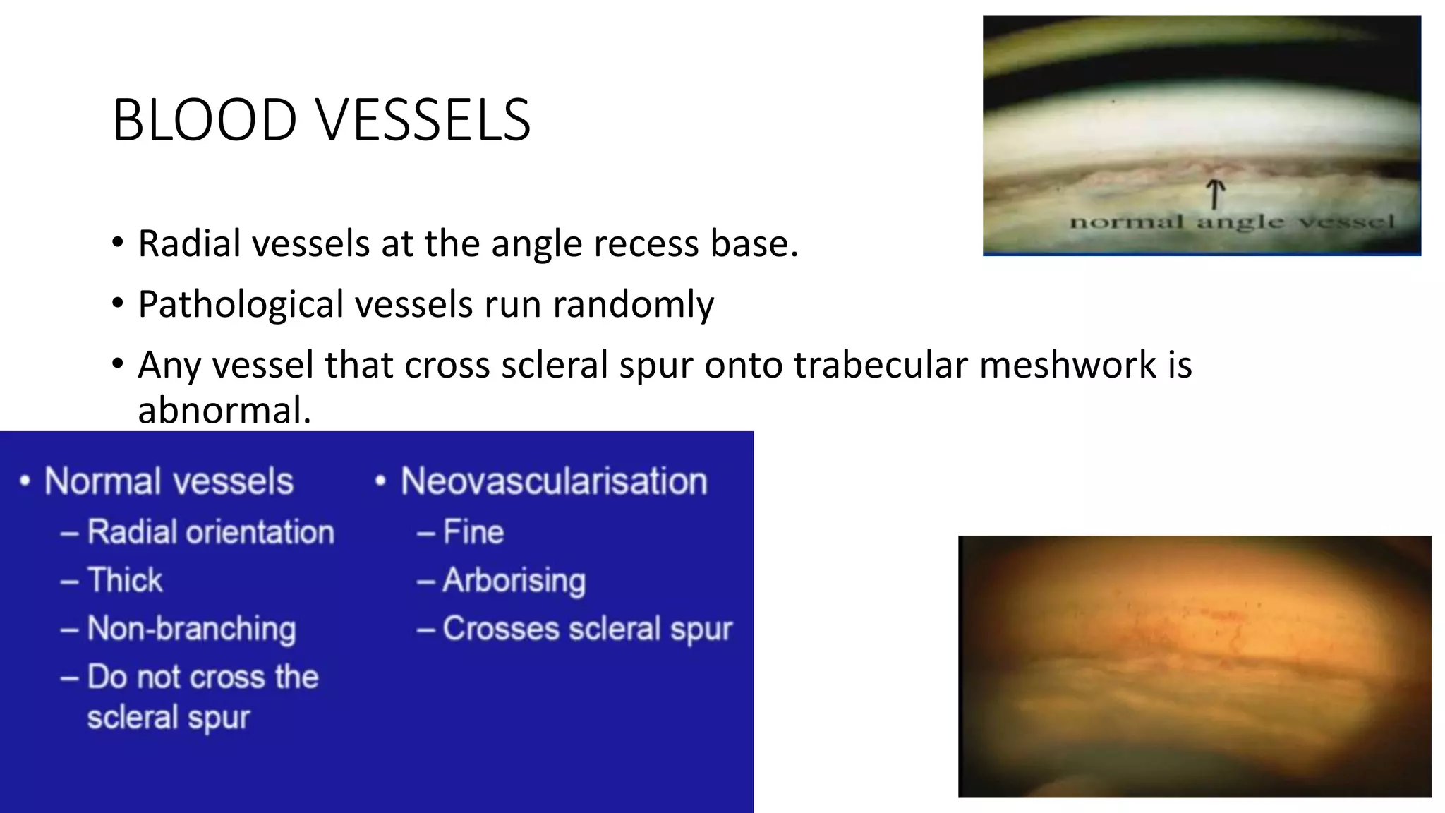
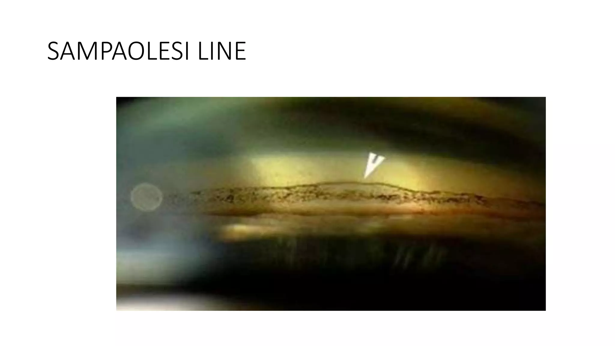
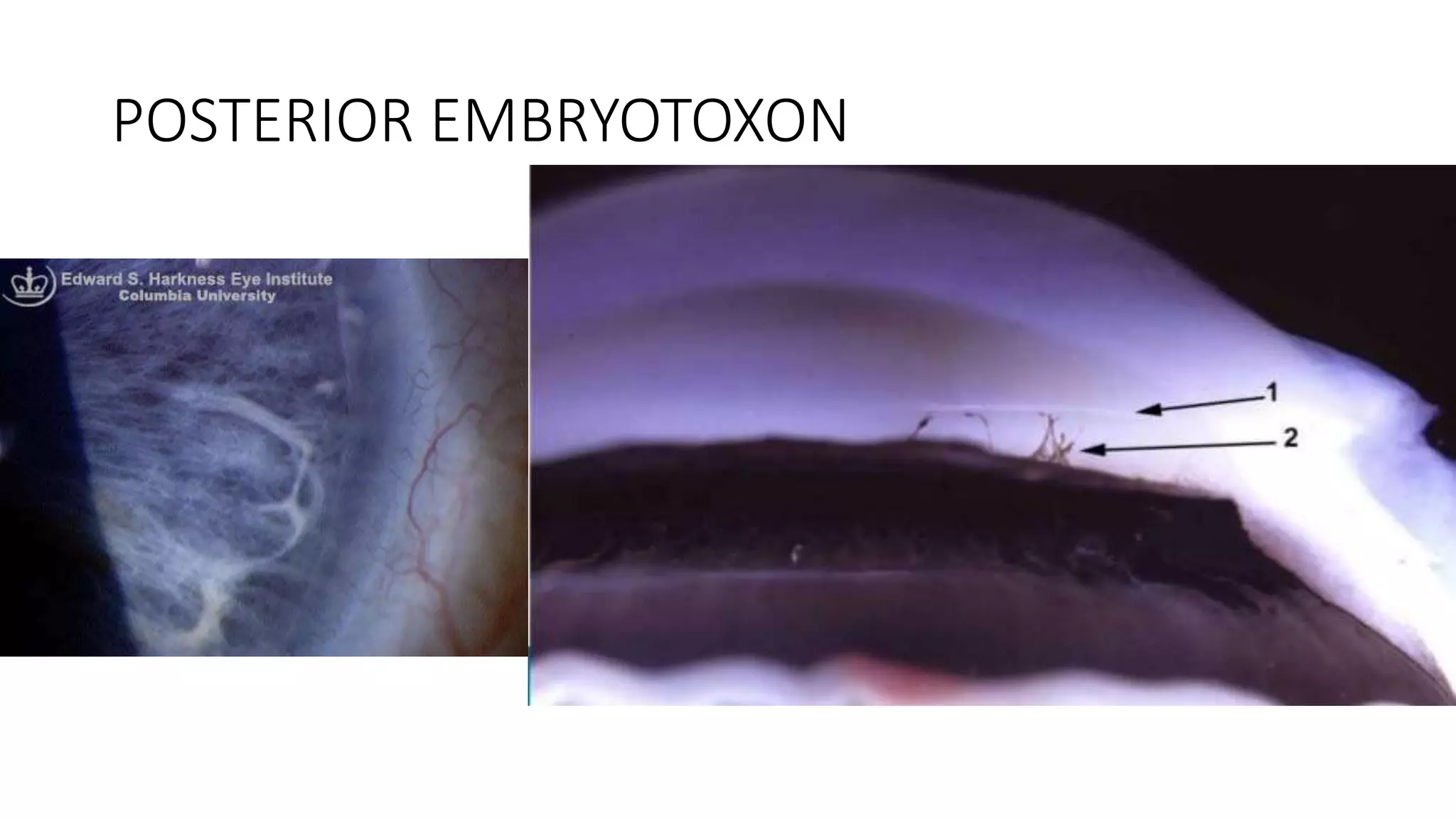
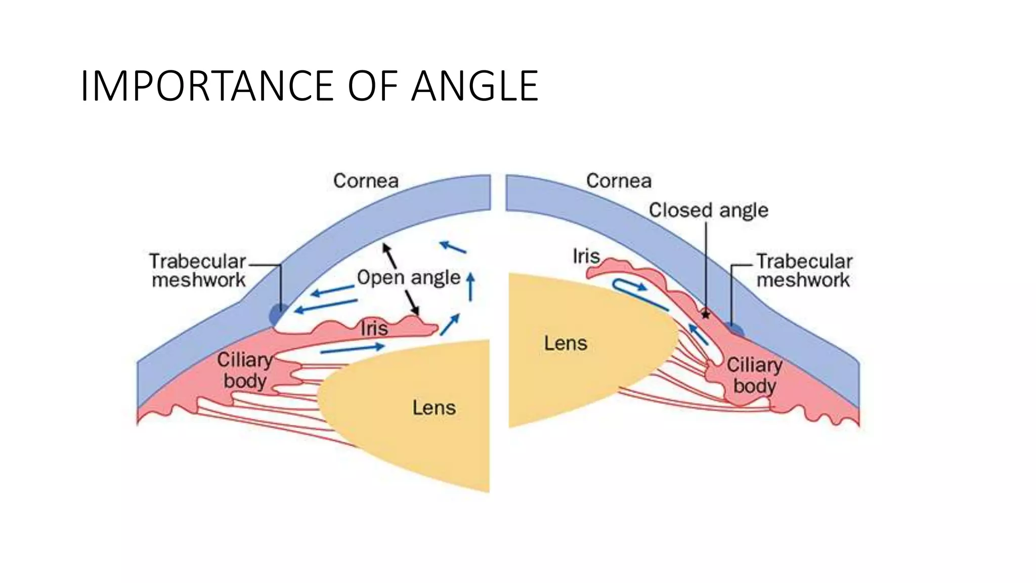
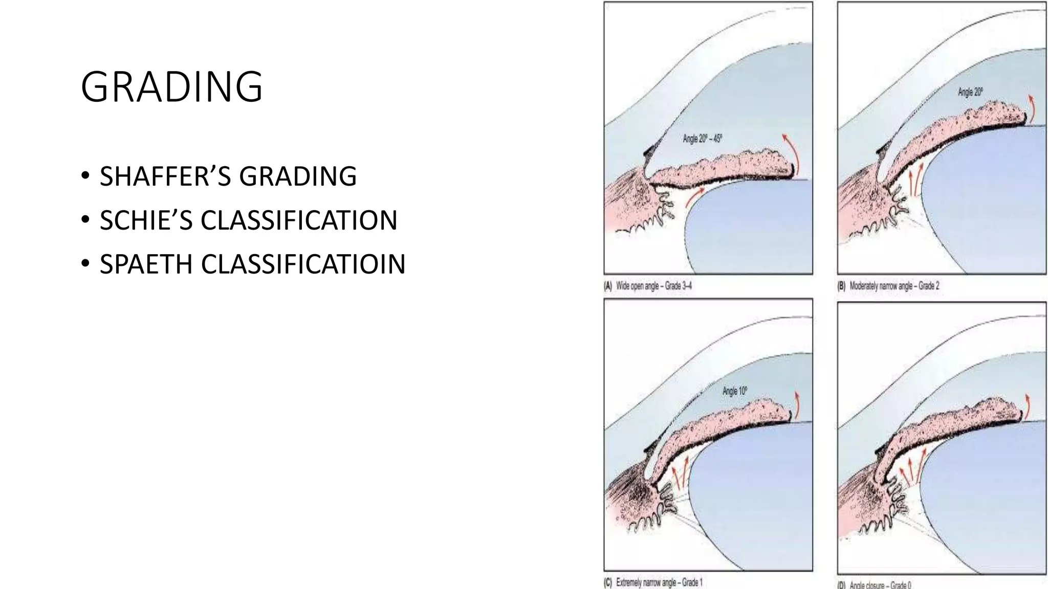
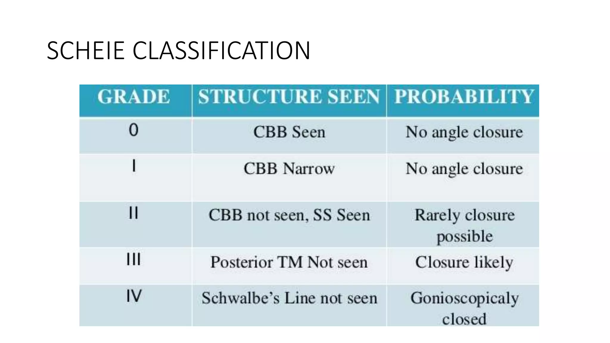
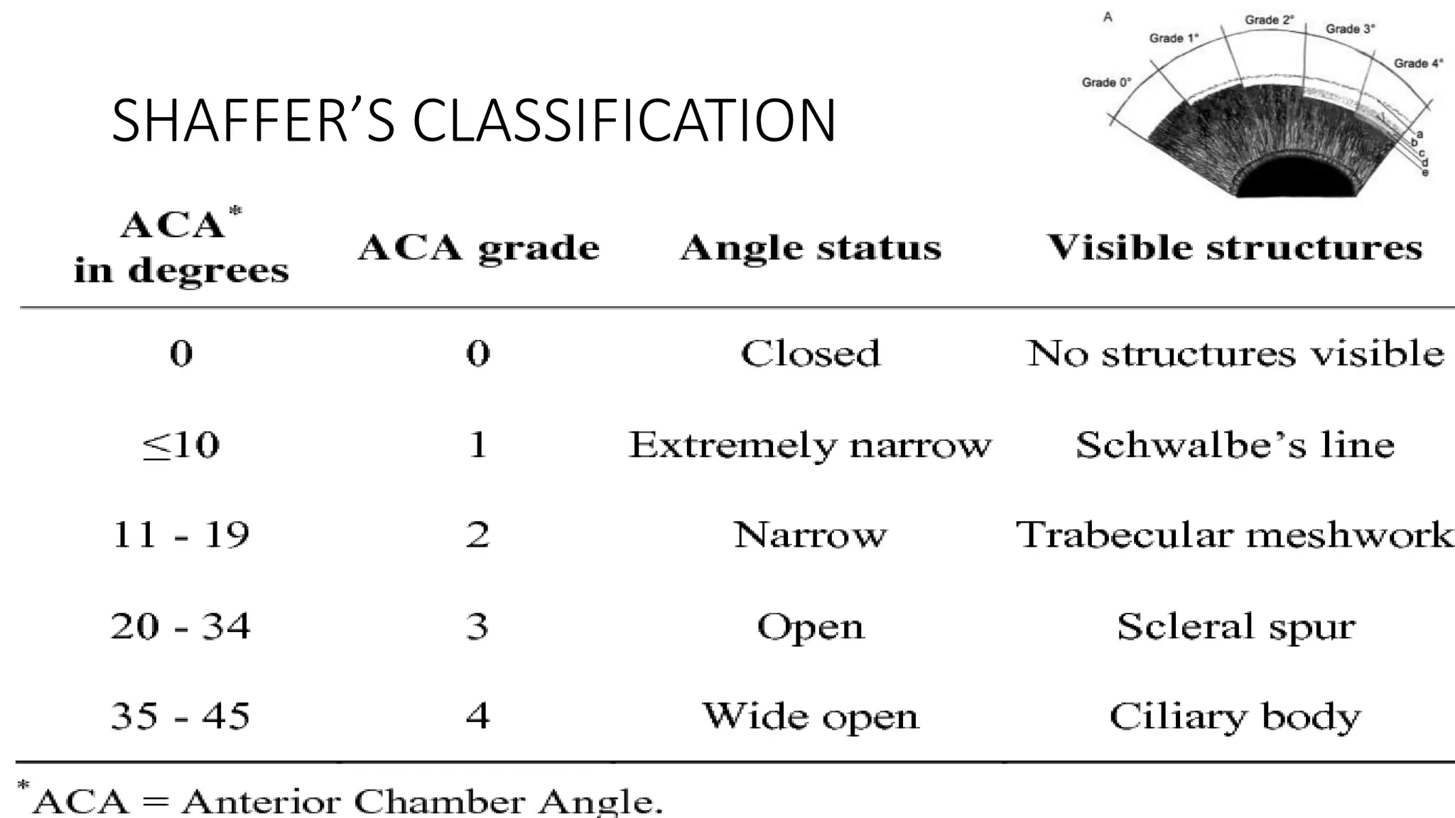
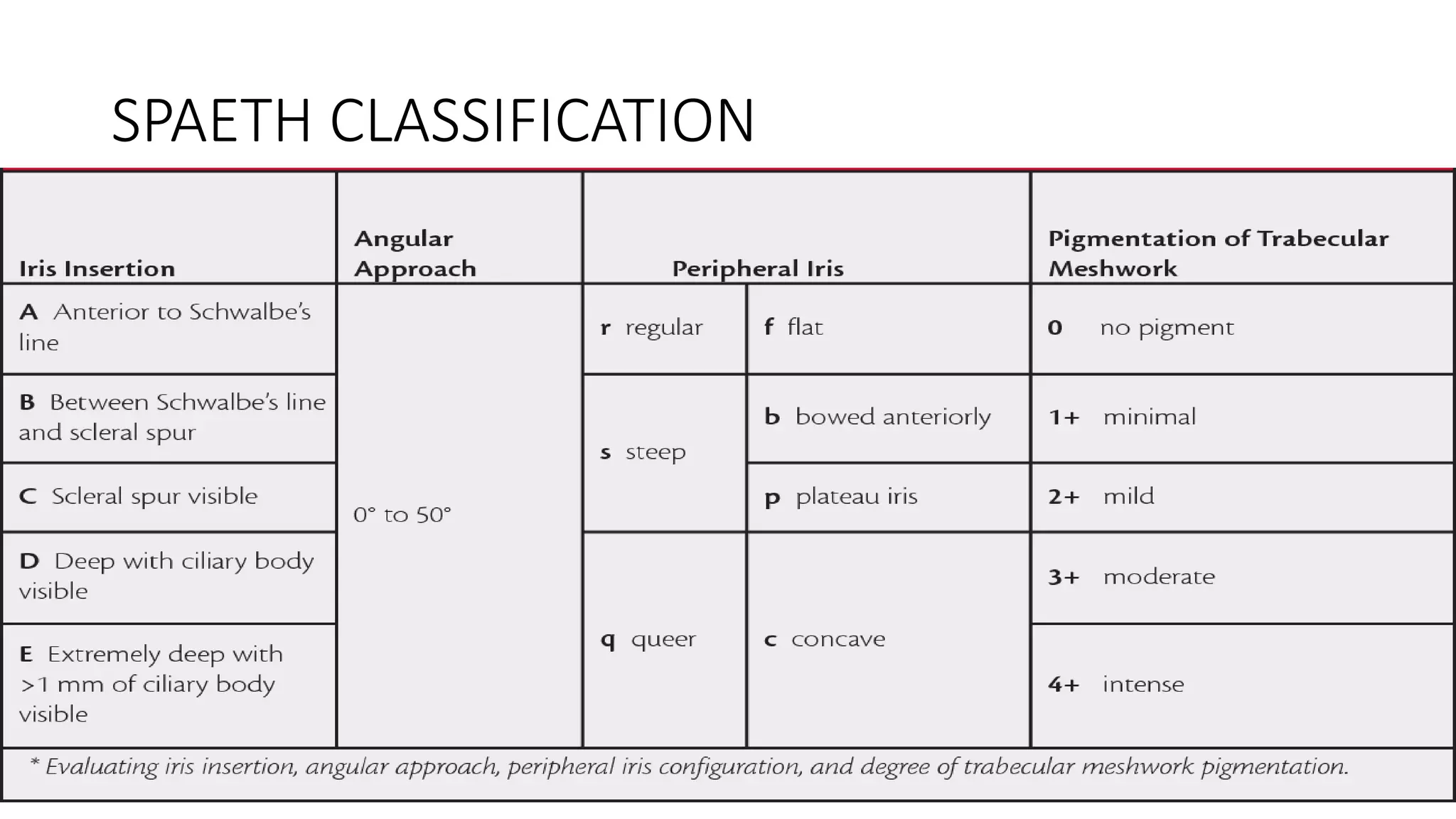
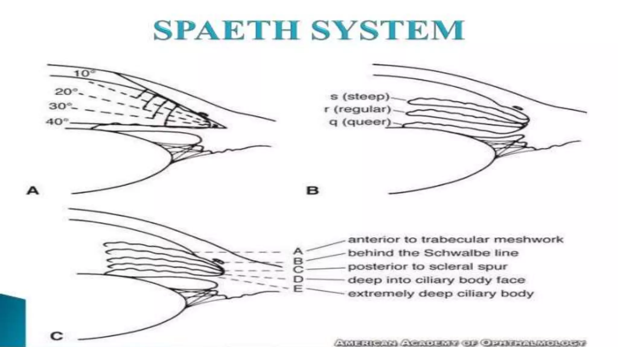
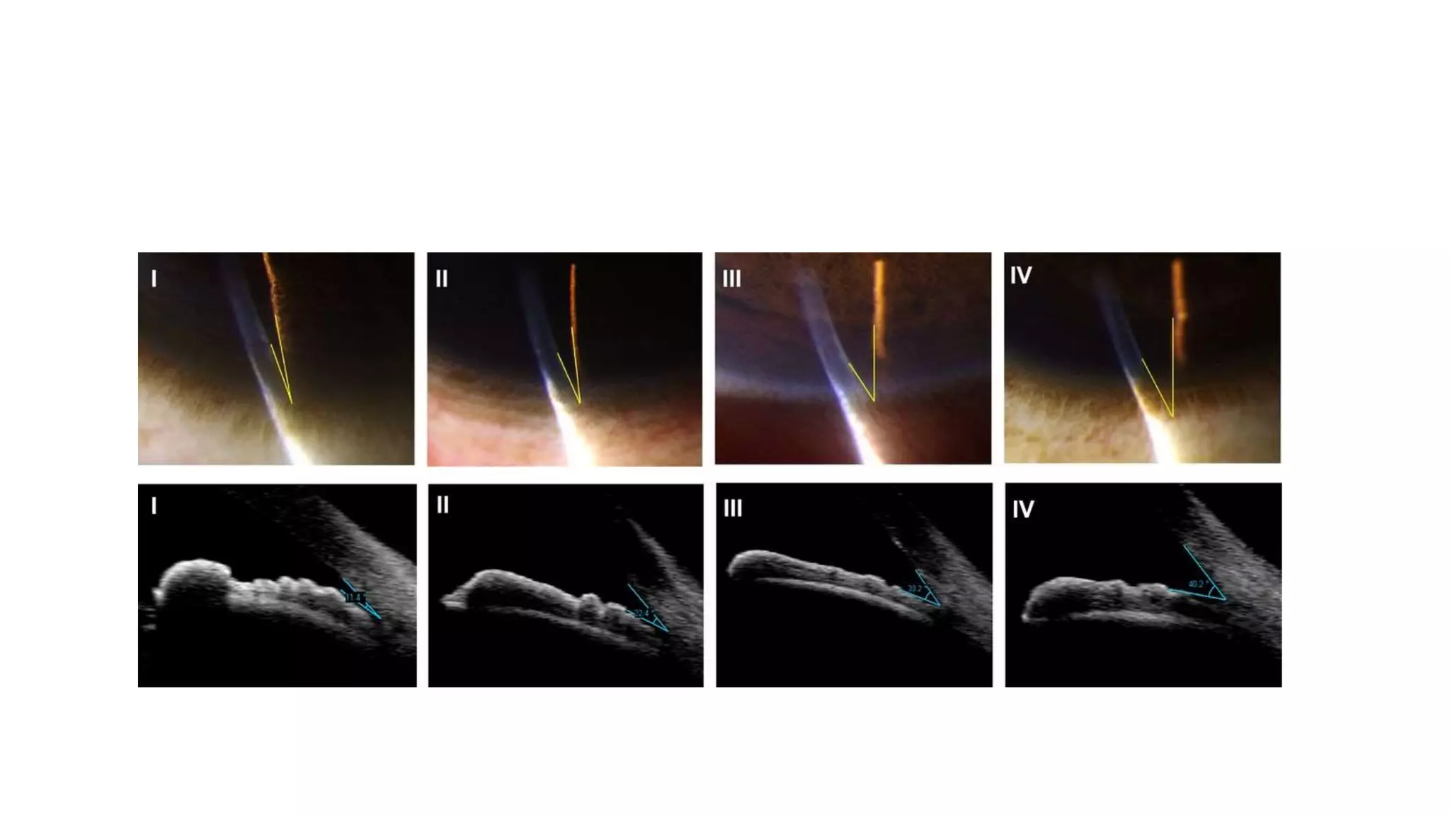
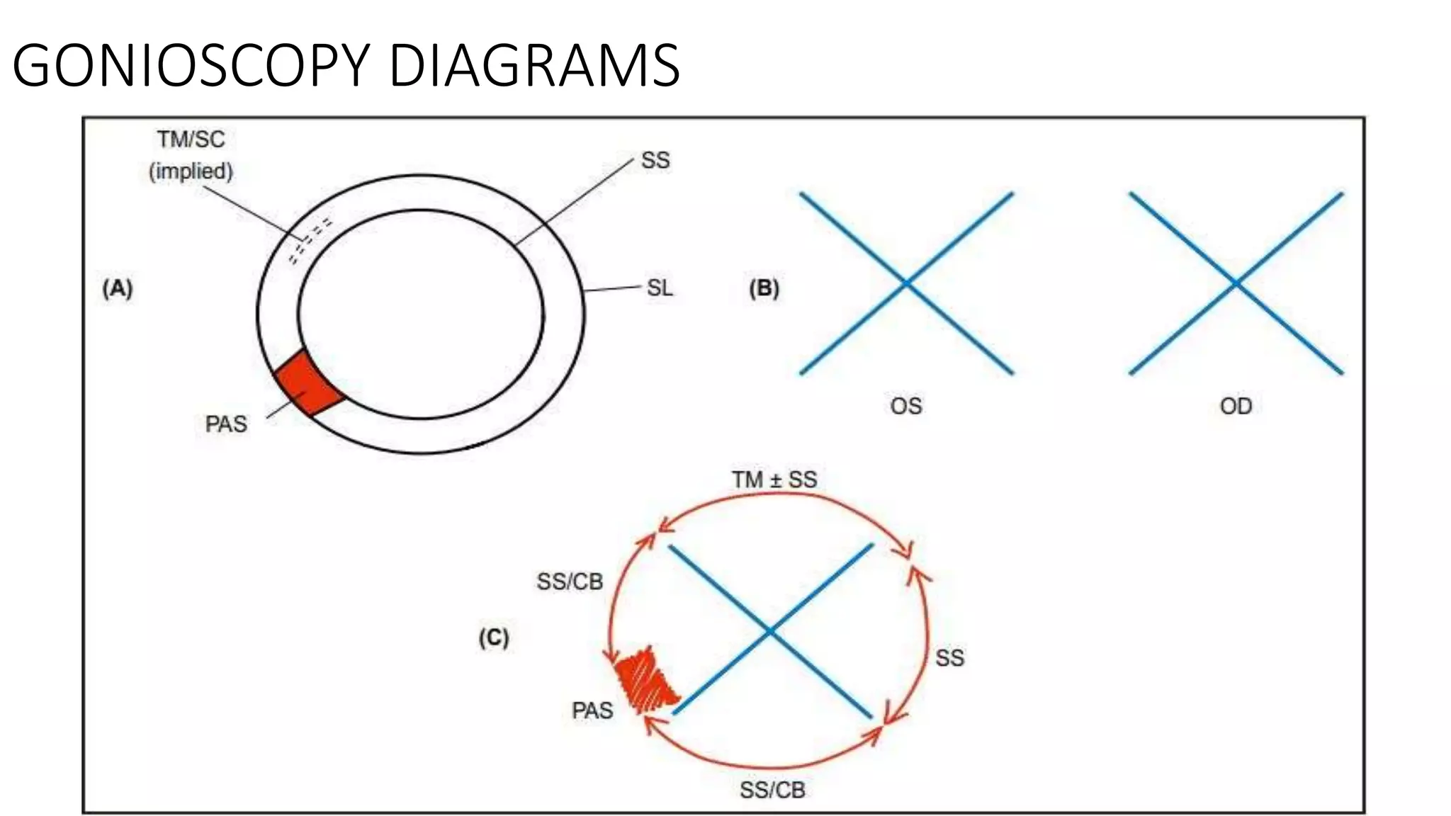
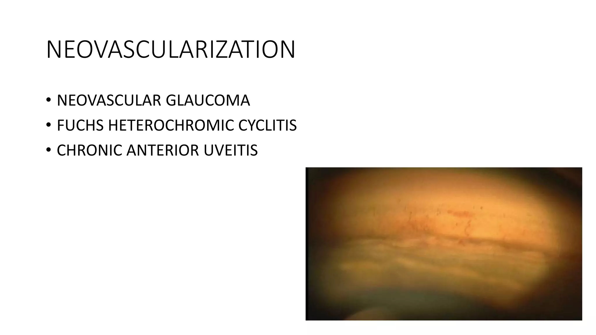
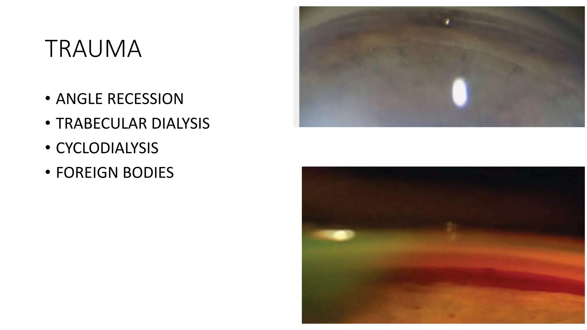
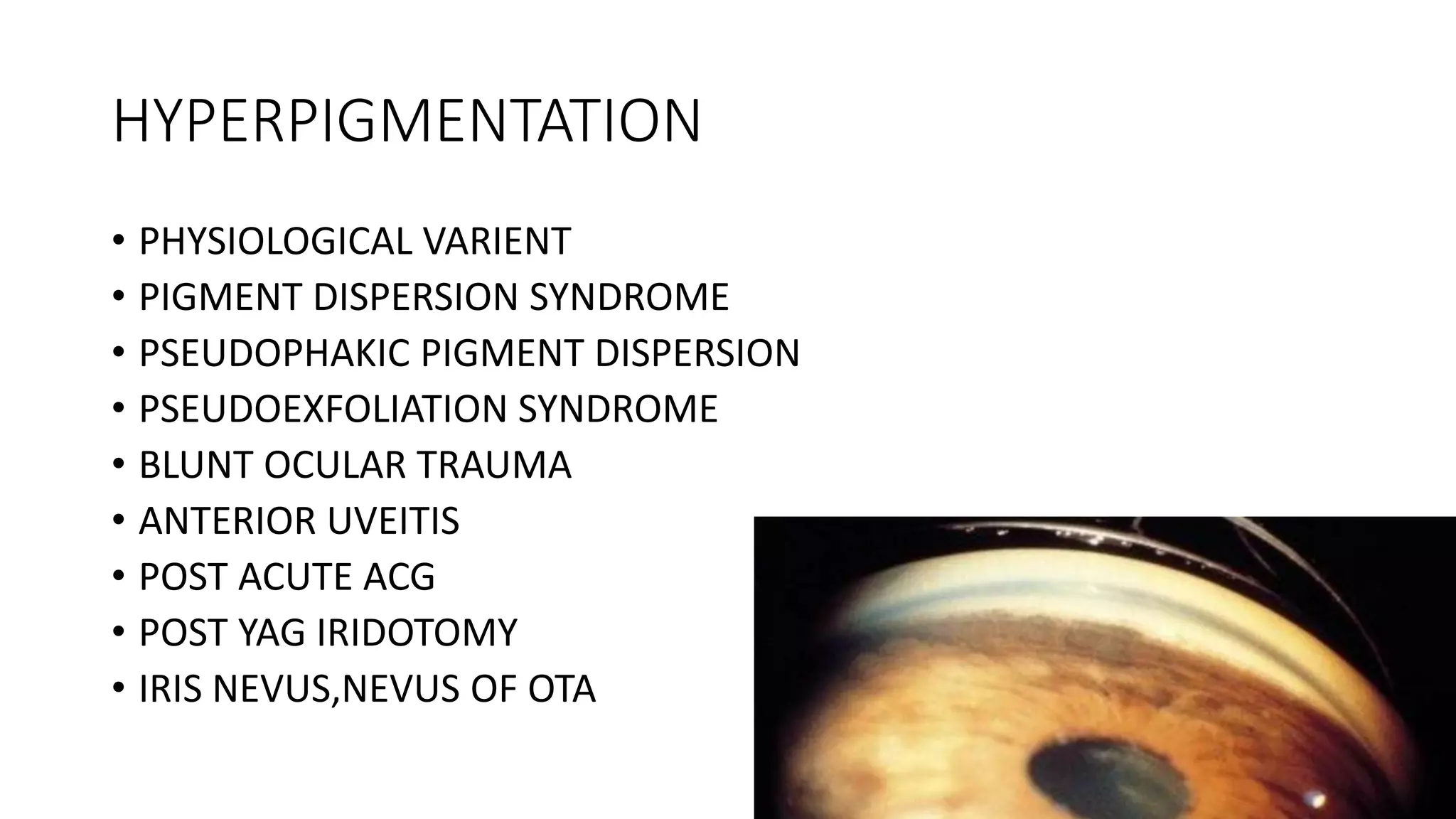
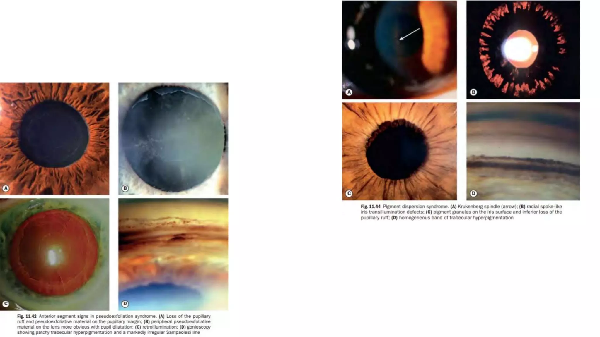
ANGLE ANATOMY
PRESENTOR : DR R GENICKSON JEYARAJ, JR
MODERATOR : DR BHAGABAT NAYAK, CONSULTANT
DR GARIMA, SR
ANGLE OF EYE
STRUCTURES IN ANGLE
• POSTERIOR TO ANTERIOR
• CILIARY BAND
• SCLERAL SPUR
• TRABECULAR MESHWORK
• SCHWALBE’S LINE
CILIARY BAND
• This structural portion of ciliary body is visible in the A.C. as a
result of iris insertion into ciliary body
• Width depends on level of iris insertion
• Wider in myopes and narrow in
hyperopia
• Color: grey to dark brown
SCLERAL SPUR
• Most anterior projection of the sclera aand site of attachment of
longitudinal muscles of the ciliary body
• This is the posterior Lip of scleral sulcus which is attached to
the ciliary body posteriorly and
corneo-scleral meshwork anteriorly
• Color : prominent white line
TRABECULAR MESHWORK
• Pigmented band anterior to scleral spur
• Although extent of TMW is from root of iris to Schwalbe's line it
is considered as 2 portions
• a) Anterior - between Schwalbe's line and anterior Edge of Schlemm's
canal Involved in lesser degree of aqueous out flow
• b) Posterior – Functional part , primary site of aqueous out flow
• Appearance of functional TMW depends on amount of pigment
deposition
• At birth no pigment and with age from faint to dark brown
• Pigment deposition may be homogeneous or irregular
• When lightly pigmented blood reflex in Schlemm's canal may be
seen as a red band
SCHWALBE’S LINE
• Junction between anterior chamber angle structures and cornea
where the Descemet's membrane terminates
• Fine ridge anterior to TMW identified by a small built up of
pigment
• Landmark for TMW in narrow angle
IRIS PROCESSES
• Small tenuous extensions of anterior surface of the iris that insert at
the level of scleral spur and cover the ciliary boy to a variable extend.
BLOOD VESSELS
• Radial vessels at the angle recess base.
• Pathological vessels run randomly
• Any vessel that cross scleral spur onto trabecular meshwork is
abnormal.
SAMPAOLESI LINE
POSTERIOR EMBRYOTOXON
IMPORTANCE OF ANGLE
Open angle closed angle
GONIOSCOPY
GRADING
• SHAFFER’S GRADING
• SCHIE’S CLASSIFICATION
• SPAETH CLASSIFICATIOIN
SCHEIE CLASSIFICATION
SHAFFER’S CLASSIFICATION
SPAETH CLASSIFICATION
VAN HERICK SYSEM OF CLASSIFICATION
GONIOSCOPY DIAGRAMS
PATHOLOGICAL FINDINGS IN GONIOSCOPY
PERIPHERAL ANTERIOR SYNECHIAE
• PRIMARY ANGLE CLOSURE GLAUCOMA
• ANTERIOR UVEITIS
• ICE SYNDROME
PERIPHERAL ANTERIOR SYNECHIAE IRIS PROCESSES
• Free
• Extend into scleral spur
• Follow concavity of recess
• Underlying structures are seen
• Iris moves with indentation
• Breaks with angle recession
• Broad
• Extending beyond scleral spur
• Bridge concavity of recess
• obscure structures
• Resist movement
• intact
NEOVASCULARIZATION
• NEOVASCULAR GLAUCOMA
• FUCHS HETEROCHROMIC CYCLITIS
• CHRONIC ANTERIOR UVEITIS
TRAUMA
• ANGLE RECESSION
• TRABECULAR DIALYSIS
• CYCLODIALYSIS
• FOREIGN BODIES
HYPERPIGMENTATION
• PHYSIOLOGICAL VARIENT
• PIGMENT DISPERSION SYNDROME
• PSEUDOPHAKIC PIGMENT DISPERSION
• PSEUDOEXFOLIATION SYNDROME
• BLUNT OCULAR TRAUMA
• ANTERIOR UVEITIS
• POST ACUTE ACG
• POST YAG IRIDOTOMY
• IRIS NEVUS,NEVUS OF OTA
BLOOD IN SCHLEMM CANAL
• PHYSIOLOGICAL VARIENT
• CAROTID ACAVERNOUS FISTULA AND DURAL SHUNT
• STURGE WEBER SYNDROME
• SVC OBSTRUCTION
THANK YOU…

ANGLE ANATOMY

  • 1.
    ANGLE ANATOMY PRESENTOR :DR R GENICKSON JEYARAJ, JR MODERATOR : DR BHAGABAT NAYAK, CONSULTANT DR GARIMA, SR
  • 2.
  • 3.
    STRUCTURES IN ANGLE •POSTERIOR TO ANTERIOR • CILIARY BAND • SCLERAL SPUR • TRABECULAR MESHWORK • SCHWALBE’S LINE
  • 4.
    CILIARY BAND • Thisstructural portion of ciliary body is visible in the A.C. as a result of iris insertion into ciliary body • Width depends on level of iris insertion • Wider in myopes and narrow in hyperopia • Color: grey to dark brown
  • 5.
    SCLERAL SPUR • Mostanterior projection of the sclera aand site of attachment of longitudinal muscles of the ciliary body • This is the posterior Lip of scleral sulcus which is attached to the ciliary body posteriorly and corneo-scleral meshwork anteriorly • Color : prominent white line
  • 6.
    TRABECULAR MESHWORK • Pigmentedband anterior to scleral spur • Although extent of TMW is from root of iris to Schwalbe's line it is considered as 2 portions • a) Anterior - between Schwalbe's line and anterior Edge of Schlemm's canal Involved in lesser degree of aqueous out flow • b) Posterior – Functional part , primary site of aqueous out flow • Appearance of functional TMW depends on amount of pigment deposition
  • 7.
    • At birthno pigment and with age from faint to dark brown • Pigment deposition may be homogeneous or irregular • When lightly pigmented blood reflex in Schlemm's canal may be seen as a red band
  • 8.
    SCHWALBE’S LINE • Junctionbetween anterior chamber angle structures and cornea where the Descemet's membrane terminates • Fine ridge anterior to TMW identified by a small built up of pigment • Landmark for TMW in narrow angle
  • 9.
    IRIS PROCESSES • Smalltenuous extensions of anterior surface of the iris that insert at the level of scleral spur and cover the ciliary boy to a variable extend.
  • 10.
    BLOOD VESSELS • Radialvessels at the angle recess base. • Pathological vessels run randomly • Any vessel that cross scleral spur onto trabecular meshwork is abnormal.
  • 11.
  • 12.
  • 13.
  • 14.
  • 15.
  • 17.
    GRADING • SHAFFER’S GRADING •SCHIE’S CLASSIFICATION • SPAETH CLASSIFICATIOIN
  • 18.
  • 19.
  • 20.
  • 22.
    VAN HERICK SYSEMOF CLASSIFICATION
  • 24.
  • 25.
  • 26.
    PERIPHERAL ANTERIOR SYNECHIAE •PRIMARY ANGLE CLOSURE GLAUCOMA • ANTERIOR UVEITIS • ICE SYNDROME
  • 27.
    PERIPHERAL ANTERIOR SYNECHIAEIRIS PROCESSES • Free • Extend into scleral spur • Follow concavity of recess • Underlying structures are seen • Iris moves with indentation • Breaks with angle recession • Broad • Extending beyond scleral spur • Bridge concavity of recess • obscure structures • Resist movement • intact
  • 28.
    NEOVASCULARIZATION • NEOVASCULAR GLAUCOMA •FUCHS HETEROCHROMIC CYCLITIS • CHRONIC ANTERIOR UVEITIS
  • 29.
    TRAUMA • ANGLE RECESSION •TRABECULAR DIALYSIS • CYCLODIALYSIS • FOREIGN BODIES
  • 30.
    HYPERPIGMENTATION • PHYSIOLOGICAL VARIENT •PIGMENT DISPERSION SYNDROME • PSEUDOPHAKIC PIGMENT DISPERSION • PSEUDOEXFOLIATION SYNDROME • BLUNT OCULAR TRAUMA • ANTERIOR UVEITIS • POST ACUTE ACG • POST YAG IRIDOTOMY • IRIS NEVUS,NEVUS OF OTA
  • 32.
    BLOOD IN SCHLEMMCANAL • PHYSIOLOGICAL VARIENT • CAROTID ACAVERNOUS FISTULA AND DURAL SHUNT • STURGE WEBER SYNDROME • SVC OBSTRUCTION
  • 33.

Editor's Notes

  • #8 Average width of 600micrometere Younger people groud glass translucent appearance Patchy pigmentation in a suspiciously narrow angles rasise possibility of intermittent iris contact.
  • #9 May have a double line configuration and the posterior component is mistaken for the pigmented meshwork Corneaal wedge: for locating inconspicuous SL.narrow slit and two distinct linear corneal reflections can be identified one on the inner surface and other on the outer surface. Outer reflection will arc round across the corneoscleral interface to meet the inner reflection at the apex as the sclera is opaque.
  • #10 Not to be confuse with peripheral ant synechiae which typically extend more anteriorly and are more substantial.
  • #12 Pigments along schwalbes line seen in pseudoexofoliation syndrome and pigment dispersion syndrome
  • #13 Schwalbe line usually lies in plane of posterior corneal surface, but in 15-20% may be variably hypertrophied and project as a delicate glistening ridge into anterior chamber called posterior embryotoxon Axenfeld rigler anomaly mention
  • #18 SCHEIE SYSTEM: most posterior structure visible. SHAFFER’S SYSTEM : assess geometric angle width in 4 grades . angle potential for occlusion. SPAETH SYSTEM : three dimensional structure of angle - level of iris insertion and peripheral iris configuration. RPC GRADING
  • #25 Diagramming gonioscopy with concentric circles. The inner circle represents the posterior limit of the scleral spur (SS), and the outer circle represents Schwalbe’s line (SL). The trabecular meshwork (TM) occupies the same location as Schlemm’s canal (SC). Here a peripheral anterior synechia (PAS) is drawn up to Schwalbe’s line. (B) Hand-drawn Xs can suffice for charting purposes to indicate gonioscopic quadrants. (C) In this hand-drawn example, the superior angle shows the trabecular meshwork (TM) with equivocal viewing of the scleral spur (SS), which is elsewhere seen by itself or with the ciliary body (CB); a peripheral anterior synechia (PAS) is also seen.
  • #27 ICE-IRIDOCORNEAL EPITHELIAL SYNDROME