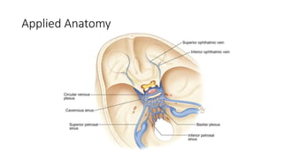
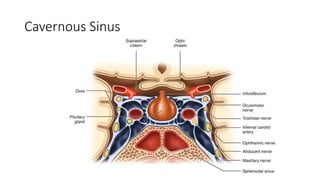
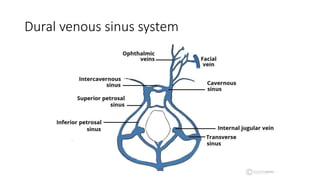
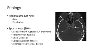
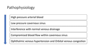
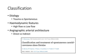
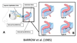
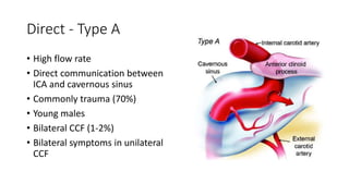
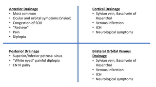
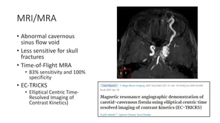
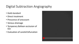
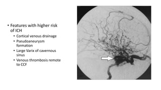
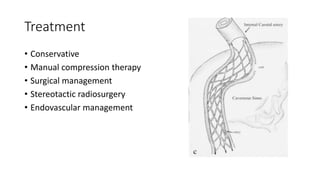
Carotid-Cavernous Fistula
Department of Neurosurgery
Dr Rejoyce Anto
Applied Anatomy
Cavernous Sinus
Dural venous sinus system
Introduction
• It is a specific type of dural arterio-venous fistula characterized by
abnormal arteriovenous shunting within the cavernous sinus.
• It results in high pressure arterial blood entering the low pressure
venous cavernous sinus
Epidemiology
• Carotid-cavernous fistulas represent approximately 12% of all dural
arteriovenous fistulas.
• Trauma (70-75%)
• Young males (HIGH FLOW DIRECT FISTULAS)
• 0.2% of cerebrocranial trauma and 4% basilar skull fractures
• Spontaneous (30%)
• Older females (LOW FLOW INDIRECT FISTULAS)
Etiology
• Head trauma (70-75%)
• Blunt
• Penetrating
• Spontaneous (30%)
• Associated with ruptured ICA aneurysms
• Fibromuscular dysplasia
• Ehlers-Danlos sx
• Collagen vascular diseases
• Atherosclerotic vascular disease
Pathophysiology
High pressure arterial blood
Low pressure cavernous sinus
Interference with normal venous drainage
Compromised blood flow within cavernous sinus
Ophthalmic venous hypertension and Orbital venous congestion
Classification
• Etiology
• Trauma vs Spontaneous
• Haemodynamic features
• High flow vs Low flow
• Angiographic arterial architecture
• Direct vs Indirect
BARROW et al. (1985)
Direct - Type A
• High flow rate
• Direct communication between
ICA and cavernous sinus
• Commonly trauma (70%)
• Young males
• Bilateral CCF (1-2%)
• Bilateral symptoms in unilateral
CCF
Indirect – Types B,C,D
Branches of ICA and CS Branches of ECA and CS
Branches of both ICA and
ECA and CS
Clinical Presentation of High Flow CCF
• Subjective bruit (80%)
• Proptosis (80%)
• Chemosis and conjunctival
injection (75%)
• Headache (60%)
• Diplopia (50%)
• Blurred vision (40%)
• Orbital pain (35 %)
• Ophthalmoplegia (40%)
Dandy’s Triad
Bruit
Proptosis
Conjunctival
chemosis
Fundoscopy
• Venous stasis retinopathy
• Dilated retinal veins
• Intraretinal haemorrhages
• Exposure keratopathy
• Corneal ulceration
• Elevated episcleral venous
pressure
• Secondary glaucoma
• Central retinal occlusion
Clinical Presentation of Low Flow CCFs
• Insidious onset
• Dural CCFs can produce specific patterns of symptoms based on the
rate of flow and the pattern(s) of venous drainage:
Anterior Drainage
• Most common
• Ocular and orbital symptoms (Vision)
• Congestion of SOV
• “Red eye”
• Pain
• Diplopia
Posterior Drainage
• Superior/Inferior petrosal sinus
• “White eyed” painful diplopia
• CN III palsy
Cortical Drainage
• Sylvian vein, Basal vein of
Rosenthal
• Venous infarction
• ICH
• Neurological symptoms
Bilateral Orbital Venous
Drainage
• Sylvian vein, Basal vein of
Rosenthal
• Venous infarction
• ICH
• Neurological symptoms
Imaging modalities – CT/CT Angio
• Proptosis
• Expansion of the cavernous sinus
• Superior ophthalmic vein
• Enlargement of extraocular muscles,
• Any associated skull fractures.
MRI/MRA
• Abnormal cavernous
sinus flow void
• Less sensitive for skull
fractures
• Time-of-Flight MRA
• 83% sensitivity and 100%
specificity
• EC-TRICKS
• Elliptical Centric Time-
Resolved Imaging of
Contrast Kinetics)
Digital Subtraction Angiography
• Gold standard
• Direct treatment
• Prescence of aneurysm
• Venous drainage
• Temporary Balloon occlusion of
ICA
• Evaluation of carotid bifurcation
• Features with higher risk
of ICH
• Cortical venous drainage
• Pseudoaneurysm
formation
• Large Varix of cavernous
sinus
• Venous thrombosis remote
to CCF
Differential Diagnosis
• Primary intracranial
tumour
• Lymphoma
• Aneurysm,
• Carotid dissection,
• Cavernous sinus thrombosis,
• Infection,
• Tolosa-Hunt syndrome,
• Orbital pseudotumor,
• Vasculitis
• Sarcoidosis.
Treatment
• Conservative
• Manual compression therapy
• Surgical management
• Stereotactic radiosurgery
• Endovascular management
Indications for emergency treatment
•Clinical features
• Increased ICP
• Rapidly progressing
proptosis
• ICH/SAH/External
haemorrhage
• TIA
•Angiographic features
• Pseudoaneurysm
• Large varix of CS
• Cortical venous
drainage
• Thrombosis of distal
venous outflow
pathways
Conservative
• Indirect CCFs may close spontaneously (20-60%)
• Direct CCFs unlikely to close spontaneously
• Indications for treatment
• Milder symptoms
• Serial examinations
• Follow up
• Betablockers and Acetazolamide
Manual compression therapy
• Controversial
• Contralateral hand to compress carotid for 10 seconds, several times
a day, for a few weeks.
Endovascular Treatment
• Preferred approach.
• Successful closure rates of 55-85%
• Complications (10-40%)
• ICA occlusion
• Cerebral infarction
• Worsened ocular palsy
• Two methods
• Transarterial embolization (DIRECT CCF)
• Transvenous embolization (INDIRECT CCF)
Transarterial Embolization
• Preferred strategy for most direct CCFs
THE GOAL IS TO CLOSE THE FISTULA
WHILE PRESERVING FLOW WITHIN THE
ICA.
1.Detachable balloon
2.Coils or other embolic material
3.Covered stent
Detachable balloon occlusion
• Done since 1980s
• Prerequisite – large fistulous tract and large cavernous sinus
• Transfemoral approach to proximal CCA
Coil and material embolization
• Mainstay for direct CCF
• Detachable platinum coils
• N-BCA
• EVOH
Covered stent graft placement
• Recent advances
• Immediate obliteration of a
direct CCF
• Maintain patency of ICA
• Failed balloon occlusion test
• Cons
• Longitudinal flexibility limited
• Difficult to navigate (tortuosity)
• Vasospasm
• Endoleak
• Coverage of vital perforators
Parent artery occlusion
• Life-saving emergency treatment
• Temporary balloon occlusion test
Transvenous Embolization
• Preferred strategy for most indirect CCFs
• May also be used for direct CCF when the transarterial approach
fails or is incompletely successful
• Most common - IPS
THE GOAL IS TO CATHETERIZE THE
ABNORMAL CS SUPERSELECTIVELY
WITHOUT REROUTING VENOUS DRAINAGE
TO CORTICAL STRUCTURES
Surgery
• Limited use
• Increased morbidity
• Cranial deficits
• Patients that undergo surgery –
higher risk patients
• Options
• Ligation of ICA
• Surgical trapping of fistula
• Surgical transvenous packing
Stereotactic Radiosurgery
• First performed by Barcia-Salorio and colleagues in 1977
• Gamma knife
• Long-term obliteration (75%)
• Latency of average 22 months
• 10-40 Gy
• Dose-related neurological injury
Prognosis
References
1. Barrow DL, Spector RH, Braun IF, Landman JA, Tindall SC, Tindall
GT. Classification and treatment of spontaneous carotid-cavernous fistulas. J
Neurosurg 1985; 62(2): 248–256.
2. Henderson AD, Miller NR. Carotid-cavernous fistula: current concepts in
aetiology, investigation, and management. Eye (Lond). 2018;32(2):164-172.
doi:10.1038/eye.2017.240
3. Stiebel-Kalish H, Setton A, Nimii Y, et al. Cavernous sinus dural arteriovenous
malformations: patterns of venous drainage are related to clinical signs and
symptoms. Ophthalmology. 2002;109(9):1685-1691.
4. Vattoth S, Cherian J, Pandey T. Magnetic resonance angiographic
demonstration of carotid-cavernous fistula using elliptical centric time
resolved imaging of contrast kinetics (EC-TRICKS). Magn Reson Imaging.
2007;25(8):1227-1231. Kai Y, Hamada J, Morioka M, Yano S, Kuratsu J.
Treatment of cavernous sinus dural arteriovenous fistulae by external manual
carotid compression. Neurosurgery. 2007;60(2):253-258.
MCQ 1
A 45-year-old woman is an unrestrained passenger in a motor vehicle accident
where she sustains a blow to the right side of her head. Other than a brief loss of
consciousness and head pain, she has no deficits. She has a negative non-contrast
head CT in an ED after the event and is discharged home. Over the next several
weeks, she notices diplopia, tearing, chemosis, conjunctival injection, and a
pulsating sensation of the left eye, prompting further evaluation. Upon seeing her
in your office, the most useful diagnostic test is:
A. Brain MRI with contrast
B. Cerebral angiogram
C. Optic nerve sheath ultrasound
D. Formal visual field assessment
MCQ 2
Which of the following features will most warrant urgent intervention
in a patient with a carotid-cavernous fistula?
A. Abducens Nerve Palsy
B. Young age
C. The presence of a skull fracture on CT
D. Cortical venous drainage
MCQ 3
In terms of the epidemiology of CCFs, what is the most correct answer?
A. Indirect CCFs occur mostly in older men
B. Most common cause for direct CCF is transsphenoidal surgery
C. Spontaneous CCFs have a female predominance
D. 10% of Basilar skull fractures result in direct CCFs
MCQ 4
A 46-year-old man is admitted to your ICU after facial assault with a
penetrating object through his orbit. You are called to the bedside on
post op day 1 after removal of the object because the patient is in
severe pain. Upon your examination, the eye is swollen, injected, and
pulsating. You suspect a cavernous carotid fistula. What Barrow
Classification type is this most likely to be?
A. Type A
B. Type B
C. Type C
D. Type D
MCQ 5
You explain to the patient while taking consent the various treatment
modalities that are available. What modality is the most likely to be
helpful in this particular patient?
A. Surgical packing of cavernous sinus
B. Endovascular – Transarterial embolization
C. Stereotactic radiosurgery
D. Endovascular – Transvenous embolization
THANK YOU

Carotid Cavernous Fistulas

  • 1.
    Carotid-Cavernous Fistula Department ofNeurosurgery Dr Rejoyce Anto
  • 2.
  • 3.
  • 4.
  • 5.
    Introduction • It isa specific type of dural arterio-venous fistula characterized by abnormal arteriovenous shunting within the cavernous sinus. • It results in high pressure arterial blood entering the low pressure venous cavernous sinus
  • 7.
    Epidemiology • Carotid-cavernous fistulasrepresent approximately 12% of all dural arteriovenous fistulas. • Trauma (70-75%) • Young males (HIGH FLOW DIRECT FISTULAS) • 0.2% of cerebrocranial trauma and 4% basilar skull fractures • Spontaneous (30%) • Older females (LOW FLOW INDIRECT FISTULAS)
  • 8.
    Etiology • Head trauma(70-75%) • Blunt • Penetrating • Spontaneous (30%) • Associated with ruptured ICA aneurysms • Fibromuscular dysplasia • Ehlers-Danlos sx • Collagen vascular diseases • Atherosclerotic vascular disease
  • 9.
    Pathophysiology High pressure arterialblood Low pressure cavernous sinus Interference with normal venous drainage Compromised blood flow within cavernous sinus Ophthalmic venous hypertension and Orbital venous congestion
  • 10.
    Classification • Etiology • Traumavs Spontaneous • Haemodynamic features • High flow vs Low flow • Angiographic arterial architecture • Direct vs Indirect
  • 11.
  • 12.
    Direct - TypeA • High flow rate • Direct communication between ICA and cavernous sinus • Commonly trauma (70%) • Young males • Bilateral CCF (1-2%) • Bilateral symptoms in unilateral CCF
  • 13.
    Indirect – TypesB,C,D Branches of ICA and CS Branches of ECA and CS Branches of both ICA and ECA and CS
  • 14.
    Clinical Presentation ofHigh Flow CCF • Subjective bruit (80%) • Proptosis (80%) • Chemosis and conjunctival injection (75%) • Headache (60%) • Diplopia (50%) • Blurred vision (40%) • Orbital pain (35 %) • Ophthalmoplegia (40%)
  • 15.
  • 16.
    Fundoscopy • Venous stasisretinopathy • Dilated retinal veins • Intraretinal haemorrhages • Exposure keratopathy • Corneal ulceration • Elevated episcleral venous pressure • Secondary glaucoma • Central retinal occlusion
  • 17.
    Clinical Presentation ofLow Flow CCFs • Insidious onset • Dural CCFs can produce specific patterns of symptoms based on the rate of flow and the pattern(s) of venous drainage:
  • 18.
    Anterior Drainage • Mostcommon • Ocular and orbital symptoms (Vision) • Congestion of SOV • “Red eye” • Pain • Diplopia Posterior Drainage • Superior/Inferior petrosal sinus • “White eyed” painful diplopia • CN III palsy Cortical Drainage • Sylvian vein, Basal vein of Rosenthal • Venous infarction • ICH • Neurological symptoms Bilateral Orbital Venous Drainage • Sylvian vein, Basal vein of Rosenthal • Venous infarction • ICH • Neurological symptoms
  • 19.
    Imaging modalities –CT/CT Angio • Proptosis • Expansion of the cavernous sinus • Superior ophthalmic vein • Enlargement of extraocular muscles, • Any associated skull fractures.
  • 21.
    MRI/MRA • Abnormal cavernous sinusflow void • Less sensitive for skull fractures • Time-of-Flight MRA • 83% sensitivity and 100% specificity • EC-TRICKS • Elliptical Centric Time- Resolved Imaging of Contrast Kinetics)
  • 22.
    Digital Subtraction Angiography •Gold standard • Direct treatment • Prescence of aneurysm • Venous drainage • Temporary Balloon occlusion of ICA • Evaluation of carotid bifurcation
  • 23.
    • Features withhigher risk of ICH • Cortical venous drainage • Pseudoaneurysm formation • Large Varix of cavernous sinus • Venous thrombosis remote to CCF
  • 24.
    Differential Diagnosis • Primaryintracranial tumour • Lymphoma • Aneurysm, • Carotid dissection, • Cavernous sinus thrombosis, • Infection, • Tolosa-Hunt syndrome, • Orbital pseudotumor, • Vasculitis • Sarcoidosis.
  • 25.
    Treatment • Conservative • Manualcompression therapy • Surgical management • Stereotactic radiosurgery • Endovascular management
  • 26.
    Indications for emergencytreatment •Clinical features • Increased ICP • Rapidly progressing proptosis • ICH/SAH/External haemorrhage • TIA •Angiographic features • Pseudoaneurysm • Large varix of CS • Cortical venous drainage • Thrombosis of distal venous outflow pathways
  • 27.
    Conservative • Indirect CCFsmay close spontaneously (20-60%) • Direct CCFs unlikely to close spontaneously • Indications for treatment • Milder symptoms • Serial examinations • Follow up • Betablockers and Acetazolamide
  • 28.
    Manual compression therapy •Controversial • Contralateral hand to compress carotid for 10 seconds, several times a day, for a few weeks.
  • 29.
    Endovascular Treatment • Preferredapproach. • Successful closure rates of 55-85% • Complications (10-40%) • ICA occlusion • Cerebral infarction • Worsened ocular palsy • Two methods • Transarterial embolization (DIRECT CCF) • Transvenous embolization (INDIRECT CCF)
  • 30.
    Transarterial Embolization • Preferredstrategy for most direct CCFs THE GOAL IS TO CLOSE THE FISTULA WHILE PRESERVING FLOW WITHIN THE ICA. 1.Detachable balloon 2.Coils or other embolic material 3.Covered stent
  • 31.
    Detachable balloon occlusion •Done since 1980s • Prerequisite – large fistulous tract and large cavernous sinus • Transfemoral approach to proximal CCA
  • 32.
    Coil and materialembolization • Mainstay for direct CCF • Detachable platinum coils • N-BCA • EVOH
  • 33.
    Covered stent graftplacement • Recent advances • Immediate obliteration of a direct CCF • Maintain patency of ICA • Failed balloon occlusion test • Cons • Longitudinal flexibility limited • Difficult to navigate (tortuosity) • Vasospasm • Endoleak • Coverage of vital perforators
  • 34.
    Parent artery occlusion •Life-saving emergency treatment • Temporary balloon occlusion test
  • 35.
    Transvenous Embolization • Preferredstrategy for most indirect CCFs • May also be used for direct CCF when the transarterial approach fails or is incompletely successful • Most common - IPS THE GOAL IS TO CATHETERIZE THE ABNORMAL CS SUPERSELECTIVELY WITHOUT REROUTING VENOUS DRAINAGE TO CORTICAL STRUCTURES
  • 36.
    Surgery • Limited use •Increased morbidity • Cranial deficits • Patients that undergo surgery – higher risk patients • Options • Ligation of ICA • Surgical trapping of fistula • Surgical transvenous packing
  • 37.
  • 38.
    Stereotactic Radiosurgery • Firstperformed by Barcia-Salorio and colleagues in 1977 • Gamma knife • Long-term obliteration (75%) • Latency of average 22 months • 10-40 Gy • Dose-related neurological injury
  • 39.
  • 40.
    References 1. Barrow DL,Spector RH, Braun IF, Landman JA, Tindall SC, Tindall GT. Classification and treatment of spontaneous carotid-cavernous fistulas. J Neurosurg 1985; 62(2): 248–256. 2. Henderson AD, Miller NR. Carotid-cavernous fistula: current concepts in aetiology, investigation, and management. Eye (Lond). 2018;32(2):164-172. doi:10.1038/eye.2017.240 3. Stiebel-Kalish H, Setton A, Nimii Y, et al. Cavernous sinus dural arteriovenous malformations: patterns of venous drainage are related to clinical signs and symptoms. Ophthalmology. 2002;109(9):1685-1691. 4. Vattoth S, Cherian J, Pandey T. Magnetic resonance angiographic demonstration of carotid-cavernous fistula using elliptical centric time resolved imaging of contrast kinetics (EC-TRICKS). Magn Reson Imaging. 2007;25(8):1227-1231. Kai Y, Hamada J, Morioka M, Yano S, Kuratsu J. Treatment of cavernous sinus dural arteriovenous fistulae by external manual carotid compression. Neurosurgery. 2007;60(2):253-258.
  • 41.
    MCQ 1 A 45-year-oldwoman is an unrestrained passenger in a motor vehicle accident where she sustains a blow to the right side of her head. Other than a brief loss of consciousness and head pain, she has no deficits. She has a negative non-contrast head CT in an ED after the event and is discharged home. Over the next several weeks, she notices diplopia, tearing, chemosis, conjunctival injection, and a pulsating sensation of the left eye, prompting further evaluation. Upon seeing her in your office, the most useful diagnostic test is: A. Brain MRI with contrast B. Cerebral angiogram C. Optic nerve sheath ultrasound D. Formal visual field assessment
  • 42.
    MCQ 2 Which ofthe following features will most warrant urgent intervention in a patient with a carotid-cavernous fistula? A. Abducens Nerve Palsy B. Young age C. The presence of a skull fracture on CT D. Cortical venous drainage
  • 43.
    MCQ 3 In termsof the epidemiology of CCFs, what is the most correct answer? A. Indirect CCFs occur mostly in older men B. Most common cause for direct CCF is transsphenoidal surgery C. Spontaneous CCFs have a female predominance D. 10% of Basilar skull fractures result in direct CCFs
  • 44.
    MCQ 4 A 46-year-oldman is admitted to your ICU after facial assault with a penetrating object through his orbit. You are called to the bedside on post op day 1 after removal of the object because the patient is in severe pain. Upon your examination, the eye is swollen, injected, and pulsating. You suspect a cavernous carotid fistula. What Barrow Classification type is this most likely to be? A. Type A B. Type B C. Type C D. Type D
  • 45.
    MCQ 5 You explainto the patient while taking consent the various treatment modalities that are available. What modality is the most likely to be helpful in this particular patient? A. Surgical packing of cavernous sinus B. Endovascular – Transarterial embolization C. Stereotactic radiosurgery D. Endovascular – Transvenous embolization
  • 46.