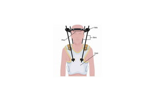
Sub axial cervical spine
DR.SHAMEEJ MUHAMED KV
SENIOR RESIDENT
NEUROSURGERY DEPARTMENT
GOVT MEDICAL COLLEGE , CALICUT
• The normal anatomy of the cervical spine consists of 7 cervical
vertebrae separated by intervertebral disks and joined by a complex
network of ligaments.
• These ligaments keep individual bony elements behaving as a single
unit.
• The anterior and posterior longitudinal ligaments maintain the
structural integrity of the anterior and middle columns.
• The posterior column is held in alignment by a complex ligamentous
system, including the nuchal ligament complex, capsular ligaments,
and the ligamentum flavum.
• If one column is disrupted, other columns may provide sufficient
stability to prevent spinal cord injury.
• If 2 columns are disrupted, the spine may move as 2 separate units,
increasing the likelihood of spinal cord injury.
• The atlas (C1) and the axis (C2) differ markedly from other cervical
vertebrae.
• The atlas has no vertebral body; however, it is composed of a thick anterior
arch with 2 prominent lateral masses and a thin posterior arch.
• The axis contains the odontoid process that represents fused remnants of
the atlas body. The odontoid process is held in tight approximation to the
posterior aspect of the anterior arch of C1 by the transverse ligament,
which stabilizes the atlanto-axial joint.
• Apical, alar, and transverse ligaments provide further stabilization by
allowing spinal column rotation; this prevents posterior displacement of
the dens in relation to the atlas.
The sub axial spine is from C3-C7 . The subaxial spine is distinct from axial
spine so it is taken as separate entity.
Osseous Anatomy
• Uncovertebral Joint(JOINT OF
LUSCHKA)
– Lateral projections of body
– Medial to vertebral artery
• Facet joints
– Sagittal orientation at 30-45
degrees
• Spinous processes
– Bifid C3-5,
prominent C7
The Facet Joints
• Also called ZYGAPOPHYSEAL JOINTS.
• The facet joints are formed by the articular processes of adjacent
vertebrae.
• The inferior articular process of a vertebra articulates with the
superior articular process of the vertebra below.
• These are synovial gliding joints
• Facet joints are oriented in different planes depending on their
anatomic location. Sagittal orientation at 30-45 degrees
Lateral Mass Anatomy
• Medial border - Lateral edge of
the lamina
• Lateral border - watch for
bleeders
• Superior/Inferior borders -
facets
• C7 frequently has abnormal
anatomy
• Vertebral artery is just anterior
to the medial border of the
lateral mass, enters at C6
• Nerve runs dorsal to the artery
and anterior to the inferior half
of the lateral mass
• 4 quadrants of the lateral mass
with the supero-lateral quadrant
being “safe”
Ligamentous Anatomy
• Anterior – ALL, PLL,
intervertebral disc
• Posterior– Nuchal Ligaments
ligamentum nuchae,
supraspinous ligament,
interspinous ligament
• Ligamentum flavum and the
facet joint capsules
Vascular Anatomy
Vertebral Artery
– Originates from subclavian
– Enters spine at C6 foramen
– At C2 it turns posterior and
lateral
– Forms Basilar Artery
Foramen Transversarium
– Gradually moves anteriorly
and medially from C6 to C2
NEUROANATOMY
• Spinal cord diameter subaxial: 17-22mm
• Occupies ~ 50% of canal
• Neural Foramen
– Pedicles above and below
– Facets posteriorly
– Disc, body and uncinate process anteriorly
Denis -3 column model
• View the cervical spine as 3 distinct columns: anterior, middle, and
posterior.
• The anterior column is of the anterior longitudinal ligament and the
anterior two thirds of the VB, the annulus fibrosus and the
intervertebral discs.
• The middle column is composed of the posterior longitudinal
ligament and the posterior one third of the VB, the annulus and
intervertebral discs.
• • The posterior column contains all of the bony elements formed by
the pedicles, transverse processes, articulating facets, laminae, and
spinous processes.
Classifications
• Although numerous classification systems have been developed over time, there is
currently no universally accepted subaxial injury classification system.
• Allen and Ferguson Spine 1982
• Harris et al OCNA 1986
• Anderson Skeletal Trauma 1998
• Stauffer and MacMillan Fractures
1996
• AO/OTA Classification
• SLIC
• Most are based on mechanism of
injury
• SLIC is not mechanism based
Mechanism of Injury
• Hyperflexion
• Axial Compression
• Hyperextension
HYPER FLEXION INJURIES
• Distraction creates tensile forces
in posterior column
• Can result in compression of
body (anterior column)
• Most commonly results from
MVC and falls
AXIAL COMPRESSION
Result from axial loading
Commonly from diving, football,
MVA
– Injury pattern
depends on initial head position
– May create burst,
wedge or compression fx’s
HYPEREXTENSION INJURIES
• Impaction of posterior arches and
facet compression causing many
types of fx’s
lamina
spinous processes
pedicles
• With distraction get disruption
of ALL
• Evaluate carefully for stability
• LOOK FOR CENTRAL CORD
SYNDROME
AO SYSTEM
• Based on two column concept
• And the concept that the stable spine acts to resist three
core forces:
axial compression
axial distraction
torsional forces
• Not specific for cervical spine
• Provides some treatment guidelines
• Type A – Axial loading;
compression; stable
• Type B – Bending type injuries
• Type C – Circumferential injuries;
multi-axial
Type A compression injuries
• result in failure of the anterior structures (vertebral body)
and/or the posterior structures (spinous processes, lamina),
with varying degrees of severity
AO
Relatively minor injuries result in
isolated lamina or spinous process
fractures
A1
Compression fractures involving a
single end plate without
involvement of the posterior
wall of the vertebral body
A2
Coronal split or pincer fractures
involving both end plates without
involvement of the posterior wall of the
vertebral body
A3 Subtype A4
Burst fracture or sagittal split injury
involving both end plates
Type B
• Tension Band Injuries /Bending type injuries
• Tension band injures involve either the anterior or posterior
tension band of the cervical spine
Subtype B2
• Complete disruption or
separation of the posterior
capsule-ligamentous or bony
structures
B3 anterior tension band injury
• Anterior tension band injuries involve physical disruption of either the vertebral body or disk, with
an intact posterior hinge that prevent complete displacement
Type C Translational Injuries
• Translational injuries result from displacement of one vertebral body
relative to another
• This can occur in any direction (anterior, posterior, lateral, vertical),
and frequently can have an associated vertebral body or posterior
element fracture as well
• highly unstable injuries
Allen and Ferguson
Allen and Ferguson
C-Spine Trauma :
Injury Mechanism Classification
• Flexion
Anterior subluxation
Bilateral interfacetal dislocation
Wedge fracture vertebral body
Flexion teardrop fracture
Clay Shoveler's fracture
• Extension
Extension teardrop fracture
Hangman's fracture
Posterior atlantal arch fracture
C-Spine Trauma :
Injury Mechanism Classification (cont.)
• Rotation
Unilateral facet dislocation
Unilateral pillar fracture
• Vertical compression
Burst fracture of vertebral body
C-Spine Injuries :Degree of Stability
A. Stable
1. Anterior subluxation
2. Unilateral facet dislocation
3. Simple wedge fracture
4. Burst fracture of lower cervical vertebrae
5. Pillar fracture
6. Clay-Shoveler's fracture
B. Unstable
1. Bilateral facet dislocation
2. teardrop fracture (unstable in flexion,stable in extension)
3. Hyperextension fracture - dislocation
Compression Fractures
• Collapse of vertebra as a result
of axial loading forces upon a
flexed spine
• # appear as wedge deformity of
VB
• Stability determines treatment
BURST COMPRESSION FRACTURES
• Axial loading forces overcome the middle column disrupting the
discoligamentous complex resulting in deformity and instability of the
cervical spine.
• high-energy compression fractures
• involve the middle column and disrupt the posterior vertebral body
wall, the posterior ligamentous complex is often intact.
• In neurologically intact patients without significant vertebral body
height loss (< 40%) or kyphosis (< 20 degrees), the injury may be
amenable to treatment with external immobilization in a semirigid or
rigid (Halo or Minerva) cervical orthosis.
• Burst fractures resulting in incomplete spinal cord injury necessitate early
closed and open reduction and decompression.
• common surgical technique to decompress the spinal canal is through an
anterior approach in order to directly access and remove the intruding
fragments.
• Achieved with a corpectomy and subsequent reconstruction and
stabilization using ventral instrumentation.
• Patients presenting with complete spinal cord injury, Early decompression
does not pertain to improved neurologic outcomes in comparison to
delayed decompression, but surgical intervention may result in
improvement of one to two root levels below the level of injury compared
to conservative management.
Clay Shoveler’s Fracture
• Spinous process fractures are
most commonly seen at C6 and
C7 and is classically known as
the “clay shoveler fracture”
• Hyperflexion associated avulsion
of the spinous process-MOI
• Stable fracture
Flexion Teardrop Fracture
• High energy flexion,compressive
force
• Flexion injury causing # of the
anteroinferior portion of the VB
• most commonly seen at the C5-6
• the anterior, middle, and
posterior columns are frequently
involved with concomitant
disruption of the PLC
• Unstable
• Treatment is generally surgical as disruption of the posterior
ligamentous complex correlates with anatomic instability.
• Surgical treatment of teardrop fractures may be performed through
an anterior approach with a corpectomy and ventral instrumentation.
• Injury to the PLC increases the risk of delayed spinal instability and
circumferential fusion is indicated in more severe forms of teardrop #.
• In circumferential fusion, the ventral approach is performed first for
direct decompression of the spinal canal and placement of a graft
subsequently followed by posterior stabilization with a long segment
instrumentation and fusion
EXTENSION TEARDROP FRACTURE
• is a less severe form of injury arising secondary to forced extension of
the neck
• resulting in avulsion of the anterior inferior edge of the VB at the
attachment of the anterior longitudinal ligament.
• present more commonly with acute central cord syndrome
FACET FRACTURES
• Unilateral Facet Dislocation
• Arise secondary to rotational injuries in combination with either
flexion or extension.
• Small and minimally displaced # are often considered stable and
most are adequately treated with an external cervical orthosis for 6 to
12 week .
• closely follow patients suffering facet fractures with frequent upright
radiographs (often at 2 to 4 week intervals).
• Spector and colleagues studied 24 unilateral facet fractures and found
that fractures involving greater than 40% of the facet or an absolute
height of 1 cm were associated with increased risk of nonoperative
treatment failure requiring surgical stabilization
• Facet fractures with significant displacement or evidence of
neurologic or anatomic instability are indications for surgical
intervention.
• Anterior procedures usually consist of a single level interbody fusion
with arthrodesis, whereas posterior constructs can be achieved with
bilateral lateral mass screws stabilized by rods.
Bilateral Facet Dislocation
• Flexion injury
• Subluxation of dislocated
vertebra of greater than ½ the
AP diameter of the VB below it
• High incidence of spinal cord
injury
• Extremely unstable
• Facet dislocations and flexion distraction injuries represent stages along a
continuum.
• In lower energy injuries, a unilateral facet subluxation may be observed
secondary to disruption of the facet capsule.
• This can occur with or without associated catastrophic posterior
ligamentous complex disruption and presents with less than 25% of VB
subluxation.
• Higher energy injuries produce bilateral facet subluxation, which is
invariably associated with severe posterior ligamentous disruption and may
result in perched or jumped facets.
• In contrast to unilateral facet dislocations, bilateral facet dislocations
present with 50% or greater VB subluxation
STABILITY
• Evaluation of stability should include
anatomic components (bony and ligamentous)
static radiographic evaluation of displacement
dynamic evaluation of displacement
(controversial)
neurologic status (unstable if neurologic injury)
future anticipated loads.
• “Clinical instability is defined as the loss of the spine’s ability under
physiologic loads to maintain its patterns of displacement, so as to
avoid initial or additional neurologic deficits, incapacitating deformity
and intractable pain.”
White and Panjabi 1987
White and Punjabi Criteria
• Guidelines for daignosing clinical instability of mid and lower C-spine
• If there is inadequate information for any item, add half of the value
for that item to the total.
• In cervical spine , posterior elements are anantomic components that
are posterior to PLL .
• Stretch test: apply incremental cervical traction loads of 10 pounds
for 5 minutes upto 33% of body weight(65pounds max). Check x.ray
and neuro examination after each.
• Positive if in seperation > 1.7mm or angle >7.5 degrees on x.ray or
change in neurological examn.
• Pavlov ratio: ratio of the spinal canal diameter to width of verteberal
body. If <0.8 it is indicative of spinal canal stenosis.
• Unstable if total points ≥5
SLICS (Subaxial Injury Classification and Severity Scale) was developed in 2007
by the Spine Trauma Study Group
Interpretation
SLIC score management
1-3 Non surgical
4 Not specified
≥5 surgical
Definite Signs of Unstable C-spine Injury
• All anterior or posterior elements fractured
> 3.5 mm horizontal vertebral body displacement
> 11 degrees of kyphotic angulation
Clinical assessment
• Advance Trauma Life Support (ATLS) guidelines
• Primary and secondary surveys
• Adequate airway and ventilation are the most important factors
• Supplemental oxygenation
• Early intubation is critical to limit secondary injury from hypoxia
• PROTECTION PRIORITY
• Detection Secondary
• Log rolling
Physical examination
• Information
• Mechanism- high /low energy
• Direction of Impact
• Associated Injuries
• Inspection and palpation
– Occiput to Coccyx
– Soft tissue swelling and bruising
– Point of spinal tenderness
– Gap or Step-off
– Spasm of associated muscles
Neurological assessment
– Motor sensation and reflexes
– PR
Neurologic assessment
• American Spinal Injury Association grade – Grade A – E
Neurologic assessment
• FRANKEL GRADING
• Plain films
AP, lateral and open mouth view
Optional: Oblique and Swimmer’s
• CT
Better for occult fractures
• MRI
Very good for spinal cord, soft tissue and ligamentous injuries
• Flexion-Extension Plain Films
to determine stability
Radiographic imaging
• Who needs an x- ray of the spine ?
• NEXUS Criteria:
1. Absence of tenderness in the posterior midline
2. Absence of a neurological deficit
3. Normal level of alertness (GCS score = 15)
4. No evidence of intoxication (drugs or alcohol)
5. No distracting injury/pain
• Patient who fulfilled all 5 of the criteria were considered low risk for C-
spine injury
No need C-spine X-ray
• For patients who had any of the 5 criteria radiographic imaging was
indicated
• Canadian C spine Rule (CCR) were developed and validated to
exclude significant injury rapidly on clinical grounds or suggest further
radiographic studies in low-risk trauma patients
• CCR more sensitive than NEXUS
Primary Management
• A - Airway control with C-spine immobilisation
• B- Avoid hypoxaemia-
• will further worsen the prognosis of an injured spinal cord
Injury above C5 will cause respiratory insufficiency
50% of C3 quadriplegics need permanent ventilation
• C- Need to minimise secondary ischemic injury to the cord -
Aim MAP > 100mmHg
SCI above C6 associated with loss of cardiac sympathetic supply leading to
hypotension & bradycardia
Loss of sympathetic vasoconstriction leads to vasodilation & venodilation
relative hypovolaemia needing plasma volume expansion& vasoconstrictors
Management of SCI
• Look for other injuries: “Life over Limb”
• Transport to appropriate SCI center once stabilized
• Consider high dose methylprednisolone
– Controversial as recent evidence questions benefit
– Must be started < 8 hours of injury
– Do not use for penetrating trauma
– 30 mg/kg bolus over 15 minute
– After bolus: infusion 5.4mg/kg IV for 23 hours
• Spinal motion restriction: immobilization devices (cervical collars,
spine boards)
• ABCs
– Increase FiO2
– Assist ventilations as needed with c-spine control
– Indications for intubation :
Acute respiratory failure
GCS <9
Increased RR with hypoxia
PCO2 > 50
-IV Access & fluids titrated to BP ~ 90-100 mmHg
Non-operative Care
• Rigid collars
– Conventional collars offer little
stability to subaxial spine and
transition zones
– May provide additional stability
with attachments
– Good for post-op immobilization
• Halo
– Many complications
– Better for upper cervical spine
injuries
Treatment Guidelines
• Anterior Approach
– Burst fx w/SCI
– Disc involvement
– Significant compression of anterior column
• Posterior Approach
– Ligamentous injuries
– Lateral mass Fx
– Dislocations
Anterior Surgery
Advantages
Anterior decompression
Trend towards improved neuro
outcome
Atraumatic approach
Supine position
Disadvantages
Limited as to number of motion
segments included
Potential for increased
morbidity
Posterior surgery
• Advantages
– Rigid fixation
– Foraminal decompression
– Deformity correction
– May extend to occiput and CT
transition zones
– Implant choices
• Disadvantages
– Minimal anterior cord
decompression
– Prone positioning
– Trend towards increased blood
loss
Anterior Approach
• Does not depend on integrity of posterior elements to achieve
stability.
1. Fractured VB with bone retropulsed into spinal canal (Burst fracture)
2. Most extension injuries
3. Severe fractures of posterior elements that precludes posterior stabilization
and fusion
4. May be used for traumatic subluxation of the cervical spine.
Usually consist of
1. Corpectomy (suggested to be done no wider than 3mm lateral to the
medial edge of longus coli muscles, this leaves 5mm margin of safety to
the foramen transversarium).
2. Bone graft or cage with plate fixation.
3. Followed by external immobilization.
4. Corpectomy of >2 levels or posterior elements injury is an indication
for augmentation with posterior instrumentation.
Posterior Approach
• Indication:
• 1. Procedure of choice for most flexion injuries.
• 2. When there is minimal injury to VB and in abscence of anterior
compression of spinal cord and nerves.
• 3. Posterior ligamentous instability, traumatic subluxation, unilateral
or bilateral locked facet, simple wedge compression fracture.
• Common techniques:
1. Open or closed reduction followed by lateral mass screws and rods
2. Interlaminar Halifax clamps are an alternative.
3. If anterior weight bearing column is significantly damaged or if there
is absence or compromise of lamina or spinous proceses, then either a
combined anterior-posterior approach is needed or posterior rigid
instrumentation(e.g lateral mass screw-plate or rod fixation) with
fusion is recomended.
Unilateral Facet DislocationTreatment
Cervicothoracic brace or halo x 12 weeks
Need anatomic reduction
closely follow patients suffering facet fractures with frequent upright
radiographs (often at 2 to 4 week intervals).
Spector and colleagues studied 24 unilateral facet fractures and found that
fractures involving greater than 40% of the facet or an absolute height of 1
cm were associated with increased risk of nonoperative treatment failure
requiring surgical stabilization
Facet fractures with significant displacement or evidence of neurologic or
anatomic instability are indications for surgical intervention.
Anterior procedures usually consist of a single level interbody fusion with
arthrodesis, whereas posterior constructs can be achieved with bilateral
lateral mass screws stabilized by rods
Bilateral Facet Dislocation
• Traction & reduction
• Timing for reduction
Spinal cord injury may be reversible at 1-3 hours
• Traction is contraindicated in occipital cervical dissociation or severely
angulated traumatic spondylolisthesis of the axis.
• Need for MRI
– If significant cord deficits, reduce prior to MRI
– If during awake reduction, paresthesias or declining status
– Difficult closed reduction
– If neurologically stable, perform MRI prior to operative treatment (loss of reduction?)
• For patients too sick to undergo surgical stabilization or for those
without neurologic compromise, nonoperative treatment of mild
unilateral facet subluxations or dislocations may be attempted.
• External immobilization is often considered inadequate for jumped or
dislocated facets, therefore surgical stabilization is the basis of
management especially for bilateral injury.
• In the presence of a herniated disc in association with facet
dislocations, surgical reduction and decompression may be
performed through an anterior approach.
• After discectomy, a lamina spreader or Caspar pins may be used to
distract the injured segments and unlock the dislocation.
Reccomendations
• Irreducible injuries should be attempted with a posterior approach
where resection of the superior articulating facet may be required for
reduction
• Augmentation of posterior stabilization even with successful
decompression and stabilization through a ventral technique is
recommended .
• posterior reduction and stabilization over anterior approaches alone
in the absence of a traumatic herniated disc .
• Despite the biomechanical advantage of posterior techniques, in
severe cases the development of segmental kyphosis is common;
therefore, circumferential fusion must be carefully considered
especially in bilateral facet injuries
• A lateral intraoperative radiograph should always be obtained to
confirm reduction.
• Care must also be taken to avoid over distraction that can exacerbate
neurologic injury.
SLIC ALGORITHM
SLIC ALGORITHM
SLIC ALGORITHM FOR HYPER EXTENSION
INJURIES
Lateral Mass Fractures
• lateral mass fractures are Considered stable fractures
• however, they too are frequently associated with more severe
injuries.
• Secondary to a hyperextension or rotational injury
• Treated with close follow-up in an external cervical orthosis
FLOATING LATERAL MASS
• Comminuted lateral mass #, especially if the articular facet is involved
or in cases with coexistent fractures through the ipsilateral lamina
and pedicle(“floating lateral mass”)
• This isolated fragment no longer allows the zygapophyseal
articulations to contribute to overall cervical stability—unstable #.
• Significant evidence of motion or kyphosis is an indication for surgical
stabilization.
• Surgical stabilization is generally performed via an anterior approach,
SUB AXIAL SPINE FIXATION
TECHNIQUES
Subaxial Cervical Fixation Techniques
• Ventral stablization
• Posterior stabilization
• Combined (360) stabilization
Anterior Fixation Techniques
• First introduced (1955) by Smith & Robinson
• Then popularized by Cloward
1. Anterior distraction (resisting compression),
2. Anterior compression (tension band),
3. Anterior cantilever beam fixation.
Anterior Distraction implants
• 1. Interbody struts:
– Bone,
– Cages,
– Acrylic, or
– Metal implants
• 2. Screw-plate construct:
– Fixed-moment arm,
– Non-fixed-moment arm,
– Applied-moment arm, or
– dynamic mode.
Inter body implants(STRUTS)
• Bone Graft
– Autograft (illiac crest )
– Allograft
• Cages (Graft substitutes)
– Carbon fiber reinforced polymers
– Polyetheretherketone (PEEK)
– Acrylic
– Titanium
Cage
• 1. Threaded (Screw cages)
• 2. Non-threaded
– Box-shaped
– Vertical ring designs
Anterior Compression (Tension Band) Fixation
• +/- interbody struts
• Allows the application of
compression using a screw-rod
construct,
• Thereby enabling preloading of
bone graft, increasing bone
healing.
Anterior screw-plate construct
• Plates Types:
• 1. Nonconstrained Plates:First-generation
• 2. Constrained (rigid) Plates -Second-generation (static)
• 2. Semiconstrained (semirigid)- Third-generation (dynamic)
First-Generation Plates
• First anterior cervical plates
were unlocked and required
bicortical purchase.
• Bohler (1967) first use
• •Orozco and Houet (1970s) :
– One-third tubular plate
– ‘H’ and ‘HH’ plates
Second-Generation Plates
(Constrained-rigid plates)
• Screw convergence
• Ventral distraction fixation in
neutral position.
• Usually with interbody graft
• In extension, resist distraction
(tension-band)
Third-Generation Plates
(Dynamic semi-constrained)
• Prevent stress shielding
• Allow subsidence
• Mechanisms of dynamism :
1. screw toggling
2. Allowance of axial settling
Advantages of Anterior Cervical Plates
• Enhancing solid fusion
• Resisting kyphosis
• Reduce external bracing
• Mobilization of adjacent segments
• Reduce risk of graft extrusion
• Reduce rate of nonunion.
Disadvantages of
Anterior Cervical Plates
• Increased cost
• Special instruments and training
• Plate-specific complications:
– screw loosening or fracture,
– infection,
– neural injury
Posterior Cervical Fixation Techniques
• 1. Wiring: (Historical)
– Sublaminar
– Facet
– Interspinous
• 2. Laminar screw fixation,
• 3. Lateral Mass:
– Plate
– Rod
• 4. Pedicle Screws
WIRING
• Intact posterior elements-pre
requisite
• Restore posterior tension band
• After soft tissue injury
• Augment other anterior or
posterior fixation techniques
Rogers’ interspinous wiring
• Burr hole at the base of the
upper and lower spinous
processes.
• Stainless steel or titanium wire
or cable through the burr holes
in a figure eight pattern.
• Wire is tightened using a
Tensioner.
Triple-wiring
• As the Rogers’ technique.
• 2nd wire through upper burr hole
and looped around upper spinous
process.
• 3rd wire through lower burr hole
and looped around the lower
spinous process.
• These two wires are passed
through two autologous bone graft
struts, lateral to spinous processes.
• Wires are tightened under tension
Posterior cervical screw fixation
• 1. Laminar screw,
• 2. Lateral Mass Screw:
– Plate
– Rod
• 3. Pedicle Screws
LAMINAR SCREWS
(Translaminar screw fixation)
• Uncommon in subaxial spine
• C7 has larger laminar size– high
unilateral screw placement success
rate:
• 100% for 3.5 mm screw,
• 92% for 4.0 mm screw
• – moderate bilateral screw
placement success rate
• 90% for 3.5 mm,
• 68.8% for 4.0 mm.
• At C3-C6, success rates much
lower.
• Utilized in only selected cases
• Deficient lateral masses
• Failure to place a lateral mass screw
• Requires intact posterior elements, specifically intact laminae
Complications of laminar screw
• laminar cortical breach:
– medial cortex (thecal and
cord injury)
• Violation of the facet joint
• Screw loosening
• hardware failure
Lateral mass screw fixation
• widely considered the mainstay technique for posterior fixation of the
subaxial spine
• With high fusion rates, (85-100%)
• ADVANTAGES
• Restore posterior column tension band
• Rotational & axial stability
• Greater stability in lateral bending
• Applicable C3 to C7 levels
• No need for intact lamina
METHOD OF AN
• ENTRY:-1 MM medial to the
midpoint of the lateral mass.
• in the cranio-caudal direction ,
mid point is used
• Trajectory:-15 degree cephalad&
30 degree laterally
• Screws 3.5mm diameter , 14-16
mm length
• Rod size 3.5mm rods
LATERAL MASS SCREW- TECHNIQUES
360 Stabilization
• Provides very strong construct in severe cervical instability
• An anterior standalone bone graft will not be sufficient for fixation….
WHY?
– Graft extrusion,
– Kyphotic deformity
– Significant risk of neural injury.
• To avoid dislocation and graft extrusion :
• 1. Anterior plating
• 2. Supplemental posterior fixation,
• 3. Rigid external orthosis (halo vest)
• As a rule Any stand-alone posterior fixation technique, is insufficient
to restore stability in cases involving the anterior and/or middle
columns.
COMPLICATIONS
• EXPOSURE INJURIES
• Perforation of aero digestive tract
• Vocal cord paresis
• Vertebral artery injury
• Carotid injury
• CSF fistula
• Horner’s syndrome
• Thoracic duct injury
• Spinal cord injury or nerve root injury
• Bone fusion problems
• Failure of fusion
• Kyphotic deformity
• Graft extrusion
• Donor site complications(hematoma, seroma, infection)
• Miscellaneous:
• Wound infection
• Post of hematoma
• Dysphagia and hoarseness
REFERENCES
• BENZEL’S SPINE SURGERY – FOURTH EDITION
• SCHMIDEK & SWEET OPERATIVE NEUROSURGICAL TECHNIQUES
• GREENBERG –HANDBOOK OF NEUROSURGERY
• Thank u

Subaxial spine

  • 1.
    Sub axial cervicalspine DR.SHAMEEJ MUHAMED KV SENIOR RESIDENT NEUROSURGERY DEPARTMENT GOVT MEDICAL COLLEGE , CALICUT
  • 2.
    • The normalanatomy of the cervical spine consists of 7 cervical vertebrae separated by intervertebral disks and joined by a complex network of ligaments. • These ligaments keep individual bony elements behaving as a single unit.
  • 3.
    • The anteriorand posterior longitudinal ligaments maintain the structural integrity of the anterior and middle columns. • The posterior column is held in alignment by a complex ligamentous system, including the nuchal ligament complex, capsular ligaments, and the ligamentum flavum. • If one column is disrupted, other columns may provide sufficient stability to prevent spinal cord injury. • If 2 columns are disrupted, the spine may move as 2 separate units, increasing the likelihood of spinal cord injury.
  • 4.
    • The atlas(C1) and the axis (C2) differ markedly from other cervical vertebrae. • The atlas has no vertebral body; however, it is composed of a thick anterior arch with 2 prominent lateral masses and a thin posterior arch. • The axis contains the odontoid process that represents fused remnants of the atlas body. The odontoid process is held in tight approximation to the posterior aspect of the anterior arch of C1 by the transverse ligament, which stabilizes the atlanto-axial joint. • Apical, alar, and transverse ligaments provide further stabilization by allowing spinal column rotation; this prevents posterior displacement of the dens in relation to the atlas.
  • 5.
    The sub axialspine is from C3-C7 . The subaxial spine is distinct from axial spine so it is taken as separate entity.
  • 6.
    Osseous Anatomy • UncovertebralJoint(JOINT OF LUSCHKA) – Lateral projections of body – Medial to vertebral artery • Facet joints – Sagittal orientation at 30-45 degrees • Spinous processes – Bifid C3-5, prominent C7
  • 7.
    The Facet Joints •Also called ZYGAPOPHYSEAL JOINTS. • The facet joints are formed by the articular processes of adjacent vertebrae. • The inferior articular process of a vertebra articulates with the superior articular process of the vertebra below. • These are synovial gliding joints • Facet joints are oriented in different planes depending on their anatomic location. Sagittal orientation at 30-45 degrees
  • 8.
    Lateral Mass Anatomy •Medial border - Lateral edge of the lamina • Lateral border - watch for bleeders • Superior/Inferior borders - facets • C7 frequently has abnormal anatomy • Vertebral artery is just anterior to the medial border of the lateral mass, enters at C6 • Nerve runs dorsal to the artery and anterior to the inferior half of the lateral mass • 4 quadrants of the lateral mass with the supero-lateral quadrant being “safe”
  • 9.
    Ligamentous Anatomy • Anterior– ALL, PLL, intervertebral disc • Posterior– Nuchal Ligaments ligamentum nuchae, supraspinous ligament, interspinous ligament • Ligamentum flavum and the facet joint capsules
  • 10.
    Vascular Anatomy Vertebral Artery –Originates from subclavian – Enters spine at C6 foramen – At C2 it turns posterior and lateral – Forms Basilar Artery Foramen Transversarium – Gradually moves anteriorly and medially from C6 to C2
  • 11.
    NEUROANATOMY • Spinal corddiameter subaxial: 17-22mm • Occupies ~ 50% of canal • Neural Foramen – Pedicles above and below – Facets posteriorly – Disc, body and uncinate process anteriorly
  • 12.
    Denis -3 columnmodel • View the cervical spine as 3 distinct columns: anterior, middle, and posterior. • The anterior column is of the anterior longitudinal ligament and the anterior two thirds of the VB, the annulus fibrosus and the intervertebral discs. • The middle column is composed of the posterior longitudinal ligament and the posterior one third of the VB, the annulus and intervertebral discs. • • The posterior column contains all of the bony elements formed by the pedicles, transverse processes, articulating facets, laminae, and spinous processes.
  • 14.
    Classifications • Although numerousclassification systems have been developed over time, there is currently no universally accepted subaxial injury classification system. • Allen and Ferguson Spine 1982 • Harris et al OCNA 1986 • Anderson Skeletal Trauma 1998 • Stauffer and MacMillan Fractures 1996 • AO/OTA Classification • SLIC • Most are based on mechanism of injury • SLIC is not mechanism based
  • 15.
    Mechanism of Injury •Hyperflexion • Axial Compression • Hyperextension
  • 16.
    HYPER FLEXION INJURIES •Distraction creates tensile forces in posterior column • Can result in compression of body (anterior column) • Most commonly results from MVC and falls
  • 17.
    AXIAL COMPRESSION Result fromaxial loading Commonly from diving, football, MVA – Injury pattern depends on initial head position – May create burst, wedge or compression fx’s
  • 18.
    HYPEREXTENSION INJURIES • Impactionof posterior arches and facet compression causing many types of fx’s lamina spinous processes pedicles • With distraction get disruption of ALL • Evaluate carefully for stability • LOOK FOR CENTRAL CORD SYNDROME
  • 19.
    AO SYSTEM • Basedon two column concept • And the concept that the stable spine acts to resist three core forces: axial compression axial distraction torsional forces • Not specific for cervical spine • Provides some treatment guidelines
  • 20.
    • Type A– Axial loading; compression; stable • Type B – Bending type injuries • Type C – Circumferential injuries; multi-axial
  • 21.
    Type A compressioninjuries • result in failure of the anterior structures (vertebral body) and/or the posterior structures (spinous processes, lamina), with varying degrees of severity
  • 22.
    AO Relatively minor injuriesresult in isolated lamina or spinous process fractures
  • 23.
    A1 Compression fractures involvinga single end plate without involvement of the posterior wall of the vertebral body
  • 24.
    A2 Coronal split orpincer fractures involving both end plates without involvement of the posterior wall of the vertebral body
  • 25.
    A3 Subtype A4 Burstfracture or sagittal split injury involving both end plates
  • 26.
    Type B • TensionBand Injuries /Bending type injuries • Tension band injures involve either the anterior or posterior tension band of the cervical spine
  • 27.
    Subtype B2 • Completedisruption or separation of the posterior capsule-ligamentous or bony structures
  • 28.
    B3 anterior tensionband injury • Anterior tension band injuries involve physical disruption of either the vertebral body or disk, with an intact posterior hinge that prevent complete displacement
  • 29.
    Type C TranslationalInjuries • Translational injuries result from displacement of one vertebral body relative to another • This can occur in any direction (anterior, posterior, lateral, vertical), and frequently can have an associated vertebral body or posterior element fracture as well • highly unstable injuries
  • 31.
  • 32.
  • 33.
    C-Spine Trauma : InjuryMechanism Classification • Flexion Anterior subluxation Bilateral interfacetal dislocation Wedge fracture vertebral body Flexion teardrop fracture Clay Shoveler's fracture • Extension Extension teardrop fracture Hangman's fracture Posterior atlantal arch fracture
  • 34.
    C-Spine Trauma : InjuryMechanism Classification (cont.) • Rotation Unilateral facet dislocation Unilateral pillar fracture • Vertical compression Burst fracture of vertebral body
  • 35.
    C-Spine Injuries :Degreeof Stability A. Stable 1. Anterior subluxation 2. Unilateral facet dislocation 3. Simple wedge fracture 4. Burst fracture of lower cervical vertebrae 5. Pillar fracture 6. Clay-Shoveler's fracture B. Unstable 1. Bilateral facet dislocation 2. teardrop fracture (unstable in flexion,stable in extension) 3. Hyperextension fracture - dislocation
  • 36.
    Compression Fractures • Collapseof vertebra as a result of axial loading forces upon a flexed spine • # appear as wedge deformity of VB • Stability determines treatment
  • 37.
    BURST COMPRESSION FRACTURES •Axial loading forces overcome the middle column disrupting the discoligamentous complex resulting in deformity and instability of the cervical spine. • high-energy compression fractures • involve the middle column and disrupt the posterior vertebral body wall, the posterior ligamentous complex is often intact. • In neurologically intact patients without significant vertebral body height loss (< 40%) or kyphosis (< 20 degrees), the injury may be amenable to treatment with external immobilization in a semirigid or rigid (Halo or Minerva) cervical orthosis.
  • 38.
    • Burst fracturesresulting in incomplete spinal cord injury necessitate early closed and open reduction and decompression. • common surgical technique to decompress the spinal canal is through an anterior approach in order to directly access and remove the intruding fragments. • Achieved with a corpectomy and subsequent reconstruction and stabilization using ventral instrumentation. • Patients presenting with complete spinal cord injury, Early decompression does not pertain to improved neurologic outcomes in comparison to delayed decompression, but surgical intervention may result in improvement of one to two root levels below the level of injury compared to conservative management.
  • 40.
    Clay Shoveler’s Fracture •Spinous process fractures are most commonly seen at C6 and C7 and is classically known as the “clay shoveler fracture” • Hyperflexion associated avulsion of the spinous process-MOI • Stable fracture
  • 41.
    Flexion Teardrop Fracture •High energy flexion,compressive force • Flexion injury causing # of the anteroinferior portion of the VB • most commonly seen at the C5-6 • the anterior, middle, and posterior columns are frequently involved with concomitant disruption of the PLC • Unstable
  • 42.
    • Treatment isgenerally surgical as disruption of the posterior ligamentous complex correlates with anatomic instability. • Surgical treatment of teardrop fractures may be performed through an anterior approach with a corpectomy and ventral instrumentation. • Injury to the PLC increases the risk of delayed spinal instability and circumferential fusion is indicated in more severe forms of teardrop #. • In circumferential fusion, the ventral approach is performed first for direct decompression of the spinal canal and placement of a graft subsequently followed by posterior stabilization with a long segment instrumentation and fusion
  • 43.
    EXTENSION TEARDROP FRACTURE •is a less severe form of injury arising secondary to forced extension of the neck • resulting in avulsion of the anterior inferior edge of the VB at the attachment of the anterior longitudinal ligament. • present more commonly with acute central cord syndrome
  • 44.
    FACET FRACTURES • UnilateralFacet Dislocation • Arise secondary to rotational injuries in combination with either flexion or extension. • Small and minimally displaced # are often considered stable and most are adequately treated with an external cervical orthosis for 6 to 12 week . • closely follow patients suffering facet fractures with frequent upright radiographs (often at 2 to 4 week intervals).
  • 45.
    • Spector andcolleagues studied 24 unilateral facet fractures and found that fractures involving greater than 40% of the facet or an absolute height of 1 cm were associated with increased risk of nonoperative treatment failure requiring surgical stabilization • Facet fractures with significant displacement or evidence of neurologic or anatomic instability are indications for surgical intervention. • Anterior procedures usually consist of a single level interbody fusion with arthrodesis, whereas posterior constructs can be achieved with bilateral lateral mass screws stabilized by rods.
  • 46.
    Bilateral Facet Dislocation •Flexion injury • Subluxation of dislocated vertebra of greater than ½ the AP diameter of the VB below it • High incidence of spinal cord injury • Extremely unstable
  • 47.
    • Facet dislocationsand flexion distraction injuries represent stages along a continuum. • In lower energy injuries, a unilateral facet subluxation may be observed secondary to disruption of the facet capsule. • This can occur with or without associated catastrophic posterior ligamentous complex disruption and presents with less than 25% of VB subluxation. • Higher energy injuries produce bilateral facet subluxation, which is invariably associated with severe posterior ligamentous disruption and may result in perched or jumped facets. • In contrast to unilateral facet dislocations, bilateral facet dislocations present with 50% or greater VB subluxation
  • 48.
    STABILITY • Evaluation ofstability should include anatomic components (bony and ligamentous) static radiographic evaluation of displacement dynamic evaluation of displacement (controversial) neurologic status (unstable if neurologic injury) future anticipated loads.
  • 49.
    • “Clinical instabilityis defined as the loss of the spine’s ability under physiologic loads to maintain its patterns of displacement, so as to avoid initial or additional neurologic deficits, incapacitating deformity and intractable pain.” White and Panjabi 1987
  • 50.
    White and PunjabiCriteria • Guidelines for daignosing clinical instability of mid and lower C-spine
  • 52.
    • If thereis inadequate information for any item, add half of the value for that item to the total. • In cervical spine , posterior elements are anantomic components that are posterior to PLL . • Stretch test: apply incremental cervical traction loads of 10 pounds for 5 minutes upto 33% of body weight(65pounds max). Check x.ray and neuro examination after each. • Positive if in seperation > 1.7mm or angle >7.5 degrees on x.ray or change in neurological examn.
  • 53.
    • Pavlov ratio:ratio of the spinal canal diameter to width of verteberal body. If <0.8 it is indicative of spinal canal stenosis. • Unstable if total points ≥5
  • 54.
    SLICS (Subaxial InjuryClassification and Severity Scale) was developed in 2007 by the Spine Trauma Study Group
  • 55.
    Interpretation SLIC score management 1-3Non surgical 4 Not specified ≥5 surgical
  • 56.
    Definite Signs ofUnstable C-spine Injury • All anterior or posterior elements fractured > 3.5 mm horizontal vertebral body displacement > 11 degrees of kyphotic angulation
  • 57.
    Clinical assessment • AdvanceTrauma Life Support (ATLS) guidelines • Primary and secondary surveys • Adequate airway and ventilation are the most important factors • Supplemental oxygenation • Early intubation is critical to limit secondary injury from hypoxia
  • 58.
    • PROTECTION PRIORITY •Detection Secondary • Log rolling
  • 59.
    Physical examination • Information •Mechanism- high /low energy • Direction of Impact • Associated Injuries • Inspection and palpation – Occiput to Coccyx – Soft tissue swelling and bruising – Point of spinal tenderness – Gap or Step-off – Spasm of associated muscles Neurological assessment – Motor sensation and reflexes – PR
  • 60.
    Neurologic assessment • AmericanSpinal Injury Association grade – Grade A – E
  • 61.
  • 62.
    • Plain films AP,lateral and open mouth view Optional: Oblique and Swimmer’s • CT Better for occult fractures • MRI Very good for spinal cord, soft tissue and ligamentous injuries • Flexion-Extension Plain Films to determine stability
  • 63.
    Radiographic imaging • Whoneeds an x- ray of the spine ? • NEXUS Criteria: 1. Absence of tenderness in the posterior midline 2. Absence of a neurological deficit 3. Normal level of alertness (GCS score = 15) 4. No evidence of intoxication (drugs or alcohol) 5. No distracting injury/pain • Patient who fulfilled all 5 of the criteria were considered low risk for C- spine injury No need C-spine X-ray • For patients who had any of the 5 criteria radiographic imaging was indicated
  • 64.
    • Canadian Cspine Rule (CCR) were developed and validated to exclude significant injury rapidly on clinical grounds or suggest further radiographic studies in low-risk trauma patients • CCR more sensitive than NEXUS
  • 66.
    Primary Management • A- Airway control with C-spine immobilisation • B- Avoid hypoxaemia- • will further worsen the prognosis of an injured spinal cord Injury above C5 will cause respiratory insufficiency 50% of C3 quadriplegics need permanent ventilation • C- Need to minimise secondary ischemic injury to the cord - Aim MAP > 100mmHg SCI above C6 associated with loss of cardiac sympathetic supply leading to hypotension & bradycardia Loss of sympathetic vasoconstriction leads to vasodilation & venodilation relative hypovolaemia needing plasma volume expansion& vasoconstrictors
  • 67.
    Management of SCI •Look for other injuries: “Life over Limb” • Transport to appropriate SCI center once stabilized • Consider high dose methylprednisolone – Controversial as recent evidence questions benefit – Must be started < 8 hours of injury – Do not use for penetrating trauma – 30 mg/kg bolus over 15 minute – After bolus: infusion 5.4mg/kg IV for 23 hours
  • 68.
    • Spinal motionrestriction: immobilization devices (cervical collars, spine boards) • ABCs – Increase FiO2 – Assist ventilations as needed with c-spine control – Indications for intubation : Acute respiratory failure GCS <9 Increased RR with hypoxia PCO2 > 50 -IV Access & fluids titrated to BP ~ 90-100 mmHg
  • 69.
    Non-operative Care • Rigidcollars – Conventional collars offer little stability to subaxial spine and transition zones – May provide additional stability with attachments – Good for post-op immobilization • Halo – Many complications – Better for upper cervical spine injuries
  • 71.
    Treatment Guidelines • AnteriorApproach – Burst fx w/SCI – Disc involvement – Significant compression of anterior column • Posterior Approach – Ligamentous injuries – Lateral mass Fx – Dislocations
  • 72.
    Anterior Surgery Advantages Anterior decompression Trendtowards improved neuro outcome Atraumatic approach Supine position Disadvantages Limited as to number of motion segments included Potential for increased morbidity
  • 73.
    Posterior surgery • Advantages –Rigid fixation – Foraminal decompression – Deformity correction – May extend to occiput and CT transition zones – Implant choices • Disadvantages – Minimal anterior cord decompression – Prone positioning – Trend towards increased blood loss
  • 74.
    Anterior Approach • Doesnot depend on integrity of posterior elements to achieve stability. 1. Fractured VB with bone retropulsed into spinal canal (Burst fracture) 2. Most extension injuries 3. Severe fractures of posterior elements that precludes posterior stabilization and fusion 4. May be used for traumatic subluxation of the cervical spine.
  • 75.
    Usually consist of 1.Corpectomy (suggested to be done no wider than 3mm lateral to the medial edge of longus coli muscles, this leaves 5mm margin of safety to the foramen transversarium). 2. Bone graft or cage with plate fixation. 3. Followed by external immobilization. 4. Corpectomy of >2 levels or posterior elements injury is an indication for augmentation with posterior instrumentation.
  • 76.
    Posterior Approach • Indication: •1. Procedure of choice for most flexion injuries. • 2. When there is minimal injury to VB and in abscence of anterior compression of spinal cord and nerves. • 3. Posterior ligamentous instability, traumatic subluxation, unilateral or bilateral locked facet, simple wedge compression fracture.
  • 77.
    • Common techniques: 1.Open or closed reduction followed by lateral mass screws and rods 2. Interlaminar Halifax clamps are an alternative. 3. If anterior weight bearing column is significantly damaged or if there is absence or compromise of lamina or spinous proceses, then either a combined anterior-posterior approach is needed or posterior rigid instrumentation(e.g lateral mass screw-plate or rod fixation) with fusion is recomended.
  • 78.
    Unilateral Facet DislocationTreatment Cervicothoracicbrace or halo x 12 weeks Need anatomic reduction closely follow patients suffering facet fractures with frequent upright radiographs (often at 2 to 4 week intervals). Spector and colleagues studied 24 unilateral facet fractures and found that fractures involving greater than 40% of the facet or an absolute height of 1 cm were associated with increased risk of nonoperative treatment failure requiring surgical stabilization Facet fractures with significant displacement or evidence of neurologic or anatomic instability are indications for surgical intervention. Anterior procedures usually consist of a single level interbody fusion with arthrodesis, whereas posterior constructs can be achieved with bilateral lateral mass screws stabilized by rods
  • 79.
    Bilateral Facet Dislocation •Traction & reduction • Timing for reduction Spinal cord injury may be reversible at 1-3 hours • Traction is contraindicated in occipital cervical dissociation or severely angulated traumatic spondylolisthesis of the axis. • Need for MRI – If significant cord deficits, reduce prior to MRI – If during awake reduction, paresthesias or declining status – Difficult closed reduction – If neurologically stable, perform MRI prior to operative treatment (loss of reduction?)
  • 80.
    • For patientstoo sick to undergo surgical stabilization or for those without neurologic compromise, nonoperative treatment of mild unilateral facet subluxations or dislocations may be attempted. • External immobilization is often considered inadequate for jumped or dislocated facets, therefore surgical stabilization is the basis of management especially for bilateral injury. • In the presence of a herniated disc in association with facet dislocations, surgical reduction and decompression may be performed through an anterior approach. • After discectomy, a lamina spreader or Caspar pins may be used to distract the injured segments and unlock the dislocation.
  • 81.
    Reccomendations • Irreducible injuriesshould be attempted with a posterior approach where resection of the superior articulating facet may be required for reduction • Augmentation of posterior stabilization even with successful decompression and stabilization through a ventral technique is recommended . • posterior reduction and stabilization over anterior approaches alone in the absence of a traumatic herniated disc .
  • 82.
    • Despite thebiomechanical advantage of posterior techniques, in severe cases the development of segmental kyphosis is common; therefore, circumferential fusion must be carefully considered especially in bilateral facet injuries • A lateral intraoperative radiograph should always be obtained to confirm reduction. • Care must also be taken to avoid over distraction that can exacerbate neurologic injury.
  • 83.
  • 84.
  • 85.
    SLIC ALGORITHM FORHYPER EXTENSION INJURIES
  • 86.
    Lateral Mass Fractures •lateral mass fractures are Considered stable fractures • however, they too are frequently associated with more severe injuries. • Secondary to a hyperextension or rotational injury • Treated with close follow-up in an external cervical orthosis
  • 87.
    FLOATING LATERAL MASS •Comminuted lateral mass #, especially if the articular facet is involved or in cases with coexistent fractures through the ipsilateral lamina and pedicle(“floating lateral mass”) • This isolated fragment no longer allows the zygapophyseal articulations to contribute to overall cervical stability—unstable #. • Significant evidence of motion or kyphosis is an indication for surgical stabilization. • Surgical stabilization is generally performed via an anterior approach,
  • 88.
    SUB AXIAL SPINEFIXATION TECHNIQUES
  • 89.
    Subaxial Cervical FixationTechniques • Ventral stablization • Posterior stabilization • Combined (360) stabilization
  • 90.
    Anterior Fixation Techniques •First introduced (1955) by Smith & Robinson • Then popularized by Cloward 1. Anterior distraction (resisting compression), 2. Anterior compression (tension band), 3. Anterior cantilever beam fixation.
  • 91.
    Anterior Distraction implants •1. Interbody struts: – Bone, – Cages, – Acrylic, or – Metal implants • 2. Screw-plate construct: – Fixed-moment arm, – Non-fixed-moment arm, – Applied-moment arm, or – dynamic mode.
  • 92.
    Inter body implants(STRUTS) •Bone Graft – Autograft (illiac crest ) – Allograft • Cages (Graft substitutes) – Carbon fiber reinforced polymers – Polyetheretherketone (PEEK) – Acrylic – Titanium
  • 93.
    Cage • 1. Threaded(Screw cages) • 2. Non-threaded – Box-shaped – Vertical ring designs
  • 94.
    Anterior Compression (TensionBand) Fixation • +/- interbody struts • Allows the application of compression using a screw-rod construct, • Thereby enabling preloading of bone graft, increasing bone healing.
  • 95.
    Anterior screw-plate construct •Plates Types: • 1. Nonconstrained Plates:First-generation • 2. Constrained (rigid) Plates -Second-generation (static) • 2. Semiconstrained (semirigid)- Third-generation (dynamic)
  • 96.
    First-Generation Plates • Firstanterior cervical plates were unlocked and required bicortical purchase. • Bohler (1967) first use • •Orozco and Houet (1970s) : – One-third tubular plate – ‘H’ and ‘HH’ plates
  • 97.
    Second-Generation Plates (Constrained-rigid plates) •Screw convergence • Ventral distraction fixation in neutral position. • Usually with interbody graft • In extension, resist distraction (tension-band)
  • 98.
    Third-Generation Plates (Dynamic semi-constrained) •Prevent stress shielding • Allow subsidence • Mechanisms of dynamism : 1. screw toggling 2. Allowance of axial settling
  • 99.
    Advantages of AnteriorCervical Plates • Enhancing solid fusion • Resisting kyphosis • Reduce external bracing • Mobilization of adjacent segments • Reduce risk of graft extrusion • Reduce rate of nonunion.
  • 100.
    Disadvantages of Anterior CervicalPlates • Increased cost • Special instruments and training • Plate-specific complications: – screw loosening or fracture, – infection, – neural injury
  • 101.
    Posterior Cervical FixationTechniques • 1. Wiring: (Historical) – Sublaminar – Facet – Interspinous • 2. Laminar screw fixation, • 3. Lateral Mass: – Plate – Rod • 4. Pedicle Screws
  • 102.
    WIRING • Intact posteriorelements-pre requisite • Restore posterior tension band • After soft tissue injury • Augment other anterior or posterior fixation techniques
  • 103.
    Rogers’ interspinous wiring •Burr hole at the base of the upper and lower spinous processes. • Stainless steel or titanium wire or cable through the burr holes in a figure eight pattern. • Wire is tightened using a Tensioner.
  • 104.
    Triple-wiring • As theRogers’ technique. • 2nd wire through upper burr hole and looped around upper spinous process. • 3rd wire through lower burr hole and looped around the lower spinous process. • These two wires are passed through two autologous bone graft struts, lateral to spinous processes. • Wires are tightened under tension
  • 105.
    Posterior cervical screwfixation • 1. Laminar screw, • 2. Lateral Mass Screw: – Plate – Rod • 3. Pedicle Screws
  • 106.
    LAMINAR SCREWS (Translaminar screwfixation) • Uncommon in subaxial spine • C7 has larger laminar size– high unilateral screw placement success rate: • 100% for 3.5 mm screw, • 92% for 4.0 mm screw • – moderate bilateral screw placement success rate • 90% for 3.5 mm, • 68.8% for 4.0 mm. • At C3-C6, success rates much lower.
  • 107.
    • Utilized inonly selected cases • Deficient lateral masses • Failure to place a lateral mass screw • Requires intact posterior elements, specifically intact laminae
  • 108.
    Complications of laminarscrew • laminar cortical breach: – medial cortex (thecal and cord injury) • Violation of the facet joint • Screw loosening • hardware failure
  • 109.
    Lateral mass screwfixation • widely considered the mainstay technique for posterior fixation of the subaxial spine • With high fusion rates, (85-100%) • ADVANTAGES • Restore posterior column tension band • Rotational & axial stability • Greater stability in lateral bending • Applicable C3 to C7 levels • No need for intact lamina
  • 110.
    METHOD OF AN •ENTRY:-1 MM medial to the midpoint of the lateral mass. • in the cranio-caudal direction , mid point is used • Trajectory:-15 degree cephalad& 30 degree laterally • Screws 3.5mm diameter , 14-16 mm length • Rod size 3.5mm rods
  • 111.
  • 112.
    360 Stabilization • Providesvery strong construct in severe cervical instability • An anterior standalone bone graft will not be sufficient for fixation…. WHY? – Graft extrusion, – Kyphotic deformity – Significant risk of neural injury. • To avoid dislocation and graft extrusion : • 1. Anterior plating • 2. Supplemental posterior fixation, • 3. Rigid external orthosis (halo vest)
  • 113.
    • As arule Any stand-alone posterior fixation technique, is insufficient to restore stability in cases involving the anterior and/or middle columns.
  • 114.
    COMPLICATIONS • EXPOSURE INJURIES •Perforation of aero digestive tract • Vocal cord paresis • Vertebral artery injury • Carotid injury • CSF fistula • Horner’s syndrome • Thoracic duct injury
  • 115.
    • Spinal cordinjury or nerve root injury • Bone fusion problems • Failure of fusion • Kyphotic deformity • Graft extrusion • Donor site complications(hematoma, seroma, infection) • Miscellaneous: • Wound infection • Post of hematoma • Dysphagia and hoarseness
  • 116.
    REFERENCES • BENZEL’S SPINESURGERY – FOURTH EDITION • SCHMIDEK & SWEET OPERATIVE NEUROSURGICAL TECHNIQUES • GREENBERG –HANDBOOK OF NEUROSURGERY
  • 117.

Editor's Notes

  • #6 Disc at every level Vertebral structures are similar
  • #7 Uncovertebral joints are formed by a bony protuberance known as uncinate process , on the lateral aspect of the superior VB which articulates with a convex area in the lateral aspect of the inferior VB The facet joints are formed by the articular processes of adjacent vertebrae. The inferior articular process of a vertebra articulates with the superior articular process of the vertebra below.
  • #8 Superior articular facets are posteromedial at c3 posterolateral at c7
  • #37 Simple compression #involve the anterior superior & inferior end plates &presents with greater VB height loss anteriorly than posteriorly as anterior coumn fails in compression .middle colomn & posterior coloumn usually intact Generally rx with external immobilization in cervical orthosis
  • #38 injury to the posterior cortex seen in burst fractures can result in retropulsion of bone into the spinal canal. Widening between the pedicles is also frequently observed. but instability may ensue from significant kyphosis, vertebral body height loss, and spinal canal compromis
  • #41 In the cervical spine, however, they are often associated with more severe injuries and thus are treated initially with external cervical immobilization
  • #42 PLC- POST LIGAMENTOUS COMPLEX Unique radiographic findings of a teardrop fracture include injury to the anterior inferior edge of the vertebral body, associated subluxation and kyphosis, and widening of the facet joints or the spinous processes. Because usually associated with posterior ligamentous injury
  • #45 occult ligamentous disruption to the anterior longitudinal ligament, disc space, or posterior ligamentous complex can exist. In this scenario patients present late with evidence of kyphosis or subluxation despite lack of initial translational deformity on presentation.
  • #46 In cases of severe dorsal disruption, circumferential fusion may be indicated, but this is uncommon with facet fractures without dislocation Large facet fractures may preclude the use of a lateral mass screw at the level of injury in which case the screw is placed in the adjacent nonfractured lateral mass.
  • #53 PLL- POSterior longitudinal lig The test is contraindicated if obvious instability.
  • #79 occult ligamentous disruption to the anterior longitudinal ligament, disc space, or posterior ligamentous complex can exist. In this scenario patients present late with evidence of kyphosis or subluxation despite lack of initial translational deformity on presentation.
  • #80 The initial management of facet dislocations is irrespective of unilateral or bilateral injury. Traction and closed reduction may be indicated for initial stabilization and early reduction in both cases when the facets are jumped
  • #99 Rounded screw head/cup configuration allows the screw to rotate in the sagittal plane with respect to the plate as subsidence occurs. AXIAL SETTLING -The screws allowed to slide along the long axis of the plate for a limited distance
  • #111 Penfield 4 may be used to palpate the medial wall of pars to help determineentry &trajectory For c7 entry is same but traj slightly less lateral at 15 degree & little less at cephalad approx. 10 degree