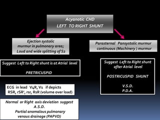
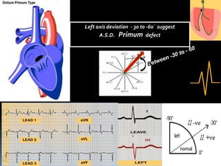
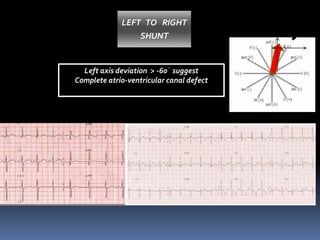
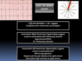
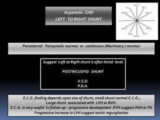
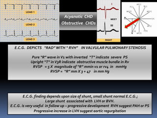
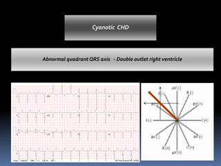
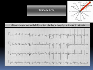
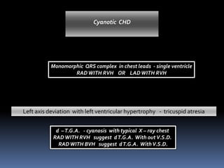
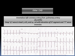
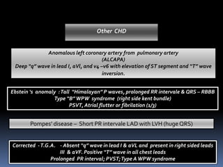
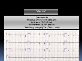
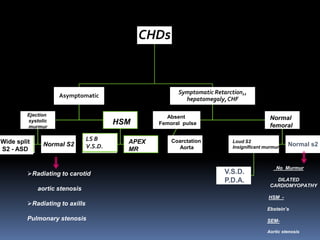
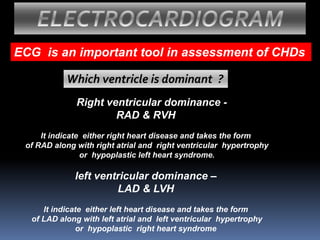
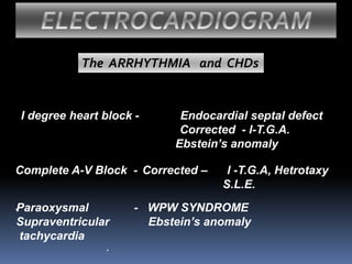
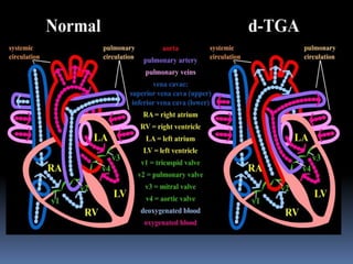
First step Suspicion of CHDs Nada’s criteria
2nd step SP O2 Acyanotic / cyanotic CHDs
3rd step
4th step
CVS examination
X ray Chest PA View
Fifth step
▪ ECG is a simple .and useful bedside
investigation which helps in the diagnosis and
management of patients with CHDs.
▪ In specific setting ECG can give specific
diagnostic informations.
E. C. G.
E. C. G.
▪ ECG reflects the underlying structural and
hemodynamic changes that occur in the
disease state. Hence a number of conditions
with similar profiles may have similar ECG
patterns.
At glance you may feel all E.C.G. look
alike. Look Closely and you will see the
differences!
How to diagnose CHDs
by ECG ?
Acyanotic CHD
LEFT TO RIGHT SHUNT
ECG in lead V4R,V1 if depicts
RSR, rSR’, rsr, RsR (volume over load)
Suggest Left to Right shunt is at Atrial level
PRETRICUSPID
Normal or Right axis deviation suggest
A.S.D.
Partial anomalous pulmonary
venous drainage (PAPVD)
Ejection systolic
murmur in pulmonary area;
Loud and wide splitting of S2
Parasternal Pansystolic murmur
continuous (Machinery ) murmur
Suggest Left to Right shunt
after Atrial level
POSTRICUSPID SHUNT
V.S.D.
P.D.A.
Acyanotic CHD
LEFT TO RIGHT SHUNT
ECG in lead V4R,V1 if depicts
RSR, rSR’, rsr, RsR (volume over load)
Suggest Left to Right shunt is at Atrial level
PRETRICUSPID
Normal or Right axis deviation suggest
A.S.D.
Partial anomalous pulmonary venous drainage (PAPVD)
Ejection systolic murmur in pulmonary area; Loud and wide splitting of S2
Left axis deviation - 30 t0 -60᷁ suggest
A.S.D. Primum defect
LEFT TO RIGHT
SHUNT
Left axis deviation > -60᷁ suggest
Complete atrio-ventricular canal defect
LEFT TO RIGHT
SHUNT
Left axis deviation > -60᷁ suggest
Complete atrio-ventricular canal defect
Associated RightVentricular Hypertrophy suggest
Lesion is associated with Pulmonary arterial
hypertension(PAH)
Or Pulmonary stenosis
Associated leftVentricular Hypertrophy suggest
Lesion is associated with
Mitral regurgitation
Rupture of sinus of valsalva into right atrium
Shunt from left ventricle to right atrium
Suggest Left to Right shunt is after Atrial level
POSTRICUSPID SHUNT
V.S.D.
P.D.A.
Parasternal Pansystolic murmur or continuous (Machinery ) murmur
E.C.G. finding depends upon size of shunt, small shunt normal E.C.G.;
Large shunt associated with LVH or BVH.
E.C.G. is very useful in follow up – progressive development RVH suggest PAH or PS
Progressive increase in LVH suggest aortic regurgitation
Acyanotic CHD
LEFT TO RIGHT SHUNT
E.C.G. DEPICTS “RAD”WITH “ RVH” IN VALVULAR PULMONARY STENOSIS
Pure “R” wave inV1 with inverted “T” indicate severe PS
Upright “T” inV3R indicate obstructive muscle bundle in Rv
RVSP = 5 X magnitude of “R” mmin v1 or rv4 in mmHg
RVSP = “R” mm X 3 + 47 in mm Hg
E.C.G. finding depends upon size of shunt, small shunt normal E.C.G.;
Large shunt associated with LVH or BVH.
E.C.G. is very useful in follow up – progressive development RVH suggest PAH or PS
Progressive increase in LVH suggest aortic regurgitation
Acyanotic CHD
Obstructive CHDs
One must decide nature CHDs
CHDs
Acyanotic CHD’s Cyanotic CHD’s
Abnormal quadrant QRS axis - Double outlet right ventricle
Cyanotic CHD
Left axis deviation with left ventricular hypertrophy - tricuspid atresia
Cyanotic CHD
Monomorphic QRS complex in chest leads - single ventricle
RADWITH RVH OR LADWITH RVH
Left axis deviation with left ventricular hypertrophy - tricuspid atresia
Cyanotic CHD
d –T.G.A. - cyanosis with typical X – ray chest
RADWITH RVH suggest dT.G.A. With outV.S.D.
RADWITH BVH suggest dT.G.A. WithV.S.D.
Ebstein ‘s anomaly :Tall “Himalayan” P waves, prolonged RR intervale & QRS – RBBB
Type “B”WPW syndrome (right side kent bundle)
PSVT, Atrial flutter or fibrilation (1/3)
Anomalous left coronary artery from pulmonary artery
(ALCAPA)
Deep “q” wave in lead I, aVl, and v4 –v6 with elevation of ST segment and “T” wave
inversion.
Pompes’ disease – Short PR intervale LAD with LVH (huge QRS)
Other CHD
Corrected -T.G.A. - Absent “q” wave in lead I & aVL and present in right sided leads
III & aVF. Positive “T” wave in all chest leads
Prolonged PR interval; PVST;Type AWPW syndrome
Dextro-cardia
Negative “P” wave in lead I & aVL
Positive “P” in lead aVR
“Q” wave inV4R,V6R and aVR
Diminishing voltage of QRS from V1 toV6
Other CHD
CHDs
Asymptomatic
Symptomatic Retarction,,
hepatomegaly,CHF
Absent
Femoral pulse
Coarctation
Aorta
Normal
femoral
Loud S2
Insignificant murmur
V.S.D.
P.D.A.
Normal s2
No Murmur
DILATED
CARDIOMYOPATHY
HSM -
Ebstein’s
SEM-
Aortic stenosis
Ejection
systolic
murmur
HSM
LS B
V.S.D.
APEX
MR
Wide split
S2 - ASD
Normal S2
➢Radiating to carotid
aortic stenosis
➢Radiating to axills
Pulmonary stenosis
Acyanotic CHD
LEFT TO RIGHT SHUNT
ECG in lead V4R,V1 if depicts
RSR, rSR’, rsr, RsR (volume over load)
Suggest Left to Right shunt is at Atrial level
PRETRICUSPID
Normal or Right axis deviation suggest
A.S.D.
Partial anomalous pulmonary
venous drainage (PAPVD)
Ejection systolic
murmur in pulmonary area;
Loud and wide splitting of S2
Parasternal Pansystolic murmur
continuous (Machinery ) murmur
Suggest Left to Right shunt
after Atrial level
POSTRICUSPID SHUNT
V.S.D.
P.D.A.
ECG is an important tool in assessment of CHDs
ELECTROCARDIOGRAM
Which ventricle is dominant ?
Right ventricular dominance -
RAD & RVH
It indicate either right heart disease and takes the form
of RAD along with right atrial and right ventricular hypertrophy
or hypoplastic left heart syndrome.
left ventricular dominance –
LAD & LVH
It indicate either left heart disease and takes the form
of LAD along with left atrial and left ventricular hypertrophy
or hypoplastic right heart syndrome
ELECTROCARDIOGRAM
I degree heart block - Endocardial septal defect
Corrected - l-T.G.A.
Ebstein’s anomaly
The ARRHYTHMIA and CHDs
Complete A-V Block - Corrected – l -T.G.A, Hetrotaxy
S.L.E.
.Paraoxysmal - WPW SYNDROME
Supraventricular Ebstein’s anomaly
tachycardia
.
ECG& CHDs.pdf
ECG& CHDs.pdf

ECG& CHDs.pdf

  • 4.
    First step Suspicionof CHDs Nada’s criteria 2nd step SP O2 Acyanotic / cyanotic CHDs 3rd step 4th step CVS examination X ray Chest PA View
  • 5.
  • 6.
    ▪ ECG isa simple .and useful bedside investigation which helps in the diagnosis and management of patients with CHDs. ▪ In specific setting ECG can give specific diagnostic informations. E. C. G.
  • 7.
    E. C. G. ▪ECG reflects the underlying structural and hemodynamic changes that occur in the disease state. Hence a number of conditions with similar profiles may have similar ECG patterns.
  • 8.
    At glance youmay feel all E.C.G. look alike. Look Closely and you will see the differences!
  • 9.
    How to diagnoseCHDs by ECG ?
  • 10.
    Acyanotic CHD LEFT TORIGHT SHUNT ECG in lead V4R,V1 if depicts RSR, rSR’, rsr, RsR (volume over load) Suggest Left to Right shunt is at Atrial level PRETRICUSPID Normal or Right axis deviation suggest A.S.D. Partial anomalous pulmonary venous drainage (PAPVD) Ejection systolic murmur in pulmonary area; Loud and wide splitting of S2 Parasternal Pansystolic murmur continuous (Machinery ) murmur Suggest Left to Right shunt after Atrial level POSTRICUSPID SHUNT V.S.D. P.D.A.
  • 11.
    Acyanotic CHD LEFT TORIGHT SHUNT ECG in lead V4R,V1 if depicts RSR, rSR’, rsr, RsR (volume over load) Suggest Left to Right shunt is at Atrial level PRETRICUSPID Normal or Right axis deviation suggest A.S.D. Partial anomalous pulmonary venous drainage (PAPVD) Ejection systolic murmur in pulmonary area; Loud and wide splitting of S2
  • 12.
    Left axis deviation- 30 t0 -60᷁ suggest A.S.D. Primum defect
  • 13.
    LEFT TO RIGHT SHUNT Leftaxis deviation > -60᷁ suggest Complete atrio-ventricular canal defect
  • 14.
    LEFT TO RIGHT SHUNT Leftaxis deviation > -60᷁ suggest Complete atrio-ventricular canal defect Associated RightVentricular Hypertrophy suggest Lesion is associated with Pulmonary arterial hypertension(PAH) Or Pulmonary stenosis Associated leftVentricular Hypertrophy suggest Lesion is associated with Mitral regurgitation Rupture of sinus of valsalva into right atrium Shunt from left ventricle to right atrium
  • 15.
    Suggest Left toRight shunt is after Atrial level POSTRICUSPID SHUNT V.S.D. P.D.A. Parasternal Pansystolic murmur or continuous (Machinery ) murmur E.C.G. finding depends upon size of shunt, small shunt normal E.C.G.; Large shunt associated with LVH or BVH. E.C.G. is very useful in follow up – progressive development RVH suggest PAH or PS Progressive increase in LVH suggest aortic regurgitation Acyanotic CHD LEFT TO RIGHT SHUNT
  • 16.
    E.C.G. DEPICTS “RAD”WITH“ RVH” IN VALVULAR PULMONARY STENOSIS Pure “R” wave inV1 with inverted “T” indicate severe PS Upright “T” inV3R indicate obstructive muscle bundle in Rv RVSP = 5 X magnitude of “R” mmin v1 or rv4 in mmHg RVSP = “R” mm X 3 + 47 in mm Hg E.C.G. finding depends upon size of shunt, small shunt normal E.C.G.; Large shunt associated with LVH or BVH. E.C.G. is very useful in follow up – progressive development RVH suggest PAH or PS Progressive increase in LVH suggest aortic regurgitation Acyanotic CHD Obstructive CHDs
  • 17.
    One must decidenature CHDs CHDs Acyanotic CHD’s Cyanotic CHD’s
  • 18.
    Abnormal quadrant QRSaxis - Double outlet right ventricle Cyanotic CHD
  • 19.
    Left axis deviationwith left ventricular hypertrophy - tricuspid atresia Cyanotic CHD
  • 20.
    Monomorphic QRS complexin chest leads - single ventricle RADWITH RVH OR LADWITH RVH Left axis deviation with left ventricular hypertrophy - tricuspid atresia Cyanotic CHD d –T.G.A. - cyanosis with typical X – ray chest RADWITH RVH suggest dT.G.A. With outV.S.D. RADWITH BVH suggest dT.G.A. WithV.S.D.
  • 21.
    Ebstein ‘s anomaly:Tall “Himalayan” P waves, prolonged RR intervale & QRS – RBBB Type “B”WPW syndrome (right side kent bundle) PSVT, Atrial flutter or fibrilation (1/3) Anomalous left coronary artery from pulmonary artery (ALCAPA) Deep “q” wave in lead I, aVl, and v4 –v6 with elevation of ST segment and “T” wave inversion. Pompes’ disease – Short PR intervale LAD with LVH (huge QRS) Other CHD Corrected -T.G.A. - Absent “q” wave in lead I & aVL and present in right sided leads III & aVF. Positive “T” wave in all chest leads Prolonged PR interval; PVST;Type AWPW syndrome
  • 22.
    Ebstein ‘s anomaly:Tall “Himalayan” P waves, prolonged RR intervale & QRS – RBBB Type “B”WPW syndrome (right side kent bundle) PSVT, Atrial flutter or fibrilation (1/3) Anomalous left coronary artery from pulmonary artery (ALCAPA) Deep “q” wave in lead I, aVl, and v4 –v6 with elevation of ST segment and “T” wave inversion. Pompes’ disease – Short PR intervale LAD with LVH (huge QRS) Other CHD Corrected -T.G.A. - Absent “q” wave in lead I & aVL and present in right sided leads III & aVF. Positive “T” wave in all chest leads Prolonged PR interval; PVST;Type AWPW syndrome
  • 23.
    Ebstein ‘s anomaly:Tall “Himalayan” P waves, prolonged RR intervale & QRS – RBBB Type “B”WPW syndrome (right side kent bundle) PSVT, Atrial flutter or fibrilation (1/3) Anomalous left coronary artery from pulmonary artery (ALCAPA) Deep “q” wave in lead I, aVl, and v4 –v6 with elevation of ST segment and “T” wave inversion. Pompes’ disease – Short PR intervale LAD with LVH (huge QRS) Other CHD Corrected -T.G.A. - Absent “q” wave in lead I & aVL and present in right sided leads III & aVF. Positive “T” wave in all chest leads Prolonged PR interval; PVST;Type AWPW syndrome
  • 24.
    Ebstein ‘s anomaly:Tall “Himalayan” P waves, prolonged RR intervale & QRS – RBBB Type “B”WPW syndrome (right side kent bundle) PSVT, Atrial flutter or fibrilation (1/3) Anomalous left coronary artery from pulmonary artery (ALCAPA) Deep “q” wave in lead I, aVl, and v4 –v6 with elevation of ST segment and “T” wave inversion. Pompes’ disease – Short PR intervale LAD with LVH (huge QRS) Other CHD Corrected -T.G.A. - Absent “q” wave in lead I & aVL and present in right sided leads III & aVF. Positive “T” wave in all chest leads Prolonged PR interval; PVST;Type AWPW syndrome
  • 25.
    Dextro-cardia Negative “P” wavein lead I & aVL Positive “P” in lead aVR “Q” wave inV4R,V6R and aVR Diminishing voltage of QRS from V1 toV6 Other CHD
  • 26.
    CHDs Asymptomatic Symptomatic Retarction,, hepatomegaly,CHF Absent Femoral pulse Coarctation Aorta Normal femoral LoudS2 Insignificant murmur V.S.D. P.D.A. Normal s2 No Murmur DILATED CARDIOMYOPATHY HSM - Ebstein’s SEM- Aortic stenosis Ejection systolic murmur HSM LS B V.S.D. APEX MR Wide split S2 - ASD Normal S2 ➢Radiating to carotid aortic stenosis ➢Radiating to axills Pulmonary stenosis
  • 27.
    Acyanotic CHD LEFT TORIGHT SHUNT ECG in lead V4R,V1 if depicts RSR, rSR’, rsr, RsR (volume over load) Suggest Left to Right shunt is at Atrial level PRETRICUSPID Normal or Right axis deviation suggest A.S.D. Partial anomalous pulmonary venous drainage (PAPVD) Ejection systolic murmur in pulmonary area; Loud and wide splitting of S2 Parasternal Pansystolic murmur continuous (Machinery ) murmur Suggest Left to Right shunt after Atrial level POSTRICUSPID SHUNT V.S.D. P.D.A.
  • 28.
    ECG is animportant tool in assessment of CHDs ELECTROCARDIOGRAM Which ventricle is dominant ? Right ventricular dominance - RAD & RVH It indicate either right heart disease and takes the form of RAD along with right atrial and right ventricular hypertrophy or hypoplastic left heart syndrome. left ventricular dominance – LAD & LVH It indicate either left heart disease and takes the form of LAD along with left atrial and left ventricular hypertrophy or hypoplastic right heart syndrome
  • 29.
    ELECTROCARDIOGRAM I degree heartblock - Endocardial septal defect Corrected - l-T.G.A. Ebstein’s anomaly The ARRHYTHMIA and CHDs Complete A-V Block - Corrected – l -T.G.A, Hetrotaxy S.L.E. .Paraoxysmal - WPW SYNDROME Supraventricular Ebstein’s anomaly tachycardia .