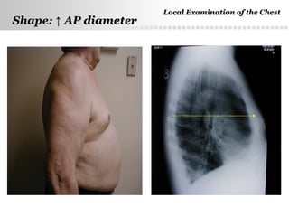
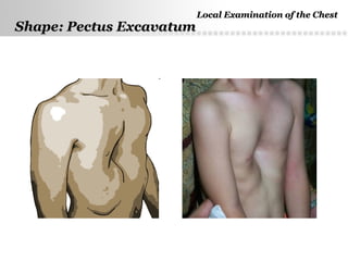
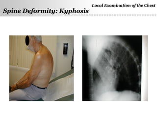
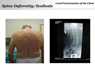
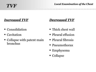
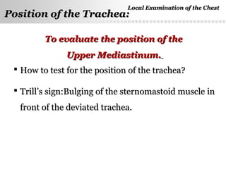
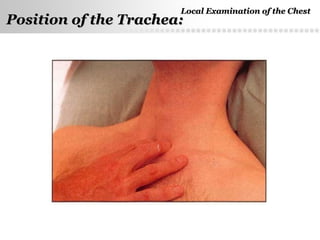
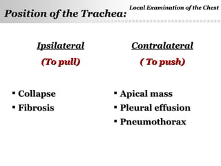
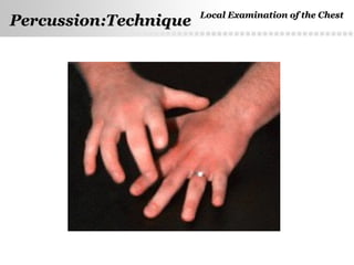
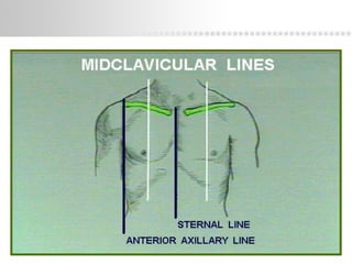
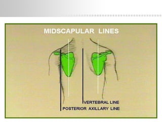
SlideShare a Scribd company logo
Local ExaminationLocal Examination
Of The ChestOf The Chest
Iman GalalIman Galal
Assistant Professor Pulmonary MedicineAssistant Professor Pulmonary Medicine
Ain Shams UniversityAin Shams University
E-mail: dr.imangalal@gmail.comE-mail: dr.imangalal@gmail.com
Page  2
InspectionInspection
PalpationPalpation
PercussionPercussion
AuscultationAuscultation
Local Examination of the Chest:Local Examination of the Chest:
Page  3
Local Examination of the ChestLocal Examination of the Chest
1- Shape of the chest & Spine Deformity1- Shape of the chest & Spine Deformity
2-MovementMovement
3-Symmetry3-Symmetry
4-Pulsations4-Pulsations
5-Respiratory movements5-Respiratory movements
6-Skin6-Skin
7-Subcostal angel7-Subcostal angel
8-Special signs8-Special signs
Inspection:Inspection:
Page  4
Normal ShapeNormal Shape
Barrel shaped chestBarrel shaped chest
Pigeon chestPigeon chest
Rachitic chestRachitic chest
Funnel-shaped chest (Pectus Excavatum)Funnel-shaped chest (Pectus Excavatum)
Shape of the Chest:Shape of the Chest:
Local Examination of the ChestLocal Examination of the Chest
Page  5
Shape:Shape: ↑↑ AP diameterAP diameter
Local Examination of the ChestLocal Examination of the Chest
Page  6
Shape: Pectus ExcavatumShape: Pectus Excavatum
Local Examination of the ChestLocal Examination of the Chest
Page  7
Spine Deformity: KyphosisSpine Deformity: Kyphosis
Local Examination of the ChestLocal Examination of the Chest
Page  8
Spine Deformity: ScoliosisSpine Deformity: Scoliosis
Local Examination of the ChestLocal Examination of the Chest
Page  9
Both sides of normal chest areBoth sides of normal chest are
symmetrical in shape and mobility.symmetrical in shape and mobility.
The diseased side or part is less mobileThe diseased side or part is less mobile
than the healthy one.than the healthy one.
Movement & Symmetry :
Local Examination of the ChestLocal Examination of the Chest
Page  10
Shape: Pigeon ChestShape: Pigeon Chest
Local Examination of the ChestLocal Examination of the Chest
Page  11
Significance ofSignificance of ↓↓ respiratory movements:respiratory movements:
 UnilateralUnilateral ↓↓ of chest wall movements:of chest wall movements:
•Pleural effusionPleural effusion
•EmpyemaEmpyema
•PneumothoraxPneumothorax
•Pulmonary consolidationPulmonary consolidation
•Pulmonary collapsePulmonary collapse
•Pleural or parenchymatous pulmonary fibrosisPleural or parenchymatous pulmonary fibrosis
 BilateralBilateral ↓↓ of chest wall movements:of chest wall movements:
•Bronchial asthmaBronchial asthma
•EmphysemaEmphysema
•Diffuse pulmonary fibrosisDiffuse pulmonary fibrosis
Local Examination of the ChestLocal Examination of the Chest
Page  12
BulgingBulging RetractionRetraction
 Pleural effusionPleural effusion
 PneumothoraxPneumothorax
 HydropneumothoraxHydropneumothorax
 EmpyemaEmpyema
 Precordial bulgePrecordial bulge
 Chest wall causesChest wall causes
 Pulmonary collapsePulmonary collapse
 Pulm. FibrosisPulm. Fibrosis
 Pleural fibrosisPleural fibrosis
Local Examination of the ChestLocal Examination of the Chest
Movement & Symmetry :
Page  13
 Respiratory rate
 Type of respiration
 Regularity of respiration
 Pulse Rate: Respiratory Rate Ratio
Respiratory Movements:Respiratory Movements:
Local Examination of the ChestLocal Examination of the Chest
Page  14
Skin eruptionSkin eruption e.g HZe.g HZ
NodulesNodules (inflammatory,metastatic,lipoma, neurofibroma…)(inflammatory,metastatic,lipoma, neurofibroma…)
Subcutaneous emphysemaSubcutaneous emphysema
Purpuric spots,Vascular spiders, BruisesPurpuric spots,Vascular spiders, Bruises
Dilated prominent blood vesselsDilated prominent blood vessels (SVC obstruction)(SVC obstruction)
ScarsScars (previous operation,trauma, intercostal tube…)(previous operation,trauma, intercostal tube…)
Discharging sinusesDischarging sinuses
Lesions of the breastsLesions of the breasts
Skin:Skin:
Local Examination of the ChestLocal Examination of the Chest
Page  15
Skin:Skin: SVC ObstructionSVC Obstruction
Local Examination of the ChestLocal Examination of the Chest
Page  16
 ApicalApical
 ParasternalParasternal
 EpigastricEpigastric
Pulsations:Pulsations:
Local Examination of the ChestLocal Examination of the Chest
Page  17
Palpation:Palpation:
 To confirm Respiratory Movements +/- ExpansionTo confirm Respiratory Movements +/- Expansion
 Pulsations (see before)Pulsations (see before)
 Palpable Adventitious SoundsPalpable Adventitious Sounds
 TTactileactile VVocalocal FFremitus (TVF)remitus (TVF)
 Position of the TracheaPosition of the Trachea
Local Examination of the ChestLocal Examination of the Chest
Page  18
Palpation of Respiratory MovementsPalpation of Respiratory Movements
1.1. Respiratory movements in the infraclavicular regionsRespiratory movements in the infraclavicular regions
2.2. Respiratory movements at the costal marginsRespiratory movements at the costal margins
3.3. Respiratory movements of the lower ribs posteriorlyRespiratory movements of the lower ribs posteriorly
Local Examination of the ChestLocal Examination of the Chest
Page  19
Symmetry & Movement:
Local Examination of the ChestLocal Examination of the Chest
Page  20
Palpation: Chest ExcursionPalpation: Chest Excursion
Local Examination of the ChestLocal Examination of the Chest
Page  21
TVFTVF
Increased TVFIncreased TVF Decreased TVFDecreased TVF
 Consolidation
 Cavitation
 Collapse with patent main
bronchus
 Thick chest wall
 Pleural effusion
 Pleural fibrosis
 Pneumothorax
 Emphysema
 Collapse
Local Examination of the ChestLocal Examination of the Chest
Page  22
Palpable Adventitious SoundsPalpable Adventitious Sounds
Palpable RhonchiPalpable Rhonchi
•Diffuse
•Localized and Persistent
Palpable Pleural Rub
Local Examination of the ChestLocal Examination of the Chest
Page  23
Position of the Trachea:Position of the Trachea:
 How to test for the position of the trachea?How to test for the position of the trachea?
 Trill’s sign:Bulging of the sternomastoid muscle inTrill’s sign:Bulging of the sternomastoid muscle in
front of the deviated trachea.front of the deviated trachea.
To evaluate the position of theTo evaluate the position of the
Upper Mediastinum.Upper Mediastinum.
Local Examination of the ChestLocal Examination of the Chest
Page  24
Position of the Trachea:Position of the Trachea:
Local Examination of the ChestLocal Examination of the Chest
Page  25
Causes of deviation of the trachea
IpsilateralIpsilateral
(To pull)(To pull)
ContralateralContralateral
( To push)( To push)
 CollapseCollapse
 FibrosisFibrosis
 Apical massApical mass
 Pleural effusionPleural effusion
 PneumothoraxPneumothorax
Local Examination of the ChestLocal Examination of the Chest
Position of the Trachea:Position of the Trachea:
Page  26
Percussion:TechniquePercussion:Technique Local Examination of the ChestLocal Examination of the Chest
Page  27
Page  28
Page  29
Page  30
Percussion
Cut your nails
Page  31
Percussion: Anterior ChestPercussion: Anterior Chest
1.1. Percuss from side to side andPercuss from side to side and
top to bottom using thetop to bottom using the
pattern shown in thepattern shown in the
illustration.illustration.
2.2. Compare one side to the otherCompare one side to the other
looking for asymmetry.looking for asymmetry.
3.3. Note the location and qualityNote the location and quality
of the percussion sounds youof the percussion sounds you
hear.hear.
Page  32
Percussion: Posterior ChestPercussion: Posterior Chest
1.1. Percuss from side to side and top toPercuss from side to side and top to
bottom using this pattern. Omit thebottom using this pattern. Omit the
areas covered by the scapulae.areas covered by the scapulae.
2.2. Compare one side to the otherCompare one side to the other
looking for asymmetry.looking for asymmetry.
3.3. Note the location and quality of theNote the location and quality of the
percussion sounds you hear.percussion sounds you hear.
4.4. Find the level of the diaphragmaticFind the level of the diaphragmatic
dullness on both sides.dullness on both sides.
Page  33
Traube’s area:Traube’s area:
4 points:4 points:
Left 6Left 6thth
rib in the MCL to 8rib in the MCL to 8thth
costal cartilage in thecostal cartilage in the
parasternal line ,then along the left costal margin to the 11parasternal line ,then along the left costal margin to the 11thth
rib in the MAL, then the 9rib in the MAL, then the 9thth
rib in the MAL.rib in the MAL.
Page  34
Kronig’s Isthmus :Kronig’s Isthmus :
 It is a band of resonance representing lung apex.It is a band of resonance representing lung apex.
 LaterallyLaterally it is marked by a line joining 2 points:it is marked by a line joining 2 points:
1.1. The junction of the medial 2/3 of the clavicle with theThe junction of the medial 2/3 of the clavicle with the
lateral 1/3.lateral 1/3.
2.2. The junction of the medial 1/3 of the scapular spine withThe junction of the medial 1/3 of the scapular spine with
the lateral 2/3.the lateral 2/3.
 MediallyMedially marked by a line between the sternal end ofmarked by a line between the sternal end of
clavicle and the 7clavicle and the 7thth
cervical spine.cervical spine.
Page  35
Bare area of the heart :Bare area of the heart :
 Medial border: left lateral border of the sternumMedial border: left lateral border of the sternum
 Lateral border: left parasternal lineLateral border: left parasternal line
 Superior border: lower border of Lt 4Superior border: lower border of Lt 4thth
rib.rib.
 Inferior border: upper border of Lt 6Inferior border: upper border of Lt 6thth
ribrib
Page  36
Surface anatomy of liver:Surface anatomy of liver:
Upper border:Upper border:
It starts from the left 6It starts from the left 6thth
ribrib just inside the MCL, passing tojust inside the MCL, passing to
the Rt and slightly upwards to the 5the Rt and slightly upwards to the 5thth
rib in the MCL, thenrib in the MCL, then
the 7the 7thth
rib in anterior axillary line, to the 9rib in anterior axillary line, to the 9thth
rib in mid-axillaryrib in mid-axillary
line.line.
Page  37
1.1. Find the level of the diaphragmatic dullness on both sides.Find the level of the diaphragmatic dullness on both sides.
2.2. Ask the patient to inspire deeply.Ask the patient to inspire deeply.
3.3. The level of dullness (diaphragmatic excursion) should goThe level of dullness (diaphragmatic excursion) should go
down 3-5cmdown 3-5cm symmetricallysymmetrically..
4.4. Decreased or asymmetric diaphragmatic excursion mayDecreased or asymmetric diaphragmatic excursion may
indicate paralysis or emphysema.indicate paralysis or emphysema.
Diaphragmatic ExcursionDiaphragmatic Excursion
Local Examination of the ChestLocal Examination of the Chest
Page  38
1. It is used to differentiate supra-diaphragmatic from infra-
diaphragmatic dullness.
2. While the patient seated find the upper level of dullness
3. Ask the patient to take deep inspiration and to hold it then
percuss again.
4. If the note becomes resonant  infra-diaphragmatic cause.
5. If there is no change of the note  supra-diaphragmatic
cause as pleural effusion.
Tidal percussion:Tidal percussion: Local Examination of the ChestLocal Examination of the Chest
Page  39
Auscultation:Auscultation:
 Intensity of breath soundsIntensity of breath sounds
 Type of breath soundsType of breath sounds
 Adventitious soundsAdventitious sounds
 Voice sounds (vocal resonance)Voice sounds (vocal resonance)
Local Examination of the ChestLocal Examination of the Chest
Page  40
Technique of Auscultation
• While the patient relaxed and breathes normally with mouthWhile the patient relaxed and breathes normally with mouth
open, auscultate the lungs, making sure to auscultate theopen, auscultate the lungs, making sure to auscultate the
apices and middle and lower lung fields posteriorly, laterallyapices and middle and lower lung fields posteriorly, laterally
and anteriorly.and anteriorly.
• Alternate and compare both sides at each site.Alternate and compare both sides at each site.
• Listen to at least one complete respiratory cycle at each site.Listen to at least one complete respiratory cycle at each site.
• First listen with quiet respiration. If breath sounds areFirst listen with quiet respiration. If breath sounds are
inaudible, then have him take deep breaths.inaudible, then have him take deep breaths.
• First describe the breath sounds and then the adventitiousFirst describe the breath sounds and then the adventitious
sounds.sounds.
Local Examination of the ChestLocal Examination of the Chest
Page  41
Technique of AuscultationTechnique of Auscultation
• Note the intensity of breath sounds and make a comparisonNote the intensity of breath sounds and make a comparison
with the opposite side.with the opposite side.
• Assess length of inspiration and expiration. Listen for a pauseAssess length of inspiration and expiration. Listen for a pause
between inspiration, expiration and the quality of pitch of thebetween inspiration, expiration and the quality of pitch of the
soundsound
• Also compare the intensity of breath sounds between upperAlso compare the intensity of breath sounds between upper
and lower chest in upright position. Compare the intensity ofand lower chest in upright position. Compare the intensity of
breath sounds from dependent to top lung in the decubitusbreath sounds from dependent to top lung in the decubitus
position.position.
• Note the presence or absence of adventitious sounds.Note the presence or absence of adventitious sounds.
Local Examination of the ChestLocal Examination of the Chest
Page  42
Bronchial breathing may be heard in:Bronchial breathing may be heard in:
 ConsolidationConsolidation
 Collapse with patent large airwaysCollapse with patent large airways
 Compressed lung by a large pl effusion or a tensionCompressed lung by a large pl effusion or a tension
pneumothoraxpneumothorax
 Pulmonary fibrosisPulmonary fibrosis
 CavitationCavitation
Local Examination of the ChestLocal Examination of the Chest
Auscultation:Auscultation: Bronchial BreathingBronchial Breathing
Page  43
Auscultation:Adventitious soundsAuscultation:Adventitious sounds
 Crepitations: typesCrepitations: types
 Rhonchi: sibilant and sonorousRhonchi: sibilant and sonorous
 Pleural rubPleural rub
Local Examination of the ChestLocal Examination of the Chest
Page  44
Auscultation: Voice soundsAuscultation: Voice sounds
Voice Transmission Tests: are only used in special
situations. All these tests become abnormal in
consolidation. They include:
Bronchophony
Whispered Pectoriloquy
Egophony
Local Examination of the ChestLocal Examination of the Chest
Page  45
Auscultation: Voice sounds- BronchophonyAuscultation: Voice sounds- Bronchophony
1. Ask the patient to say "ninety-nine“ or 44 in arabic several
times in a normal voice.
2. Auscultate several symmetrical areas over each lung.
3. The sounds you hear should be muffled and indistinct.
Louder, clearer sounds are called bronchophony.bronchophony.
Local Examination of the ChestLocal Examination of the Chest
Page  46
Voice sounds: Whispered PectoriloquyVoice sounds: Whispered Pectoriloquy
1. Ask the patient to whisper "ninety-nine“ or 44 in arabic
several times.
2. Auscultate several symmetrical areas over each lung.
3. You should hear only faint sounds or nothing at all. If you
hear the sounds clearly this is referred to as whisperedwhispered
pectoriloquy.pectoriloquy.
Local Examination of the ChestLocal Examination of the Chest
Page  47
Voice sounds: EgophonyVoice sounds: Egophony
1.1. Ask the patient to say "ee" continuously.Ask the patient to say "ee" continuously.
2.2. Auscultate several symmetrical areas over each lung.Auscultate several symmetrical areas over each lung.
3.3. You should hear a muffled "ee" sound. If you hear an "ay"You should hear a muffled "ee" sound. If you hear an "ay"
sound this is referred to assound this is referred to as "E -> A" or"E -> A" or egophony.egophony.
Local Examination of the ChestLocal Examination of the Chest
Page  48

More Related Content

What's hot

Examination of gastrointestinal system by HX
Examination of gastrointestinal system by HXExamination of gastrointestinal system by HX
Examination of gastrointestinal system by HXDr. Rubz
 
Percussion in respiratory system
Percussion in respiratory systemPercussion in respiratory system
Percussion in respiratory systemKurian Joseph
 
Local chest examination
Local chest examinationLocal chest examination
Local chest examination
abeerabdulkareem
 
Examination of motor system
Examination of motor systemExamination of motor system
Examination of motor system8224080546
 
Dysphagia
DysphagiaDysphagia
Dysphagia
Kundan Singh
 
Auscultation
AuscultationAuscultation
Auscultation
Dr.Priyanka Das
 
Exam Of Abdomen.
Exam Of Abdomen.Exam Of Abdomen.
Exam Of Abdomen.
Shaikhani.
 
Cyanosis ppt by dr girish jain
Cyanosis ppt by dr girish jainCyanosis ppt by dr girish jain
Cyanosis ppt by dr girish jain
Girish jain
 
Clubbing
ClubbingClubbing
Systemic examination of respiratory system
Systemic examination of respiratory systemSystemic examination of respiratory system
Systemic examination of respiratory system
alok thakur
 
Cranial nerve examination
Cranial nerve examinationCranial nerve examination
Cranial nerve examinationGagan Gupta
 
Abdominal examination
Abdominal examinationAbdominal examination
Abdominal examination
shashank agrawal
 
Cardiovascular system examination
Cardiovascular system examinationCardiovascular system examination
Cardiovascular system examination
Ashish Dhandare
 
Part 5 examination of reflex
Part 5 examination of reflexPart 5 examination of reflex
Part 5 examination of reflex
Atul Saswat
 
General examination
General examinationGeneral examination
General examination
Chiranjeevi JIPMER Puducherry
 

What's hot (20)

Respiratory Exam
Respiratory ExamRespiratory Exam
Respiratory Exam
 
Examination of gastrointestinal system by HX
Examination of gastrointestinal system by HXExamination of gastrointestinal system by HX
Examination of gastrointestinal system by HX
 
Percussion in respiratory system
Percussion in respiratory systemPercussion in respiratory system
Percussion in respiratory system
 
Musculoskeletal Exam
Musculoskeletal ExamMusculoskeletal Exam
Musculoskeletal Exam
 
Local chest examination
Local chest examinationLocal chest examination
Local chest examination
 
Examination of motor system
Examination of motor systemExamination of motor system
Examination of motor system
 
Abdominal Exam
Abdominal ExamAbdominal Exam
Abdominal Exam
 
Dysphagia
DysphagiaDysphagia
Dysphagia
 
Jerrin's
Jerrin'sJerrin's
Jerrin's
 
Auscultation
AuscultationAuscultation
Auscultation
 
Exam Of Abdomen.
Exam Of Abdomen.Exam Of Abdomen.
Exam Of Abdomen.
 
Cyanosis ppt by dr girish jain
Cyanosis ppt by dr girish jainCyanosis ppt by dr girish jain
Cyanosis ppt by dr girish jain
 
Clubbing
ClubbingClubbing
Clubbing
 
Systemic examination of respiratory system
Systemic examination of respiratory systemSystemic examination of respiratory system
Systemic examination of respiratory system
 
Cranial nerve examination
Cranial nerve examinationCranial nerve examination
Cranial nerve examination
 
Abdominal examination
Abdominal examinationAbdominal examination
Abdominal examination
 
Sensory Examination
Sensory ExaminationSensory Examination
Sensory Examination
 
Cardiovascular system examination
Cardiovascular system examinationCardiovascular system examination
Cardiovascular system examination
 
Part 5 examination of reflex
Part 5 examination of reflexPart 5 examination of reflex
Part 5 examination of reflex
 
General examination
General examinationGeneral examination
General examination
 

Similar to Local chest examination

Chest trauma PTT
Chest trauma PTTChest trauma PTT
Chest trauma PTT
Ansam Azzam
 
Chest examination
Chest examination Chest examination
Chest examination
Allen Rojer
 
Abdominal anatomy
Abdominal anatomyAbdominal anatomy
Abdominal anatomyJSlinkyNY
 
Basics Of CXR interpretation www.radiologydefinition.com
Basics Of CXR interpretation   www.radiologydefinition.comBasics Of CXR interpretation   www.radiologydefinition.com
Basics Of CXR interpretation www.radiologydefinition.com
Radiology Definition
 
Anatomy of lung & pleura
Anatomy of lung & pleuraAnatomy of lung & pleura
Anatomy of lung & pleuraimangalal
 
Viewing of the chest film
Viewing of the chest filmViewing of the chest film
Viewing of the chest film
Tarequl Shawrab
 
X ray chest
X ray chest X ray chest
X ray chest
yasseryasserahmed
 
5 Chest and lung.pdf
5 Chest and lung.pdf5 Chest and lung.pdf
5 Chest and lung.pdf
Sani191640
 
Chest x ray dasic approach 2015 - dr magdi sasi
Chest  x ray  dasic approach 2015 - dr magdi sasiChest  x ray  dasic approach 2015 - dr magdi sasi
Chest x ray dasic approach 2015 - dr magdi sasi
cardilogy
 
chest trauma management
 chest trauma management chest trauma management
chest trauma management
Sumer Yadav
 
Mediastinal tumors
Mediastinal tumorsMediastinal tumors
Mediastinal tumors
Isha Jaiswal
 
6-Radiological Anatomy Of The Chest..pdf
6-Radiological Anatomy Of The Chest..pdf6-Radiological Anatomy Of The Chest..pdf
6-Radiological Anatomy Of The Chest..pdf
hdhdufyfuei78
 
Anatomy of lung ppt
Anatomy of lung pptAnatomy of lung ppt
Anatomy of lung ppt
Shubham Rathod
 
lungs pdf.pdf
lungs pdf.pdflungs pdf.pdf
lungs pdf.pdf
Pankaj Bhagat
 
Neurosonogram.. Dr.Padmesh
Neurosonogram.. Dr.PadmeshNeurosonogram.. Dr.Padmesh
Neurosonogram.. Dr.Padmesh
Dr Padmesh Vadakepat
 
Wayang kulit, no 1 a fundamentals
Wayang kulit, no 1 a fundamentalsWayang kulit, no 1 a fundamentals
Wayang kulit, no 1 a fundamentals
ycche19
 
Chest trauma presentation
Chest trauma  presentationChest trauma  presentation
Chest trauma presentation
OM VERMA
 
CXR Simple by DrRaghu Ram
CXR Simple by DrRaghu RamCXR Simple by DrRaghu Ram
CXR Simple by DrRaghu Ram
Punnami Chandra
 
Examination of Respiratory system
Examination of Respiratory systemExamination of Respiratory system
Examination of Respiratory system
AltynaiTabyldyeva1
 

Similar to Local chest examination (20)

Chest trauma PTT
Chest trauma PTTChest trauma PTT
Chest trauma PTT
 
Chest examination
Chest examination Chest examination
Chest examination
 
Abdominal anatomy
Abdominal anatomyAbdominal anatomy
Abdominal anatomy
 
Basics Of CXR interpretation www.radiologydefinition.com
Basics Of CXR interpretation   www.radiologydefinition.comBasics Of CXR interpretation   www.radiologydefinition.com
Basics Of CXR interpretation www.radiologydefinition.com
 
Anatomy of lung & pleura
Anatomy of lung & pleuraAnatomy of lung & pleura
Anatomy of lung & pleura
 
Viewing of the chest film
Viewing of the chest filmViewing of the chest film
Viewing of the chest film
 
X ray chest
X ray chest X ray chest
X ray chest
 
5 Chest and lung.pdf
5 Chest and lung.pdf5 Chest and lung.pdf
5 Chest and lung.pdf
 
Chest x ray dasic approach 2015 - dr magdi sasi
Chest  x ray  dasic approach 2015 - dr magdi sasiChest  x ray  dasic approach 2015 - dr magdi sasi
Chest x ray dasic approach 2015 - dr magdi sasi
 
Chest examination
Chest examinationChest examination
Chest examination
 
chest trauma management
 chest trauma management chest trauma management
chest trauma management
 
Mediastinal tumors
Mediastinal tumorsMediastinal tumors
Mediastinal tumors
 
6-Radiological Anatomy Of The Chest..pdf
6-Radiological Anatomy Of The Chest..pdf6-Radiological Anatomy Of The Chest..pdf
6-Radiological Anatomy Of The Chest..pdf
 
Anatomy of lung ppt
Anatomy of lung pptAnatomy of lung ppt
Anatomy of lung ppt
 
lungs pdf.pdf
lungs pdf.pdflungs pdf.pdf
lungs pdf.pdf
 
Neurosonogram.. Dr.Padmesh
Neurosonogram.. Dr.PadmeshNeurosonogram.. Dr.Padmesh
Neurosonogram.. Dr.Padmesh
 
Wayang kulit, no 1 a fundamentals
Wayang kulit, no 1 a fundamentalsWayang kulit, no 1 a fundamentals
Wayang kulit, no 1 a fundamentals
 
Chest trauma presentation
Chest trauma  presentationChest trauma  presentation
Chest trauma presentation
 
CXR Simple by DrRaghu Ram
CXR Simple by DrRaghu RamCXR Simple by DrRaghu Ram
CXR Simple by DrRaghu Ram
 
Examination of Respiratory system
Examination of Respiratory systemExamination of Respiratory system
Examination of Respiratory system
 

More from imangalal

Lung & pregnancy
Lung & pregnancyLung & pregnancy
Lung & pregnancyimangalal
 
OSA is a systemic disease
OSA is a systemic diseaseOSA is a systemic disease
OSA is a systemic diseaseimangalal
 
Chronic obstructive pulmonary disease
Chronic obstructive pulmonary diseaseChronic obstructive pulmonary disease
Chronic obstructive pulmonary diseaseimangalal
 
Basic life support
Basic life supportBasic life support
Basic life supportimangalal
 
Sarcoidosis & orphan lung disease
Sarcoidosis & orphan lung diseaseSarcoidosis & orphan lung disease
Sarcoidosis & orphan lung diseaseimangalal
 
Behcet’s disease
Behcet’s diseaseBehcet’s disease
Behcet’s diseaseimangalal
 
Chest history taking
Chest history takingChest history taking
Chest history takingimangalal
 
Celiac disease
Celiac diseaseCeliac disease
Celiac diseaseimangalal
 

More from imangalal (9)

Lung & pregnancy
Lung & pregnancyLung & pregnancy
Lung & pregnancy
 
Hemoptysis
HemoptysisHemoptysis
Hemoptysis
 
OSA is a systemic disease
OSA is a systemic diseaseOSA is a systemic disease
OSA is a systemic disease
 
Chronic obstructive pulmonary disease
Chronic obstructive pulmonary diseaseChronic obstructive pulmonary disease
Chronic obstructive pulmonary disease
 
Basic life support
Basic life supportBasic life support
Basic life support
 
Sarcoidosis & orphan lung disease
Sarcoidosis & orphan lung diseaseSarcoidosis & orphan lung disease
Sarcoidosis & orphan lung disease
 
Behcet’s disease
Behcet’s diseaseBehcet’s disease
Behcet’s disease
 
Chest history taking
Chest history takingChest history taking
Chest history taking
 
Celiac disease
Celiac diseaseCeliac disease
Celiac disease
 

Recently uploaded

GBSN - Biochemistry (Unit 5) Chemistry of Lipids
GBSN - Biochemistry (Unit 5) Chemistry of LipidsGBSN - Biochemistry (Unit 5) Chemistry of Lipids
GBSN - Biochemistry (Unit 5) Chemistry of Lipids
Areesha Ahmad
 
Orion Air Quality Monitoring Systems - CWS
Orion Air Quality Monitoring Systems - CWSOrion Air Quality Monitoring Systems - CWS
Orion Air Quality Monitoring Systems - CWS
Columbia Weather Systems
 
Predicting property prices with machine learning algorithms.pdf
Predicting property prices with machine learning algorithms.pdfPredicting property prices with machine learning algorithms.pdf
Predicting property prices with machine learning algorithms.pdf
binhminhvu04
 
Structures and textures of metamorphic rocks
Structures and textures of metamorphic rocksStructures and textures of metamorphic rocks
Structures and textures of metamorphic rocks
kumarmathi863
 
Body fluids_tonicity_dehydration_hypovolemia_hypervolemia.pptx
Body fluids_tonicity_dehydration_hypovolemia_hypervolemia.pptxBody fluids_tonicity_dehydration_hypovolemia_hypervolemia.pptx
Body fluids_tonicity_dehydration_hypovolemia_hypervolemia.pptx
muralinath2
 
insect morphology and physiology of insect
insect morphology and physiology of insectinsect morphology and physiology of insect
insect morphology and physiology of insect
anitaento25
 
Large scale production of streptomycin.pptx
Large scale production of streptomycin.pptxLarge scale production of streptomycin.pptx
Large scale production of streptomycin.pptx
Cherry
 
erythropoiesis-I_mechanism& clinical significance.pptx
erythropoiesis-I_mechanism& clinical significance.pptxerythropoiesis-I_mechanism& clinical significance.pptx
erythropoiesis-I_mechanism& clinical significance.pptx
muralinath2
 
platelets_clotting_biogenesis.clot retractionpptx
platelets_clotting_biogenesis.clot retractionpptxplatelets_clotting_biogenesis.clot retractionpptx
platelets_clotting_biogenesis.clot retractionpptx
muralinath2
 
RNA INTERFERENCE: UNRAVELING GENETIC SILENCING
RNA INTERFERENCE: UNRAVELING GENETIC SILENCINGRNA INTERFERENCE: UNRAVELING GENETIC SILENCING
RNA INTERFERENCE: UNRAVELING GENETIC SILENCING
AADYARAJPANDEY1
 
Observation of Io’s Resurfacing via Plume Deposition Using Ground-based Adapt...
Observation of Io’s Resurfacing via Plume Deposition Using Ground-based Adapt...Observation of Io’s Resurfacing via Plume Deposition Using Ground-based Adapt...
Observation of Io’s Resurfacing via Plume Deposition Using Ground-based Adapt...
Sérgio Sacani
 
Structural Classification Of Protein (SCOP)
Structural Classification Of Protein  (SCOP)Structural Classification Of Protein  (SCOP)
Structural Classification Of Protein (SCOP)
aishnasrivastava
 
Comparative structure of adrenal gland in vertebrates
Comparative structure of adrenal gland in vertebratesComparative structure of adrenal gland in vertebrates
Comparative structure of adrenal gland in vertebrates
sachin783648
 
Circulatory system_ Laplace law. Ohms law.reynaults law,baro-chemo-receptors-...
Circulatory system_ Laplace law. Ohms law.reynaults law,baro-chemo-receptors-...Circulatory system_ Laplace law. Ohms law.reynaults law,baro-chemo-receptors-...
Circulatory system_ Laplace law. Ohms law.reynaults law,baro-chemo-receptors-...
muralinath2
 
platelets- lifespan -Clot retraction-disorders.pptx
platelets- lifespan -Clot retraction-disorders.pptxplatelets- lifespan -Clot retraction-disorders.pptx
platelets- lifespan -Clot retraction-disorders.pptx
muralinath2
 
Richard's entangled aventures in wonderland
Richard's entangled aventures in wonderlandRichard's entangled aventures in wonderland
Richard's entangled aventures in wonderland
Richard Gill
 
SCHIZOPHRENIA Disorder/ Brain Disorder.pdf
SCHIZOPHRENIA Disorder/ Brain Disorder.pdfSCHIZOPHRENIA Disorder/ Brain Disorder.pdf
SCHIZOPHRENIA Disorder/ Brain Disorder.pdf
SELF-EXPLANATORY
 
Mammalian Pineal Body Structure and Also Functions
Mammalian Pineal Body Structure and Also FunctionsMammalian Pineal Body Structure and Also Functions
Mammalian Pineal Body Structure and Also Functions
YOGESH DOGRA
 
filosofia boliviana introducción jsjdjd.pptx
filosofia boliviana introducción jsjdjd.pptxfilosofia boliviana introducción jsjdjd.pptx
filosofia boliviana introducción jsjdjd.pptx
IvanMallco1
 
FAIR & AI Ready KGs for Explainable Predictions
FAIR & AI Ready KGs for Explainable PredictionsFAIR & AI Ready KGs for Explainable Predictions
FAIR & AI Ready KGs for Explainable Predictions
Michel Dumontier
 

Recently uploaded (20)

GBSN - Biochemistry (Unit 5) Chemistry of Lipids
GBSN - Biochemistry (Unit 5) Chemistry of LipidsGBSN - Biochemistry (Unit 5) Chemistry of Lipids
GBSN - Biochemistry (Unit 5) Chemistry of Lipids
 
Orion Air Quality Monitoring Systems - CWS
Orion Air Quality Monitoring Systems - CWSOrion Air Quality Monitoring Systems - CWS
Orion Air Quality Monitoring Systems - CWS
 
Predicting property prices with machine learning algorithms.pdf
Predicting property prices with machine learning algorithms.pdfPredicting property prices with machine learning algorithms.pdf
Predicting property prices with machine learning algorithms.pdf
 
Structures and textures of metamorphic rocks
Structures and textures of metamorphic rocksStructures and textures of metamorphic rocks
Structures and textures of metamorphic rocks
 
Body fluids_tonicity_dehydration_hypovolemia_hypervolemia.pptx
Body fluids_tonicity_dehydration_hypovolemia_hypervolemia.pptxBody fluids_tonicity_dehydration_hypovolemia_hypervolemia.pptx
Body fluids_tonicity_dehydration_hypovolemia_hypervolemia.pptx
 
insect morphology and physiology of insect
insect morphology and physiology of insectinsect morphology and physiology of insect
insect morphology and physiology of insect
 
Large scale production of streptomycin.pptx
Large scale production of streptomycin.pptxLarge scale production of streptomycin.pptx
Large scale production of streptomycin.pptx
 
erythropoiesis-I_mechanism& clinical significance.pptx
erythropoiesis-I_mechanism& clinical significance.pptxerythropoiesis-I_mechanism& clinical significance.pptx
erythropoiesis-I_mechanism& clinical significance.pptx
 
platelets_clotting_biogenesis.clot retractionpptx
platelets_clotting_biogenesis.clot retractionpptxplatelets_clotting_biogenesis.clot retractionpptx
platelets_clotting_biogenesis.clot retractionpptx
 
RNA INTERFERENCE: UNRAVELING GENETIC SILENCING
RNA INTERFERENCE: UNRAVELING GENETIC SILENCINGRNA INTERFERENCE: UNRAVELING GENETIC SILENCING
RNA INTERFERENCE: UNRAVELING GENETIC SILENCING
 
Observation of Io’s Resurfacing via Plume Deposition Using Ground-based Adapt...
Observation of Io’s Resurfacing via Plume Deposition Using Ground-based Adapt...Observation of Io’s Resurfacing via Plume Deposition Using Ground-based Adapt...
Observation of Io’s Resurfacing via Plume Deposition Using Ground-based Adapt...
 
Structural Classification Of Protein (SCOP)
Structural Classification Of Protein  (SCOP)Structural Classification Of Protein  (SCOP)
Structural Classification Of Protein (SCOP)
 
Comparative structure of adrenal gland in vertebrates
Comparative structure of adrenal gland in vertebratesComparative structure of adrenal gland in vertebrates
Comparative structure of adrenal gland in vertebrates
 
Circulatory system_ Laplace law. Ohms law.reynaults law,baro-chemo-receptors-...
Circulatory system_ Laplace law. Ohms law.reynaults law,baro-chemo-receptors-...Circulatory system_ Laplace law. Ohms law.reynaults law,baro-chemo-receptors-...
Circulatory system_ Laplace law. Ohms law.reynaults law,baro-chemo-receptors-...
 
platelets- lifespan -Clot retraction-disorders.pptx
platelets- lifespan -Clot retraction-disorders.pptxplatelets- lifespan -Clot retraction-disorders.pptx
platelets- lifespan -Clot retraction-disorders.pptx
 
Richard's entangled aventures in wonderland
Richard's entangled aventures in wonderlandRichard's entangled aventures in wonderland
Richard's entangled aventures in wonderland
 
SCHIZOPHRENIA Disorder/ Brain Disorder.pdf
SCHIZOPHRENIA Disorder/ Brain Disorder.pdfSCHIZOPHRENIA Disorder/ Brain Disorder.pdf
SCHIZOPHRENIA Disorder/ Brain Disorder.pdf
 
Mammalian Pineal Body Structure and Also Functions
Mammalian Pineal Body Structure and Also FunctionsMammalian Pineal Body Structure and Also Functions
Mammalian Pineal Body Structure and Also Functions
 
filosofia boliviana introducción jsjdjd.pptx
filosofia boliviana introducción jsjdjd.pptxfilosofia boliviana introducción jsjdjd.pptx
filosofia boliviana introducción jsjdjd.pptx
 
FAIR & AI Ready KGs for Explainable Predictions
FAIR & AI Ready KGs for Explainable PredictionsFAIR & AI Ready KGs for Explainable Predictions
FAIR & AI Ready KGs for Explainable Predictions
 

Local chest examination

  • 1. Local ExaminationLocal Examination Of The ChestOf The Chest Iman GalalIman Galal Assistant Professor Pulmonary MedicineAssistant Professor Pulmonary Medicine Ain Shams UniversityAin Shams University E-mail: dr.imangalal@gmail.comE-mail: dr.imangalal@gmail.com
  • 3. Page  3 Local Examination of the ChestLocal Examination of the Chest 1- Shape of the chest & Spine Deformity1- Shape of the chest & Spine Deformity 2-MovementMovement 3-Symmetry3-Symmetry 4-Pulsations4-Pulsations 5-Respiratory movements5-Respiratory movements 6-Skin6-Skin 7-Subcostal angel7-Subcostal angel 8-Special signs8-Special signs Inspection:Inspection:
  • 4. Page  4 Normal ShapeNormal Shape Barrel shaped chestBarrel shaped chest Pigeon chestPigeon chest Rachitic chestRachitic chest Funnel-shaped chest (Pectus Excavatum)Funnel-shaped chest (Pectus Excavatum) Shape of the Chest:Shape of the Chest: Local Examination of the ChestLocal Examination of the Chest
  • 5. Page  5 Shape:Shape: ↑↑ AP diameterAP diameter Local Examination of the ChestLocal Examination of the Chest
  • 6. Page  6 Shape: Pectus ExcavatumShape: Pectus Excavatum Local Examination of the ChestLocal Examination of the Chest
  • 7. Page  7 Spine Deformity: KyphosisSpine Deformity: Kyphosis Local Examination of the ChestLocal Examination of the Chest
  • 8. Page  8 Spine Deformity: ScoliosisSpine Deformity: Scoliosis Local Examination of the ChestLocal Examination of the Chest
  • 9. Page  9 Both sides of normal chest areBoth sides of normal chest are symmetrical in shape and mobility.symmetrical in shape and mobility. The diseased side or part is less mobileThe diseased side or part is less mobile than the healthy one.than the healthy one. Movement & Symmetry : Local Examination of the ChestLocal Examination of the Chest
  • 10. Page  10 Shape: Pigeon ChestShape: Pigeon Chest Local Examination of the ChestLocal Examination of the Chest
  • 11. Page  11 Significance ofSignificance of ↓↓ respiratory movements:respiratory movements:  UnilateralUnilateral ↓↓ of chest wall movements:of chest wall movements: •Pleural effusionPleural effusion •EmpyemaEmpyema •PneumothoraxPneumothorax •Pulmonary consolidationPulmonary consolidation •Pulmonary collapsePulmonary collapse •Pleural or parenchymatous pulmonary fibrosisPleural or parenchymatous pulmonary fibrosis  BilateralBilateral ↓↓ of chest wall movements:of chest wall movements: •Bronchial asthmaBronchial asthma •EmphysemaEmphysema •Diffuse pulmonary fibrosisDiffuse pulmonary fibrosis Local Examination of the ChestLocal Examination of the Chest
  • 12. Page  12 BulgingBulging RetractionRetraction  Pleural effusionPleural effusion  PneumothoraxPneumothorax  HydropneumothoraxHydropneumothorax  EmpyemaEmpyema  Precordial bulgePrecordial bulge  Chest wall causesChest wall causes  Pulmonary collapsePulmonary collapse  Pulm. FibrosisPulm. Fibrosis  Pleural fibrosisPleural fibrosis Local Examination of the ChestLocal Examination of the Chest Movement & Symmetry :
  • 13. Page  13  Respiratory rate  Type of respiration  Regularity of respiration  Pulse Rate: Respiratory Rate Ratio Respiratory Movements:Respiratory Movements: Local Examination of the ChestLocal Examination of the Chest
  • 14. Page  14 Skin eruptionSkin eruption e.g HZe.g HZ NodulesNodules (inflammatory,metastatic,lipoma, neurofibroma…)(inflammatory,metastatic,lipoma, neurofibroma…) Subcutaneous emphysemaSubcutaneous emphysema Purpuric spots,Vascular spiders, BruisesPurpuric spots,Vascular spiders, Bruises Dilated prominent blood vesselsDilated prominent blood vessels (SVC obstruction)(SVC obstruction) ScarsScars (previous operation,trauma, intercostal tube…)(previous operation,trauma, intercostal tube…) Discharging sinusesDischarging sinuses Lesions of the breastsLesions of the breasts Skin:Skin: Local Examination of the ChestLocal Examination of the Chest
  • 15. Page  15 Skin:Skin: SVC ObstructionSVC Obstruction Local Examination of the ChestLocal Examination of the Chest
  • 16. Page  16  ApicalApical  ParasternalParasternal  EpigastricEpigastric Pulsations:Pulsations: Local Examination of the ChestLocal Examination of the Chest
  • 17. Page  17 Palpation:Palpation:  To confirm Respiratory Movements +/- ExpansionTo confirm Respiratory Movements +/- Expansion  Pulsations (see before)Pulsations (see before)  Palpable Adventitious SoundsPalpable Adventitious Sounds  TTactileactile VVocalocal FFremitus (TVF)remitus (TVF)  Position of the TracheaPosition of the Trachea Local Examination of the ChestLocal Examination of the Chest
  • 18. Page  18 Palpation of Respiratory MovementsPalpation of Respiratory Movements 1.1. Respiratory movements in the infraclavicular regionsRespiratory movements in the infraclavicular regions 2.2. Respiratory movements at the costal marginsRespiratory movements at the costal margins 3.3. Respiratory movements of the lower ribs posteriorlyRespiratory movements of the lower ribs posteriorly Local Examination of the ChestLocal Examination of the Chest
  • 19. Page  19 Symmetry & Movement: Local Examination of the ChestLocal Examination of the Chest
  • 20. Page  20 Palpation: Chest ExcursionPalpation: Chest Excursion Local Examination of the ChestLocal Examination of the Chest
  • 21. Page  21 TVFTVF Increased TVFIncreased TVF Decreased TVFDecreased TVF  Consolidation  Cavitation  Collapse with patent main bronchus  Thick chest wall  Pleural effusion  Pleural fibrosis  Pneumothorax  Emphysema  Collapse Local Examination of the ChestLocal Examination of the Chest
  • 22. Page  22 Palpable Adventitious SoundsPalpable Adventitious Sounds Palpable RhonchiPalpable Rhonchi •Diffuse •Localized and Persistent Palpable Pleural Rub Local Examination of the ChestLocal Examination of the Chest
  • 23. Page  23 Position of the Trachea:Position of the Trachea:  How to test for the position of the trachea?How to test for the position of the trachea?  Trill’s sign:Bulging of the sternomastoid muscle inTrill’s sign:Bulging of the sternomastoid muscle in front of the deviated trachea.front of the deviated trachea. To evaluate the position of theTo evaluate the position of the Upper Mediastinum.Upper Mediastinum. Local Examination of the ChestLocal Examination of the Chest
  • 24. Page  24 Position of the Trachea:Position of the Trachea: Local Examination of the ChestLocal Examination of the Chest
  • 25. Page  25 Causes of deviation of the trachea IpsilateralIpsilateral (To pull)(To pull) ContralateralContralateral ( To push)( To push)  CollapseCollapse  FibrosisFibrosis  Apical massApical mass  Pleural effusionPleural effusion  PneumothoraxPneumothorax Local Examination of the ChestLocal Examination of the Chest Position of the Trachea:Position of the Trachea:
  • 26. Page  26 Percussion:TechniquePercussion:Technique Local Examination of the ChestLocal Examination of the Chest
  • 31. Page  31 Percussion: Anterior ChestPercussion: Anterior Chest 1.1. Percuss from side to side andPercuss from side to side and top to bottom using thetop to bottom using the pattern shown in thepattern shown in the illustration.illustration. 2.2. Compare one side to the otherCompare one side to the other looking for asymmetry.looking for asymmetry. 3.3. Note the location and qualityNote the location and quality of the percussion sounds youof the percussion sounds you hear.hear.
  • 32. Page  32 Percussion: Posterior ChestPercussion: Posterior Chest 1.1. Percuss from side to side and top toPercuss from side to side and top to bottom using this pattern. Omit thebottom using this pattern. Omit the areas covered by the scapulae.areas covered by the scapulae. 2.2. Compare one side to the otherCompare one side to the other looking for asymmetry.looking for asymmetry. 3.3. Note the location and quality of theNote the location and quality of the percussion sounds you hear.percussion sounds you hear. 4.4. Find the level of the diaphragmaticFind the level of the diaphragmatic dullness on both sides.dullness on both sides.
  • 33. Page  33 Traube’s area:Traube’s area: 4 points:4 points: Left 6Left 6thth rib in the MCL to 8rib in the MCL to 8thth costal cartilage in thecostal cartilage in the parasternal line ,then along the left costal margin to the 11parasternal line ,then along the left costal margin to the 11thth rib in the MAL, then the 9rib in the MAL, then the 9thth rib in the MAL.rib in the MAL.
  • 34. Page  34 Kronig’s Isthmus :Kronig’s Isthmus :  It is a band of resonance representing lung apex.It is a band of resonance representing lung apex.  LaterallyLaterally it is marked by a line joining 2 points:it is marked by a line joining 2 points: 1.1. The junction of the medial 2/3 of the clavicle with theThe junction of the medial 2/3 of the clavicle with the lateral 1/3.lateral 1/3. 2.2. The junction of the medial 1/3 of the scapular spine withThe junction of the medial 1/3 of the scapular spine with the lateral 2/3.the lateral 2/3.  MediallyMedially marked by a line between the sternal end ofmarked by a line between the sternal end of clavicle and the 7clavicle and the 7thth cervical spine.cervical spine.
  • 35. Page  35 Bare area of the heart :Bare area of the heart :  Medial border: left lateral border of the sternumMedial border: left lateral border of the sternum  Lateral border: left parasternal lineLateral border: left parasternal line  Superior border: lower border of Lt 4Superior border: lower border of Lt 4thth rib.rib.  Inferior border: upper border of Lt 6Inferior border: upper border of Lt 6thth ribrib
  • 36. Page  36 Surface anatomy of liver:Surface anatomy of liver: Upper border:Upper border: It starts from the left 6It starts from the left 6thth ribrib just inside the MCL, passing tojust inside the MCL, passing to the Rt and slightly upwards to the 5the Rt and slightly upwards to the 5thth rib in the MCL, thenrib in the MCL, then the 7the 7thth rib in anterior axillary line, to the 9rib in anterior axillary line, to the 9thth rib in mid-axillaryrib in mid-axillary line.line.
  • 37. Page  37 1.1. Find the level of the diaphragmatic dullness on both sides.Find the level of the diaphragmatic dullness on both sides. 2.2. Ask the patient to inspire deeply.Ask the patient to inspire deeply. 3.3. The level of dullness (diaphragmatic excursion) should goThe level of dullness (diaphragmatic excursion) should go down 3-5cmdown 3-5cm symmetricallysymmetrically.. 4.4. Decreased or asymmetric diaphragmatic excursion mayDecreased or asymmetric diaphragmatic excursion may indicate paralysis or emphysema.indicate paralysis or emphysema. Diaphragmatic ExcursionDiaphragmatic Excursion Local Examination of the ChestLocal Examination of the Chest
  • 38. Page  38 1. It is used to differentiate supra-diaphragmatic from infra- diaphragmatic dullness. 2. While the patient seated find the upper level of dullness 3. Ask the patient to take deep inspiration and to hold it then percuss again. 4. If the note becomes resonant  infra-diaphragmatic cause. 5. If there is no change of the note  supra-diaphragmatic cause as pleural effusion. Tidal percussion:Tidal percussion: Local Examination of the ChestLocal Examination of the Chest
  • 39. Page  39 Auscultation:Auscultation:  Intensity of breath soundsIntensity of breath sounds  Type of breath soundsType of breath sounds  Adventitious soundsAdventitious sounds  Voice sounds (vocal resonance)Voice sounds (vocal resonance) Local Examination of the ChestLocal Examination of the Chest
  • 40. Page  40 Technique of Auscultation • While the patient relaxed and breathes normally with mouthWhile the patient relaxed and breathes normally with mouth open, auscultate the lungs, making sure to auscultate theopen, auscultate the lungs, making sure to auscultate the apices and middle and lower lung fields posteriorly, laterallyapices and middle and lower lung fields posteriorly, laterally and anteriorly.and anteriorly. • Alternate and compare both sides at each site.Alternate and compare both sides at each site. • Listen to at least one complete respiratory cycle at each site.Listen to at least one complete respiratory cycle at each site. • First listen with quiet respiration. If breath sounds areFirst listen with quiet respiration. If breath sounds are inaudible, then have him take deep breaths.inaudible, then have him take deep breaths. • First describe the breath sounds and then the adventitiousFirst describe the breath sounds and then the adventitious sounds.sounds. Local Examination of the ChestLocal Examination of the Chest
  • 41. Page  41 Technique of AuscultationTechnique of Auscultation • Note the intensity of breath sounds and make a comparisonNote the intensity of breath sounds and make a comparison with the opposite side.with the opposite side. • Assess length of inspiration and expiration. Listen for a pauseAssess length of inspiration and expiration. Listen for a pause between inspiration, expiration and the quality of pitch of thebetween inspiration, expiration and the quality of pitch of the soundsound • Also compare the intensity of breath sounds between upperAlso compare the intensity of breath sounds between upper and lower chest in upright position. Compare the intensity ofand lower chest in upright position. Compare the intensity of breath sounds from dependent to top lung in the decubitusbreath sounds from dependent to top lung in the decubitus position.position. • Note the presence or absence of adventitious sounds.Note the presence or absence of adventitious sounds. Local Examination of the ChestLocal Examination of the Chest
  • 42. Page  42 Bronchial breathing may be heard in:Bronchial breathing may be heard in:  ConsolidationConsolidation  Collapse with patent large airwaysCollapse with patent large airways  Compressed lung by a large pl effusion or a tensionCompressed lung by a large pl effusion or a tension pneumothoraxpneumothorax  Pulmonary fibrosisPulmonary fibrosis  CavitationCavitation Local Examination of the ChestLocal Examination of the Chest Auscultation:Auscultation: Bronchial BreathingBronchial Breathing
  • 43. Page  43 Auscultation:Adventitious soundsAuscultation:Adventitious sounds  Crepitations: typesCrepitations: types  Rhonchi: sibilant and sonorousRhonchi: sibilant and sonorous  Pleural rubPleural rub Local Examination of the ChestLocal Examination of the Chest
  • 44. Page  44 Auscultation: Voice soundsAuscultation: Voice sounds Voice Transmission Tests: are only used in special situations. All these tests become abnormal in consolidation. They include: Bronchophony Whispered Pectoriloquy Egophony Local Examination of the ChestLocal Examination of the Chest
  • 45. Page  45 Auscultation: Voice sounds- BronchophonyAuscultation: Voice sounds- Bronchophony 1. Ask the patient to say "ninety-nine“ or 44 in arabic several times in a normal voice. 2. Auscultate several symmetrical areas over each lung. 3. The sounds you hear should be muffled and indistinct. Louder, clearer sounds are called bronchophony.bronchophony. Local Examination of the ChestLocal Examination of the Chest
  • 46. Page  46 Voice sounds: Whispered PectoriloquyVoice sounds: Whispered Pectoriloquy 1. Ask the patient to whisper "ninety-nine“ or 44 in arabic several times. 2. Auscultate several symmetrical areas over each lung. 3. You should hear only faint sounds or nothing at all. If you hear the sounds clearly this is referred to as whisperedwhispered pectoriloquy.pectoriloquy. Local Examination of the ChestLocal Examination of the Chest
  • 47. Page  47 Voice sounds: EgophonyVoice sounds: Egophony 1.1. Ask the patient to say "ee" continuously.Ask the patient to say "ee" continuously. 2.2. Auscultate several symmetrical areas over each lung.Auscultate several symmetrical areas over each lung. 3.3. You should hear a muffled "ee" sound. If you hear an "ay"You should hear a muffled "ee" sound. If you hear an "ay" sound this is referred to assound this is referred to as "E -> A" or"E -> A" or egophony.egophony. Local Examination of the ChestLocal Examination of the Chest