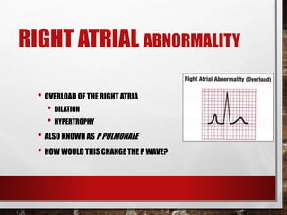
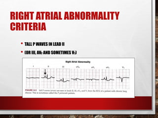
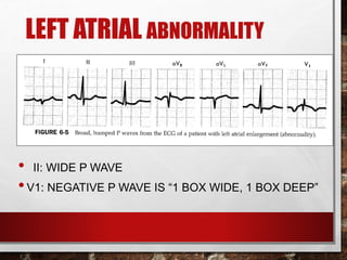
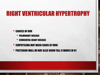
Right atrial overload (P pulmonale) and right ventricular hypertrophy. Right atrial overload (enlargement) is manifest as tall sharp P waves in lead II and V1. The cut off values are P wave amplitude more than 0.25 mV in lead II and 0.1 mV or more in V1. Dominant R waves in V1 and deep S waves in V6 indicate right ventricular hypertrophy (RVH). Sokolow-Lyon for RVH criteria mentions that R wave in V1 + S wave in V5/V6 should be 1.1 mV or more. There is also a clockwise rotation in the QRS pattern between V1 to V6. QRS axis is around +120 degrees (aVR biphasic and lead III showing tallest QRS complex). Right axis deviation is also due to right ventricular hypertrophy. T wave inversion in inferior leads and V1 could be due to right ventricular hypertrophy itself. RVH in this case is type A with dominant R in V1 and deep S in V6. This type is seen in pulmonary stenosis. Type B RVH shows dominant R waves in V1 without deep S in V6. Deep S in V6 without dominant R in V1 seen in chronic obstructive lung disease with cor-pulmonale is called type C RVH. (Strictly speaking the types are classified depending upon vector cardiographic features and not based on scalar ECG)
NVBDCP.pptx Nation vector borne disease control programSapna Thakur
NVBDCP was launched in 2003-2004 . Vector-Borne Disease: Disease that results from an infection transmitted to humans and other animals by blood-feeding arthropods, such as mosquitoes, ticks, and fleas. Examples of vector-borne diseases include Dengue fever, West Nile Virus, Lyme disease, and malaria.
These simplified slides by Dr. Sidra Arshad present an overview of the non-respiratory functions of the respiratory tract.
Learning objectives:
1. Enlist the non-respiratory functions of the respiratory tract
2. Briefly explain how these functions are carried out
3. Discuss the significance of dead space
4. Differentiate between minute ventilation and alveolar ventilation
5. Describe the cough and sneeze reflexes
Study Resources:
1. Chapter 39, Guyton and Hall Textbook of Medical Physiology, 14th edition
2. Chapter 34, Ganong’s Review of Medical Physiology, 26th edition
3. Chapter 17, Human Physiology by Lauralee Sherwood, 9th edition
4. Non-respiratory functions of the lungs https://academic.oup.com/bjaed/article/13/3/98/278874
Tom Selleck Health: A Comprehensive Look at the Iconic Actor’s Wellness Journeygreendigital
Tom Selleck, an enduring figure in Hollywood. has captivated audiences for decades with his rugged charm, iconic moustache. and memorable roles in television and film. From his breakout role as Thomas Magnum in Magnum P.I. to his current portrayal of Frank Reagan in Blue Bloods. Selleck's career has spanned over 50 years. But beyond his professional achievements. fans have often been curious about Tom Selleck Health. especially as he has aged in the public eye.
Follow us on: Pinterest
Introduction
Many have been interested in Tom Selleck health. not only because of his enduring presence on screen but also because of the challenges. and lifestyle choices he has faced and made over the years. This article delves into the various aspects of Tom Selleck health. exploring his fitness regimen, diet, mental health. and the challenges he has encountered as he ages. We'll look at how he maintains his well-being. the health issues he has faced, and his approach to ageing .
Early Life and Career
Childhood and Athletic Beginnings
Tom Selleck was born on January 29, 1945, in Detroit, Michigan, and grew up in Sherman Oaks, California. From an early age, he was involved in sports, particularly basketball. which played a significant role in his physical development. His athletic pursuits continued into college. where he attended the University of Southern California (USC) on a basketball scholarship. This early involvement in sports laid a strong foundation for his physical health and disciplined lifestyle.
Transition to Acting
Selleck's transition from an athlete to an actor came with its physical demands. His first significant role in "Magnum P.I." required him to perform various stunts and maintain a fit appearance. This role, which he played from 1980 to 1988. necessitated a rigorous fitness routine to meet the show's demands. setting the stage for his long-term commitment to health and wellness.
Fitness Regimen
Workout Routine
Tom Selleck health and fitness regimen has evolved. adapting to his changing roles and age. During his "Magnum, P.I." days. Selleck's workouts were intense and focused on building and maintaining muscle mass. His routine included weightlifting, cardiovascular exercises. and specific training for the stunts he performed on the show.
Selleck adjusted his fitness routine as he aged to suit his body's needs. Today, his workouts focus on maintaining flexibility, strength, and cardiovascular health. He incorporates low-impact exercises such as swimming, walking, and light weightlifting. This balanced approach helps him stay fit without putting undue strain on his joints and muscles.
Importance of Flexibility and Mobility
In recent years, Selleck has emphasized the importance of flexibility and mobility in his fitness regimen. Understanding the natural decline in muscle mass and joint flexibility with age. he includes stretching and yoga in his routine. These practices help prevent injuries, improve posture, and maintain mobilit
- Video recording of this lecture in English language: https://youtu.be/lK81BzxMqdo
- Video recording of this lecture in Arabic language: https://youtu.be/Ve4P0COk9OI
- Link to download the book free: https://nephrotube.blogspot.com/p/nephrotube-nephrology-books.html
- Link to NephroTube website: www.NephroTube.com
- Link to NephroTube social media accounts: https://nephrotube.blogspot.com/p/join-nephrotube-on-social-media.html
Flu Vaccine Alert in Bangalore Karnatakaaddon Scans
As flu season approaches, health officials in Bangalore, Karnataka, are urging residents to get their flu vaccinations. The seasonal flu, while common, can lead to severe health complications, particularly for vulnerable populations such as young children, the elderly, and those with underlying health conditions.
Dr. Vidisha Kumari, a leading epidemiologist in Bangalore, emphasizes the importance of getting vaccinated. "The flu vaccine is our best defense against the influenza virus. It not only protects individuals but also helps prevent the spread of the virus in our communities," he says.
This year, the flu season is expected to coincide with a potential increase in other respiratory illnesses. The Karnataka Health Department has launched an awareness campaign highlighting the significance of flu vaccinations. They have set up multiple vaccination centers across Bangalore, making it convenient for residents to receive their shots.
To encourage widespread vaccination, the government is also collaborating with local schools, workplaces, and community centers to facilitate vaccination drives. Special attention is being given to ensuring that the vaccine is accessible to all, including marginalized communities who may have limited access to healthcare.
Residents are reminded that the flu vaccine is safe and effective. Common side effects are mild and may include soreness at the injection site, mild fever, or muscle aches. These side effects are generally short-lived and far less severe than the flu itself.
Healthcare providers are also stressing the importance of continuing COVID-19 precautions. Wearing masks, practicing good hand hygiene, and maintaining social distancing are still crucial, especially in crowded places.
Protect yourself and your loved ones by getting vaccinated. Together, we can help keep Bangalore healthy and safe this flu season. For more information on vaccination centers and schedules, residents can visit the Karnataka Health Department’s official website or follow their social media pages.
Stay informed, stay safe, and get your flu shot today!
Ozempic: Preoperative Management of Patients on GLP-1 Receptor Agonists Saeid Safari
Preoperative Management of Patients on GLP-1 Receptor Agonists like Ozempic and Semiglutide
ASA GUIDELINE
NYSORA Guideline
2 Case Reports of Gastric Ultrasound
Report Back from SGO 2024: What’s the Latest in Cervical Cancer?bkling
Are you curious about what’s new in cervical cancer research or unsure what the findings mean? Join Dr. Emily Ko, a gynecologic oncologist at Penn Medicine, to learn about the latest updates from the Society of Gynecologic Oncology (SGO) 2024 Annual Meeting on Women’s Cancer. Dr. Ko will discuss what the research presented at the conference means for you and answer your questions about the new developments.
MANAGEMENT OF ATRIOVENTRICULAR CONDUCTION BLOCK.pdfJim Jacob Roy
Cardiac conduction defects can occur due to various causes.
Atrioventricular conduction blocks ( AV blocks ) are classified into 3 types.
This document describes the acute management of AV block.
New Directions in Targeted Therapeutic Approaches for Older Adults With Mantl...i3 Health
i3 Health is pleased to make the speaker slides from this activity available for use as a non-accredited self-study or teaching resource.
This slide deck presented by Dr. Kami Maddocks, Professor-Clinical in the Division of Hematology and
Associate Division Director for Ambulatory Operations
The Ohio State University Comprehensive Cancer Center, will provide insight into new directions in targeted therapeutic approaches for older adults with mantle cell lymphoma.
STATEMENT OF NEED
Mantle cell lymphoma (MCL) is a rare, aggressive B-cell non-Hodgkin lymphoma (NHL) accounting for 5% to 7% of all lymphomas. Its prognosis ranges from indolent disease that does not require treatment for years to very aggressive disease, which is associated with poor survival (Silkenstedt et al, 2021). Typically, MCL is diagnosed at advanced stage and in older patients who cannot tolerate intensive therapy (NCCN, 2022). Although recent advances have slightly increased remission rates, recurrence and relapse remain very common, leading to a median overall survival between 3 and 6 years (LLS, 2021). Though there are several effective options, progress is still needed towards establishing an accepted frontline approach for MCL (Castellino et al, 2022). Treatment selection and management of MCL are complicated by the heterogeneity of prognosis, advanced age and comorbidities of patients, and lack of an established standard approach for treatment, making it vital that clinicians be familiar with the latest research and advances in this area. In this activity chaired by Michael Wang, MD, Professor in the Department of Lymphoma & Myeloma at MD Anderson Cancer Center, expert faculty will discuss prognostic factors informing treatment, the promising results of recent trials in new therapeutic approaches, and the implications of treatment resistance in therapeutic selection for MCL.
Target Audience
Hematology/oncology fellows, attending faculty, and other health care professionals involved in the treatment of patients with mantle cell lymphoma (MCL).
Learning Objectives
1.) Identify clinical and biological prognostic factors that can guide treatment decision making for older adults with MCL
2.) Evaluate emerging data on targeted therapeutic approaches for treatment-naive and relapsed/refractory MCL and their applicability to older adults
3.) Assess mechanisms of resistance to targeted therapies for MCL and their implications for treatment selection
Ethanol (CH3CH2OH), or beverage alcohol, is a two-carbon alcohol
that is rapidly distributed in the body and brain. Ethanol alters many
neurochemical systems and has rewarding and addictive properties. It
is the oldest recreational drug and likely contributes to more morbidity,
mortality, and public health costs than all illicit drugs combined. The
5th edition of the Diagnostic and Statistical Manual of Mental Disorders
(DSM-5) integrates alcohol abuse and alcohol dependence into a single
disorder called alcohol use disorder (AUD), with mild, moderate,
and severe subclassifications (American Psychiatric Association, 2013).
In the DSM-5, all types of substance abuse and dependence have been
combined into a single substance use disorder (SUD) on a continuum
from mild to severe. A diagnosis of AUD requires that at least two of
the 11 DSM-5 behaviors be present within a 12-month period (mild
AUD: 2–3 criteria; moderate AUD: 4–5 criteria; severe AUD: 6–11 criteria).
The four main behavioral effects of AUD are impaired control over
drinking, negative social consequences, risky use, and altered physiological
effects (tolerance, withdrawal). This chapter presents an overview
of the prevalence and harmful consequences of AUD in the U.S.,
the systemic nature of the disease, neurocircuitry and stages of AUD,
comorbidities, fetal alcohol spectrum disorders, genetic risk factors, and
pharmacotherapies for AUD.
Lung Cancer: Artificial Intelligence, Synergetics, Complex System Analysis, S...Oleg Kshivets
RESULTS: Overall life span (LS) was 2252.1±1742.5 days and cumulative 5-year survival (5YS) reached 73.2%, 10 years – 64.8%, 20 years – 42.5%. 513 LCP lived more than 5 years (LS=3124.6±1525.6 days), 148 LCP – more than 10 years (LS=5054.4±1504.1 days).199 LCP died because of LC (LS=562.7±374.5 days). 5YS of LCP after bi/lobectomies was significantly superior in comparison with LCP after pneumonectomies (78.1% vs.63.7%, P=0.00001 by log-rank test). AT significantly improved 5YS (66.3% vs. 34.8%) (P=0.00000 by log-rank test) only for LCP with N1-2. Cox modeling displayed that 5YS of LCP significantly depended on: phase transition (PT) early-invasive LC in terms of synergetics, PT N0—N12, cell ratio factors (ratio between cancer cells- CC and blood cells subpopulations), G1-3, histology, glucose, AT, blood cell circuit, prothrombin index, heparin tolerance, recalcification time (P=0.000-0.038). Neural networks, genetic algorithm selection and bootstrap simulation revealed relationships between 5YS and PT early-invasive LC (rank=1), PT N0—N12 (rank=2), thrombocytes/CC (3), erythrocytes/CC (4), eosinophils/CC (5), healthy cells/CC (6), lymphocytes/CC (7), segmented neutrophils/CC (8), stick neutrophils/CC (9), monocytes/CC (10); leucocytes/CC (11). Correct prediction of 5YS was 100% by neural networks computing (area under ROC curve=1.0; error=0.0).
CONCLUSIONS: 5YS of LCP after radical procedures significantly depended on: 1) PT early-invasive cancer; 2) PT N0--N12; 3) cell ratio factors; 4) blood cell circuit; 5) biochemical factors; 6) hemostasis system; 7) AT; 8) LC characteristics; 9) LC cell dynamics; 10) surgery type: lobectomy/pneumonectomy; 11) anthropometric data. Optimal diagnosis and treatment strategies for LC are: 1) screening and early detection of LC; 2) availability of experienced thoracic surgeons because of complexity of radical procedures; 3) aggressive en block surgery and adequate lymph node dissection for completeness; 4) precise prediction; 5) adjuvant chemoimmunoradiotherapy for LCP with unfavorable prognosis.
2. CARDIAC ENLARGEMENT
1. DILATION
a. STRETCHED
b. E.G. CONGESTIVE HEART FAILURE
2. HYPERTROPHY
a. INCREASE SIZE OF HEART MUSCLE FIBERS
b. E.G. AORTIC STENOSIS
3. RIGHT ATRIAL ABNORMALITY
• OVERLOAD OF THE RIGHT ATRIA
• DILATION
• HYPERTROPHY
• ALSO KNOWN AS P PULMONALE
• HOW WOULD THIS CHANGE THE P WAVE?
4. RIGHT ATRIAL ABNORMALITY
• NORMAL P WAVE IS LESS THAN 2.5 MM TALL AND 0.10 SECONDS WIDE.
• WITH RIGHT ATRIAL HYPERTROPHY, P WAVES ARE TYPICALLY TALLER
THAN 2.5 MM BUT NOT WIDER THAN 0.10SEC.
7. LEFT ATRIAL ABNORMALITY
•ALSO KNOWN AS P MITRALE
•LEFT ATRIA NORMALLY DEPOLARIZES AFTER THE RIGHT
ATRIA.
•HOW WOULD THIS AFFECT THE P WAVE?
•WIDER; LEFT ATRIAL ENLARGEMENT SHOULD PROLONG THE
P WAVE > 0.10 SEC.
10. LEFT ATRIAL ABNORMALITY
• LEAD II (AND I) SHOW
WIDE P WAVES
• (SECOND HUMP DUE TO
DELAYED
DEPOLARIZATION OF THE
LEFT ATRIUM)
• (P MITRALE: MITRAL
VALVE DISEASE)
• V1 MAY SHOW A BI-
PHASIC P WAVE
• 1 BOX WIDE, 1 BOX DEEP
• (BIPHASIC SINCE RIGHT
ATRIA IS ANTERIOR TO
THE LEFT ATRIA)
17. RIGHT VENTRICULAR HYPERTROPHY
CRITERIA
1.IN V1, R WAVE IS GREATER THAN THE S WAVE - OR - R
IN V1 GREATER THAN 7 MM
1. RIGHT AXIS DEVIATION
2. IN V1, T WAVE INVERSION (REASON UNKNOWN)
3. S WAVES IN V5 AND V6
18. RIGHT VENTRICULAR HYPERTROPHY
• CAUSES OF RVH
• PULMONARY DISEASE
• CONGENITAL HEART DISEASE
• (EMPHYSEMA MAY MASK SIGNS OF RVH)
• POSTERIOR WALL MI MAY ALSO SHOW TALL R WAVES IN V1
20. Fig 6.9
R wave in V1.
P waves in II, III, & V1
T wave inversion
PR interval
21. LEFT VENTRICULAR HYPERTROPHY
• WITH LVH, THE ELECTRICAL BALANCE IS TIPPED EVEN FURTHER TO THE
LEFT.
• TALL R WAVES IN THE LEFT CHEST LEADS
• PREDOMINATE S WAVES IN THE RIGHT CHEST LEADS
23. LEFT VENTRICULAR
HYPERTROPHY CRITERIA
•SOKOLOW-LYON VOLTAGE CRITERIA
•IF S WAVE IN V1 + R WAVE IN V5 OR V6 ≥
35 MM (≥ 50 FOR UNDER 35 YRS OF
AGE)
•R WAVE > 11 MM IN AVL OR I...
•ALSO
•LVH IS MORE LIKELY WITH A “STRAIN PATTERN”
OR ST SEGMENT CHANGES
•LEFT AXIS DEVIATION
•LEFT ATRIAL ABNORMALITY
24. LEFT VENTRICULAR HYPERTROPHY
• CAUSES:
• HYPERTENSION
• AORTIC STENOSIS
• NOT ALWAYS PATHOLOGICAL
• RISKS OF LVH
• CONGESTIVE HEART FAILURE
• ARRHYTHMIAS
25. LEFT VENTRICULAR HYPERTROPHY
• HIGH VOLTAGE CAN BE SEEN IN NORMAL PEOPLE, ESPECIALLY ATHLETES
• WITH HYPERTROPHY IN BOTH VENTRICLES, THE ECG WILL SHOW MORE
EVIDENCE OF LVH
36. Tall R waves in V4 and V5 with down sloping ST segment depression and T wave inversion are
suggestive of left ventricular hypertrophy (LVH) with strain pattern. LVH with strain pattern usually
occurs in pressure overload of the left ventricle as in systemic hypertension or aortic stenosis.
Similar pattern may also occur in long standing severe aortic regurgitation, though the usual pattern
in aortic regurgitation is left ventricular volume overload.
Negative P waves in lead V1 is indicative of left atrial overload. Shallow T wave inversions are seen
in inferior leads. Two supra ventricular ectopic beats are also seen in the rhythm strip. They are
characterized by their premature nature, a P wave of different morphology preceding the QRS (in this
case merging with the T wave of the previous beat), narrow QRS complex and an incomplete
compensatory pause.
37. Right atrial overload (P pulmonale) and right ventricular hypertrophy. Right atrial overload (enlargement) is
manifest as tall sharp P waves in lead II and V1. The cut off values are P wave amplitude more than 0.25 mV in lead II
and 0.1 mV or more in V1. Dominant R waves in V1 and deep S waves in V6 indicate right ventricular hypertrophy (RVH).
Sokolow-Lyon for RVH criteria mentions that R wave in V1 + S wave in V5/V6 should be 1.1 mV or more. There is also a
clockwise rotation in the QRS pattern between V1 to V6. QRS axis is around +120 degrees (aVR biphasic and lead III
showing tallest QRS complex). Right axis deviation is also due to right ventricular hypertrophy. T wave inversion in inferior
leads and V1 could be due to right ventricular hypertrophy itself. RVH in this case is type A with dominant R in V1 and
deep S in V6. This type is seen in pulmonary stenosis. Type B RVH shows dominant R waves in V1 without deep S in V6.
Deep S in V6 without dominant R in V1 seen in chronic obstructive lung disease with cor-pulmonale is called type C RVH.
(Strictly speaking the types are classified depending upon vector cardiographic features and not based on scalar ECG)