Downloaded 12 times
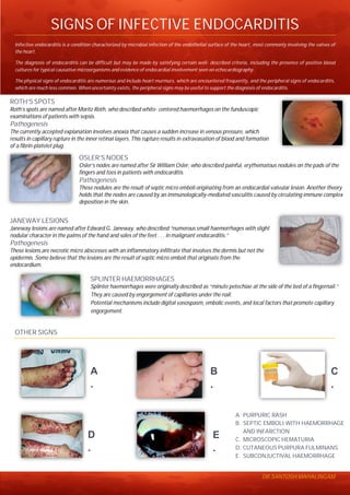

Infective endocarditis is a condition characterized by microbial infection of the heart valves. The diagnosis is made based on positive blood cultures, evidence of infection on echocardiogram, and signs such as heart murmurs. Peripheral signs like Roth's spots, Osler's nodes, Janeway lesions, and splinter hemorrhages can also support the diagnosis when uncertainty exists. Roth's spots are retinal hemorrhages seen on eye exam. Osler's nodes are painful finger and toe nodules caused by septic emboli. Janeway lesions are skin hemorrhages on the palms and soles thought to be from septic emboli. Splinter hemorrhages are nail bed hemorrhages caused


