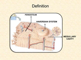
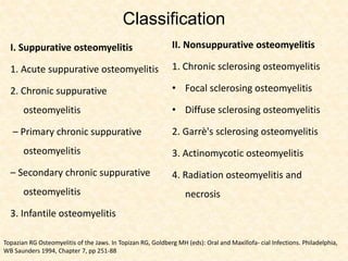
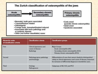
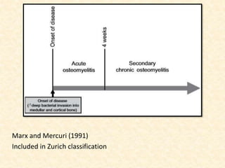
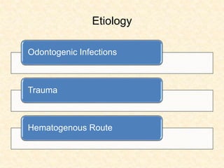
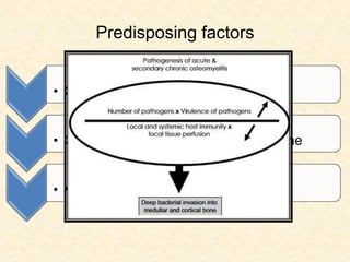
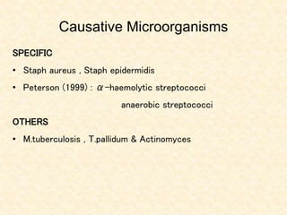
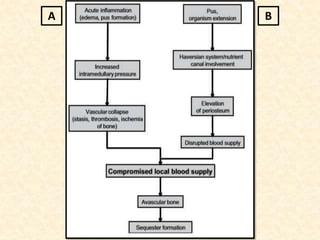
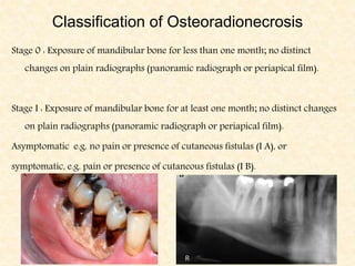
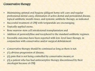
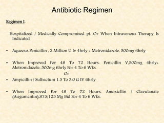
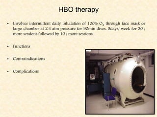
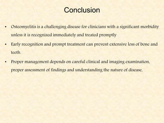
SlideShare a Scribd company logo
Osteomyelitis
Dr. Ruchika Garg
References
• Textbook of Oral & Maxillofacial Infections 4th edition : Richard G. Topazian.
• Osteomyelitis of the jaws : Marc M Baltensperger and Gerold K Eyrich
• Textbook of Oral Pathology 6rth edition : Shafer Hine Levy
• Diagnostic imaging of the jaws : Langland & Langlais
• Textbook of Oral Radiology : White & Pharoah.
• Aliya A Khan et al. Diagnosis and Management of Osteonecrosis of the Jaw: A Systematic
Review and International Consensus. Journal of Bone and Mineral Research, Vol. 30, No. 1,
January 2015, pp 3–23 DOI: 10.1002/jbmr.2405.
• Deepak Gupta et al. Role of Maxillofacial Radiology and Imaging in the diagnosis and Treatment
of Osteomyelitis of the Jaws. Dentistry, Oral Disorders & Therapy.
Contents
• Introduction
• Definition
• Etiology
• Predisposing factors
• Pathogenesis
• Classification
• Types of osteomyelitis
• Investigations
• Treatment
• Conclusion
Introduction
OSTEON
MYELOS
ITIS
OSTEOMYELITIS
Definition
PERIOSTEUM
HAVERSIAN SYSTEM
MEDULLARY
CAVITY
Classification
I. Suppurative osteomyelitis
1. Acute suppurative osteomyelitis
2. Chronic suppurative
osteomyelitis
– Primary chronic suppurative
osteomyelitis
– Secondary chronic suppurative
osteomyelitis
3. Infantile osteomyelitis
II. Nonsuppurative osteomyelitis
1. Chronic sclerosing osteomyelitis
• Focal sclerosing osteomyelitis
• Diffuse sclerosing osteomyelitis
2. Garrè's sclerosing osteomyelitis
3. Actinomycotic osteomyelitis
4. Radiation osteomyelitis and
necrosis
Topazian RG Osteomyelitis of the Jaws. In Topizan RG, Goldberg MH (eds): Oral and Maxillofa- cial Infections. Philadelphia,
WB Saunders 1994, Chapter 7, pp 251-88
Marx and Mercuri (1991)
Included in Zurich classification
Etiology
Odontogenic Infections
Trauma
Hematogenous Route
Predisposing factors
• Conditions that alter host defence
• Conditions that alter vascularity of bone
• Virulence of organisms
Causative Microorganisms
SPECIFIC
• Staph aureus , Staph epidermidis
• Peterson (1999) : α-haemolytic streptococci
anaerobic streptococci
OTHERS
• M.tuberculosis , T.pallidum & Actinomyces
A B
PATHOGENESIS
ACUTE
Tissue Necrosis
Accumulation of pus
Increased Intramedullary pressure
Vascular collapse, Venous stasis, Ischemia
Accumulation of pus beneath periosteum
Decrease Vascular supply
Avascular bone
Chronic
Regression of inflammation
Granulation tissue formation
Angiogenesis - SEQUESTRUM
Involucrum , Cloacae
Acute Suppurative Osteomyelitis
Clinical Features
Age
Site
Symptoms – Severe pain, trismus, regional lymphadenopathy,
paresthesia of the lips
RADIOLOGICAL FEATURES
No early changes
Initial changes include blurring of the bony
trabeculae
Ill defined radiolucency
Moth eaten appearance
Chronic Suppurative Osteomyelitis
TYPES
• Primary
• Secondary
CLINICAL FEATURES
• Fistulae
• “Thickened / Woody” bone
Radiological Features
• In early stage there is widening of marrow spaces giving a
mottled appearance
• Granulation tissue b/w dead & living bone gives irregular lines
& zones of radiolucency resulting in moth -eaten appearance.
• In later stages the devitalized bone appears sclerosed & called
sequestrum.
• Large areas of bone destruction seen as radiolucent areas.
Axial CT Scan showing the sequestrae (white arrow) & bone
reaction — black arrow
Five Different Radiographic Appearances Of
Chronic Osteomyelitis
1. Radiolucency with ragged borders
2. Radiolucency containing one or more radio-opaque foci
3. A salt and pepper appearance
4. A dense radio-opacity
5. A cortical radiolucency
Infantile Osteomyelitis
• Etiology –
• Forcep-delivery
• Trauma caused to oral mucosa during delivery
• Infection of maxillary sinus
• Contaminated human or artificial nipples
• Infections from nose
• Hematogenous spread
C/F --
1) Sudden onset of high grade fever , anorexia , irritability.
2) Redness & swelling over mid face.
3) Purulent discharge - below median canthus of eye.
4) Sub periosteal abscess- draining sinus tracts.
5) Poor feeding.
R/F – Minimal bone loss
In later stages sequestra & necrotic tooth germs
Chronic Sclerosing Osteomyelitis
• Also called Sclerosing osteitis
Ossifying Osteomyelitis
Sclerosing cementoma
TYPES
• Focal
• Diffuse
Chronic Focal Sclerosing Osteomyelitis
• Unusual reaction of bone
• Clinical features
Age
Site
Symptoms
Radiological features
Five patterns of Condensing Osteitis
• Radio-opaque area not surrounded by perilesional halo.
• Target lesion surrounded by perilesional halo.
• Granular or lucent pattern.
• Multifloculent
• Associated with external root resorption.
Chronic Diffuse Sclerosing Osteomyelitis
Clinical features
Age - Older
Sex – No predilection
Site – Edentulous areas
Symptoms – Mild expansion of jaw, episodes of recurrent swelling
Radiological features
Chronic Osteomyelitis with Proliferative
Periostitis
• Also called Garre’s sclerosing Osteomyelitis
Proliferative periostitis
Periostitis ossificans
• First described by CARL GARRE in 1893.
Pathogenesis
Localization of infection in
periosteum
Bony thickening visible on r/g
CLINICAL FEATURES
Age
Site
Symptoms
Radiological Features
Onion skin pattern
Radial pattern
Tuberculous Osteomyelitis
• Clinical features
i. Involvement of mandible is more
ii. Painless swelling
iii. Loosening of teeth
iv. Sequestration of bone
v. C/o chronic discharging sinus
vi. Palpable lymph nodes
RADIOGRAPHIC EXAMINATION
OPG
PA mandible
Lateral oblique view of mandible.
Well defined radiolucency with destruction of buccal or medial
cortical plates
DIAGNOSIS
o Culture for pus – Test for acid fast bacilli
o Culture for sputum—AFB
o Mantoux test
o Biopsy of lesion
Actinomycotic Osteomelitis
Site
• Angle of mandible
• Posterior aspect of body
Radiological features
• Radiolucent area at apex of one / more teeth.
• Well defined radiolucency with a sclerotic bone at periphery : CYST LIKE
• Scattered area of bone destruction separated by normal /sclerosed bone.
• Shadow of tooth socket with increasing density of adjacent bone.
Diagnosis –
• Biopsy
• Gram staining
• Immunofluorescent staining
• Treatment –
• i.v for 4 – 6 weeks
• Oral for 6 – 12 months
Nocardial Osteomyelitis
• Resembles actinomycosis
• Causative organism – Nocardia asteroids
• With or without dental injury
• Suppurative lesion with necrosis & abscess formation.
• T/t – drainage , Sulphonamides for 6 wks.
Syphilitic Osteomyelitis
• Frequently involved - hard palate.
• The gummatous destruction is painless.
• Syphilitic osteomyelitis of the jaws is difficult to distinguish from
chronic suppurative osteomyelitis since their radiographic
appearances are similar.
• Maxilla is more affected than mandible
PERIOSTITIS : several layers of new bone parallel to margin of jaw.
“Gross caricature of network / lattice”.
• May lead to oroantral , oronasal communication
• Sequestra is called as Filiary sequestra
Osteoradionecrosis
• It is a chronic, non- healing wound caused by hypoxia, hypocellularity
and hypovascularity of the irradiated tissue. (MARX)
• POST- RADIATION OSTEONECROSIS.
• Intense irradiation > 50 Gy.
Clinical features
 Remain asymptomatic for prolonged periods of weeks, months, or even years.
 Signs and symptoms - pain, tooth mobility, mucosal swelling, erythema,
ulceration, paresthesia, or even anesthesia of the associated branch of the
trigeminal nerve.
 Intraoral and extraoral fistulae may develop when necrotic mandible or maxilla
becomes secondarily infected.
 Chronic maxillary sinusitis secondary to osteonecrosis with or without an oral-
antral fistula may be the presenting feature in patients with maxillary bone
involvement.
Classification of Osteoradionecrosis
Stage 0 : Exposure of mandibular bone for less than one month; no distinct
changes on plain radiographs (panoramic radiograph or periapical film).
Stage I : Exposure of mandibular bone for at least one month; no distinct changes
on plain radiographs (panoramic radiograph or periapical film).
Asymptomatic e.g. no pain or presence of cutaneous fistulas (I A), or
symptomatic, e.g. pain or presence of cutaneous fistulas (I B).
Stage II : Exposure of mandibular bone for at least one month; distinct changes
present on plain radiographs (panoramic radiograph or periapical film), but
not involving the lower border of the mandible.
Asymptomatic otherwise, e.g. no pain or presence of cutaneous fistulas (II A), or
Symptomatic, e.g. pain or presence of cutaneous fistulas (II B).
Stage III : Exposure of mandibular bone for at least one month; distinct changes
on plain radiographs (panoramic radiograph or periapical film), involving
the lower border of the mandible, irrespective of any other signs and
symptoms. Note: In case of doubt about the presence and/or extent of
radiographical bone involvement, the lower stage should be allotted.
Conservative therapy
• Maintaining optimal oral hygiene (diligent home self-care and regular
professional dental care), elimination of active dental and periodontal disease,
topical antibiotic mouth rinses, and systemic antibiotic therapy, as indicated.
• Successful treatment of ONJ with teriparatide are encouraging
• Topically applied ozone,
• Bone marrow stem cell intralesional transplantation and
• Addition of pentoxifylline and tocopherol to the standard antibiotic regimen.
• Favorable outcomes have been reported with low-level laser therapy, in
conjunction with conservative and/or surgical debridement.
• Conservative therapy should be continued as long as there is not:
• (1) obvious progression of disease;
• (2) pain that is not being controlled by conservative means; or
• (3) a patient who has had antiresorptive therapy discontinued by their
oncologist because of ONJ.
Surgical management
• A full-thickness mucoperiosteal flap should be elevated and extended to reveal
the entire area of exposed bone and beyond to disease-free margins.
• Resection of the affected bone should be extended to reach healthy-appearing,
bleeding bone.
• Sharp edges should be smoothed
• It is also proposed that if surgery is indicated, resection with tension-free
closure affords the most positive results.
• Promising results have also been reported with surgical debridement in
combination with platelet-derived growth factor (PDGF) applied to the site
• Cases also reported that intraoperative fluorescence guidance was helpful in
identifying surgical resection margins
• It was suggested that longer-term preoperative antibiotics (23 to 54 days)
resulted in improved surgical outcomes versus short-term antibiotic therapy (1
to 8 days). Adjunctive therapy with hyperbaric oxygen (HBO) in combination
with surgery has been investigated with encouraging results.
Differential Diagnosis
 Paget’s disease
 Fibrous dysplasia
 Florid cemento-osseus dysplasia
 Osteopetrosis
 Ewing’s sarcoma
 Osteosarcoma
 Caffey’s disease
Investigations
• Gram Staining
• Culture & Sensitivity
• WBC count & Complete Hemogram
• Blood Sugar
• Montoux Test
• Radiographs
• Scintigraphy
• Computed tomography
• MRI
Decision tree for use of imaging studies in diagnosis
of Osteomyelitis
Conventional
radiography
Technetium bone scan
67Ga / In WBC scan
MRI/CT
MANAGEMENT
Conservative management
• Bed rest
• Supportive therapy
• Dehydration
• Blood transfusion
• Control of pain
• Antibiotic therapy
• HBO therapy
Antibiotic Regimen
Regimen I:
Hospitalized / Medically Compromised pt. Or When Intravenous Therapy Is
Indicated
• Aqueous Penicillin , 2 Million U Iv 4hrly + Metronidazole, 500mg 6hrly
• When Improved For 48 To 72 Hours: Penicillin V,500mg 4hrly+
Metronidazole, 500mg 6hrly For 4 To 6 Wks.
Or
• Ampicillin / Sulbactum 1.5 To 3.0 G IV 6hrly
• When Improved For 48 To 72 Hours: Amoxicllin / Clavulanate
(Augumentin),875/125 Mg Bid For 4 To 6 Wks.
Regimen II:
For Outpatient Treatment
• Penicillin V , 2gm + Metronidazole, 500mg 8hrly For 2 To 4 Wks After Last
Sequestra Removed.
Or
• Clindamycin ,600 To 900mg 6hrly Iv , Then
• Clindamycin ,300 To 450mg 6hrly
PENICILLINASE ALLERGIC PTS.
• Clindamycin
• Cefotaxim
HBO therapy
• Involves intermittent daily inhalation of 100% O2 through face mask or
large chamber at 2.4 atm pressure for 90min dives: 5days/ week for 30 /
more sessions followed by 10 / more sessions.
• Functions
• Contraindications
• Complications
• HBO therapy involves the intermittent , usually daily , inhalation of 100%
humidified oxygen under pressure , greater than one atmospheric pressure
(ATA).
• HBO reduces the hypoxia within the affected tissues and stimulates
angiogenesis in the hypo vascular tissues .
• It enhances phagocytic activity of leucocytes to stimulate fibroblast growth ,
increase in collagen formation and promote growth of new capillaries.
• Patient is placed in a chamber.
• Oxygen is given by mask or by hood .
• Each dive is 90 minute in length.
• 5 days per week for 30,60, or more dives at 2.4 ATA for 90 minutes while
breathing 100% oxygen twice daily.
• Protocol of MARX (1983)
30 initial dives , if improved then 60 dives completed.
Otherwise , SEQUESTRECTOMY + 30 dives , if wound dehiscence, RESECTION +
60 dives & 20 dives after 10 weeks
Patient with pathological # ,oro-cutaneous fistula are given 30 dives more prior
going to resection
Contraindications by Fisher (1988) & Marx(1985)
1. Pneumothorax
2. COPD
3. Optic neuritis
4. Acute viral infection
5. Acute seizures
6. URI
7. Pregnancy
8. Thoracic surgery
9. Ear surgery
Conclusion
• Osteomyelitis is a challenging disease for clinicians with a significant morbidity
unless it is recognized immediately and treated promptly
• Early recognition and prompt treatment can prevent extensive loss of bone and
teeth.
• Proper management depends on careful clinical and imaging examination,
proper assessment of findings and understanding the nature of disease,
Osteomyelitis

More Related Content

What's hot

Dental Trauma
Dental Trauma Dental Trauma
Dental Trauma
Weam Faroun
 
cysts of the oral and maxillofacial region
cysts of the oral and maxillofacial regioncysts of the oral and maxillofacial region
cysts of the oral and maxillofacial region
madhusudhan reddy
 
Tmj examination & imaging
Tmj examination & imagingTmj examination & imaging
Tmj examination & imaging
Chetan Basnet
 
Mixed radiopaque & radiolucent lesions of jaw
Mixed radiopaque & radiolucent lesions of jaw Mixed radiopaque & radiolucent lesions of jaw
Mixed radiopaque & radiolucent lesions of jaw
Dr. Samarth Johari
 
Myofacial pain dysfunction syndrome
Myofacial pain dysfunction syndromeMyofacial pain dysfunction syndrome
Myofacial pain dysfunction syndrome
Manash Bora
 
Osteomyelitis of jaw--department of oral medicine and radiology
Osteomyelitis of jaw--department of oral medicine and radiologyOsteomyelitis of jaw--department of oral medicine and radiology
Osteomyelitis of jaw--department of oral medicine and radiology
Government Dental College and Hospital, Shimla
 
Fibrousdysplasia
Fibrousdysplasia Fibrousdysplasia
Fibrousdysplasia
D Venkatesh Kumar
 
Dental mobility
Dental mobilityDental mobility
Dental mobility
Sannah Jahangir
 
Multilocular radiolucencies
Multilocular radiolucenciesMultilocular radiolucencies
Multilocular radiolucencies
Selva Arockiam
 
Radiopaque Lesions
Radiopaque LesionsRadiopaque Lesions
Radiopaque Lesions
Maryam Arbab
 
Periapical diseases and classification
Periapical diseases and classificationPeriapical diseases and classification
Periapical diseases and classification
Abhishek Verma
 
Dentin Dysplasia
Dentin DysplasiaDentin Dysplasia
Soft tissue calcification orofacial region
Soft tissue calcification orofacial regionSoft tissue calcification orofacial region
Soft tissue calcification orofacial region
Dr. Monali Prajapati
 
Peripheral and central giant cell granuloma
Peripheral and central giant cell granulomaPeripheral and central giant cell granuloma
Peripheral and central giant cell granuloma
Rijuwana77
 
Pulp diseases
Pulp diseasesPulp diseases
Pulp diseases
Panda Park
 
Neuromuscular Disorders affecting the orofacial region
Neuromuscular Disorders affecting the orofacial regionNeuromuscular Disorders affecting the orofacial region
Neuromuscular Disorders affecting the orofacial region
Vibhuti Kaul
 
Periapical pathology 22 (1)
Periapical  pathology 22 (1)Periapical  pathology 22 (1)
Periapical pathology 22 (1)lonmjjh
 

What's hot (20)

Dental Trauma
Dental Trauma Dental Trauma
Dental Trauma
 
cysts of the oral and maxillofacial region
cysts of the oral and maxillofacial regioncysts of the oral and maxillofacial region
cysts of the oral and maxillofacial region
 
Tmj examination & imaging
Tmj examination & imagingTmj examination & imaging
Tmj examination & imaging
 
Osteomyelitis
OsteomyelitisOsteomyelitis
Osteomyelitis
 
Mixed radiopaque & radiolucent lesions of jaw
Mixed radiopaque & radiolucent lesions of jaw Mixed radiopaque & radiolucent lesions of jaw
Mixed radiopaque & radiolucent lesions of jaw
 
Myofacial pain dysfunction syndrome
Myofacial pain dysfunction syndromeMyofacial pain dysfunction syndrome
Myofacial pain dysfunction syndrome
 
Osteomyelitis of jaw--department of oral medicine and radiology
Osteomyelitis of jaw--department of oral medicine and radiologyOsteomyelitis of jaw--department of oral medicine and radiology
Osteomyelitis of jaw--department of oral medicine and radiology
 
Fibrousdysplasia
Fibrousdysplasia Fibrousdysplasia
Fibrousdysplasia
 
Dental mobility
Dental mobilityDental mobility
Dental mobility
 
Multilocular radiolucencies
Multilocular radiolucenciesMultilocular radiolucencies
Multilocular radiolucencies
 
Radiopaque Lesions
Radiopaque LesionsRadiopaque Lesions
Radiopaque Lesions
 
Periapical diseases and classification
Periapical diseases and classificationPeriapical diseases and classification
Periapical diseases and classification
 
Dentin Dysplasia
Dentin DysplasiaDentin Dysplasia
Dentin Dysplasia
 
Osteomyelitis
OsteomyelitisOsteomyelitis
Osteomyelitis
 
Osteomyelitis of jaw
Osteomyelitis of jawOsteomyelitis of jaw
Osteomyelitis of jaw
 
Soft tissue calcification orofacial region
Soft tissue calcification orofacial regionSoft tissue calcification orofacial region
Soft tissue calcification orofacial region
 
Peripheral and central giant cell granuloma
Peripheral and central giant cell granulomaPeripheral and central giant cell granuloma
Peripheral and central giant cell granuloma
 
Pulp diseases
Pulp diseasesPulp diseases
Pulp diseases
 
Neuromuscular Disorders affecting the orofacial region
Neuromuscular Disorders affecting the orofacial regionNeuromuscular Disorders affecting the orofacial region
Neuromuscular Disorders affecting the orofacial region
 
Periapical pathology 22 (1)
Periapical  pathology 22 (1)Periapical  pathology 22 (1)
Periapical pathology 22 (1)
 

Similar to Osteomyelitis

osteomyelitis
osteomyelitisosteomyelitis
Osteomyelitis of jaw
Osteomyelitis of jawOsteomyelitis of jaw
Osteomyelitis of jaw
KathirvelGopalakrish
 
Periapical radiolucencies
Periapical radiolucencies Periapical radiolucencies
Periapical radiolucencies
Shabil Mohamed Mustafa
 
diseases of bone.pptx
diseases of bone.pptxdiseases of bone.pptx
diseases of bone.pptx
Hetvi23
 
Cysts of oral and maxillofacial region by dr. maryam salman
Cysts of oral and maxillofacial region by dr. maryam salmanCysts of oral and maxillofacial region by dr. maryam salman
Cysts of oral and maxillofacial region by dr. maryam salman
Dr.Maryam Salman
 
Osteomyelitis with Types and Treatment..
Osteomyelitis with Types and Treatment..Osteomyelitis with Types and Treatment..
Osteomyelitis with Types and Treatment..
AtQuiz
 
Complications in dental implant therapy
Complications in dental implant therapy Complications in dental implant therapy
Complications in dental implant therapy
EeshaMody
 
Chronic infections of jaws
Chronic infections of jaws  Chronic infections of jaws
Chronic infections of jaws
Mohammed Haneef Farooq
 
Management of apical lesions
Management of apical lesionsManagement of apical lesions
Management of apical lesions
Vikram Perakath
 
Differential diagnosis of swellings of head & neck
Differential diagnosis of swellings of head & neckDifferential diagnosis of swellings of head & neck
Differential diagnosis of swellings of head & neck
Saleh Bakry
 
pulpal and periapical lesions.pptx
pulpal and periapical lesions.pptxpulpal and periapical lesions.pptx
pulpal and periapical lesions.pptx
SamBradleyDavidson
 
Osteomyelitis
OsteomyelitisOsteomyelitis
Osteomyelitis
Ruhi Kashmiri
 
Pathology of the periapex
Pathology of the periapexPathology of the periapex
Pathology of the periapex
Saeed Bajafar
 
Differential Diagnosis Of Maxillary Sinus Pathology
Differential Diagnosis Of Maxillary Sinus Pathology  Differential Diagnosis Of Maxillary Sinus Pathology
Differential Diagnosis Of Maxillary Sinus Pathology
Shiji Antony
 
Fibro osseous lesions
Fibro osseous lesionsFibro osseous lesions
Fibro osseous lesions
IshitaSrivastava20
 
Jaw bone disaese
Jaw bone disaeseJaw bone disaese
Jaw bone disaese
Saeed Bajafar
 
FIBRO-OSSEOUS LESIONS IN ENT 1.pptx
FIBRO-OSSEOUS LESIONS IN ENT 1.pptxFIBRO-OSSEOUS LESIONS IN ENT 1.pptx
FIBRO-OSSEOUS LESIONS IN ENT 1.pptx
Sai Rama Raju
 
PERIAPICAL DISEASES
PERIAPICAL DISEASESPERIAPICAL DISEASES
PERIAPICAL DISEASES
Ashok Kumar
 
Pathologyoftheperiapex 130320105442-phpapp02
Pathologyoftheperiapex 130320105442-phpapp02Pathologyoftheperiapex 130320105442-phpapp02
Pathologyoftheperiapex 130320105442-phpapp02
Reetika Sharma
 

Similar to Osteomyelitis (20)

osteomyelitis
osteomyelitisosteomyelitis
osteomyelitis
 
Osteomyelitis of jaw
Osteomyelitis of jawOsteomyelitis of jaw
Osteomyelitis of jaw
 
Periapical radiolucencies
Periapical radiolucencies Periapical radiolucencies
Periapical radiolucencies
 
diseases of bone.pptx
diseases of bone.pptxdiseases of bone.pptx
diseases of bone.pptx
 
Cysts of oral and maxillofacial region by dr. maryam salman
Cysts of oral and maxillofacial region by dr. maryam salmanCysts of oral and maxillofacial region by dr. maryam salman
Cysts of oral and maxillofacial region by dr. maryam salman
 
Osteomyelitis with Types and Treatment..
Osteomyelitis with Types and Treatment..Osteomyelitis with Types and Treatment..
Osteomyelitis with Types and Treatment..
 
Complications in dental implant therapy
Complications in dental implant therapy Complications in dental implant therapy
Complications in dental implant therapy
 
Osteomyelitis
OsteomyelitisOsteomyelitis
Osteomyelitis
 
Chronic infections of jaws
Chronic infections of jaws  Chronic infections of jaws
Chronic infections of jaws
 
Management of apical lesions
Management of apical lesionsManagement of apical lesions
Management of apical lesions
 
Differential diagnosis of swellings of head & neck
Differential diagnosis of swellings of head & neckDifferential diagnosis of swellings of head & neck
Differential diagnosis of swellings of head & neck
 
pulpal and periapical lesions.pptx
pulpal and periapical lesions.pptxpulpal and periapical lesions.pptx
pulpal and periapical lesions.pptx
 
Osteomyelitis
OsteomyelitisOsteomyelitis
Osteomyelitis
 
Pathology of the periapex
Pathology of the periapexPathology of the periapex
Pathology of the periapex
 
Differential Diagnosis Of Maxillary Sinus Pathology
Differential Diagnosis Of Maxillary Sinus Pathology  Differential Diagnosis Of Maxillary Sinus Pathology
Differential Diagnosis Of Maxillary Sinus Pathology
 
Fibro osseous lesions
Fibro osseous lesionsFibro osseous lesions
Fibro osseous lesions
 
Jaw bone disaese
Jaw bone disaeseJaw bone disaese
Jaw bone disaese
 
FIBRO-OSSEOUS LESIONS IN ENT 1.pptx
FIBRO-OSSEOUS LESIONS IN ENT 1.pptxFIBRO-OSSEOUS LESIONS IN ENT 1.pptx
FIBRO-OSSEOUS LESIONS IN ENT 1.pptx
 
PERIAPICAL DISEASES
PERIAPICAL DISEASESPERIAPICAL DISEASES
PERIAPICAL DISEASES
 
Pathologyoftheperiapex 130320105442-phpapp02
Pathologyoftheperiapex 130320105442-phpapp02Pathologyoftheperiapex 130320105442-phpapp02
Pathologyoftheperiapex 130320105442-phpapp02
 

More from Ruchika Garg

Introduction to microscope and types and classification of protozoans.ppt
Introduction to microscope and types and classification of protozoans.pptIntroduction to microscope and types and classification of protozoans.ppt
Introduction to microscope and types and classification of protozoans.ppt
Ruchika Garg
 
Pulp and periapical pathologies
Pulp and periapical pathologiesPulp and periapical pathologies
Pulp and periapical pathologies
Ruchika Garg
 
Oral mucous membrane
Oral mucous membraneOral mucous membrane
Oral mucous membrane
Ruchika Garg
 
Forensic odontology part 1
Forensic odontology part 1Forensic odontology part 1
Forensic odontology part 1
Ruchika Garg
 
Forensic odontology part 2
Forensic odontology part 2Forensic odontology part 2
Forensic odontology part 2
Ruchika Garg
 
Benign and Malignant Tumours
Benign and Malignant TumoursBenign and Malignant Tumours
Benign and Malignant Tumours
Ruchika Garg
 
Temporomandibular joint disorders
Temporomandibular joint disordersTemporomandibular joint disorders
Temporomandibular joint disorders
Ruchika Garg
 
Intraoral radiographic processing and faults
Intraoral radiographic processing and faultsIntraoral radiographic processing and faults
Intraoral radiographic processing and faults
Ruchika Garg
 
Normal radiographic anatomy
Normal radiographic anatomyNormal radiographic anatomy
Normal radiographic anatomy
Ruchika Garg
 
Health physics
Health physicsHealth physics
Health physics
Ruchika Garg
 
Extra oral radiographic techniques
Extra oral radiographic techniquesExtra oral radiographic techniques
Extra oral radiographic techniques
Ruchika Garg
 
Inflammation part 1
Inflammation part 1Inflammation part 1
Inflammation part 1
Ruchika Garg
 
Laboratory investigations
Laboratory investigationsLaboratory investigations
Laboratory investigations
Ruchika Garg
 
Chronic inflammation
Chronic inflammationChronic inflammation
Chronic inflammation
Ruchika Garg
 

More from Ruchika Garg (14)

Introduction to microscope and types and classification of protozoans.ppt
Introduction to microscope and types and classification of protozoans.pptIntroduction to microscope and types and classification of protozoans.ppt
Introduction to microscope and types and classification of protozoans.ppt
 
Pulp and periapical pathologies
Pulp and periapical pathologiesPulp and periapical pathologies
Pulp and periapical pathologies
 
Oral mucous membrane
Oral mucous membraneOral mucous membrane
Oral mucous membrane
 
Forensic odontology part 1
Forensic odontology part 1Forensic odontology part 1
Forensic odontology part 1
 
Forensic odontology part 2
Forensic odontology part 2Forensic odontology part 2
Forensic odontology part 2
 
Benign and Malignant Tumours
Benign and Malignant TumoursBenign and Malignant Tumours
Benign and Malignant Tumours
 
Temporomandibular joint disorders
Temporomandibular joint disordersTemporomandibular joint disorders
Temporomandibular joint disorders
 
Intraoral radiographic processing and faults
Intraoral radiographic processing and faultsIntraoral radiographic processing and faults
Intraoral radiographic processing and faults
 
Normal radiographic anatomy
Normal radiographic anatomyNormal radiographic anatomy
Normal radiographic anatomy
 
Health physics
Health physicsHealth physics
Health physics
 
Extra oral radiographic techniques
Extra oral radiographic techniquesExtra oral radiographic techniques
Extra oral radiographic techniques
 
Inflammation part 1
Inflammation part 1Inflammation part 1
Inflammation part 1
 
Laboratory investigations
Laboratory investigationsLaboratory investigations
Laboratory investigations
 
Chronic inflammation
Chronic inflammationChronic inflammation
Chronic inflammation
 

Recently uploaded

HOT NEW PRODUCT! BIG SALES FAST SHIPPING NOW FROM CHINA!! EU KU DB BK substit...
HOT NEW PRODUCT! BIG SALES FAST SHIPPING NOW FROM CHINA!! EU KU DB BK substit...HOT NEW PRODUCT! BIG SALES FAST SHIPPING NOW FROM CHINA!! EU KU DB BK substit...
HOT NEW PRODUCT! BIG SALES FAST SHIPPING NOW FROM CHINA!! EU KU DB BK substit...
GL Anaacs
 
BENIGN PROSTATIC HYPERPLASIA.BPH. BPHpdf
BENIGN PROSTATIC HYPERPLASIA.BPH. BPHpdfBENIGN PROSTATIC HYPERPLASIA.BPH. BPHpdf
BENIGN PROSTATIC HYPERPLASIA.BPH. BPHpdf
DR SETH JOTHAM
 
Ozempic: Preoperative Management of Patients on GLP-1 Receptor Agonists
Ozempic: Preoperative Management of Patients on GLP-1 Receptor Agonists  Ozempic: Preoperative Management of Patients on GLP-1 Receptor Agonists
Ozempic: Preoperative Management of Patients on GLP-1 Receptor Agonists
Saeid Safari
 
How STIs Influence the Development of Pelvic Inflammatory Disease.pptx
How STIs Influence the Development of Pelvic Inflammatory Disease.pptxHow STIs Influence the Development of Pelvic Inflammatory Disease.pptx
How STIs Influence the Development of Pelvic Inflammatory Disease.pptx
FFragrant
 
ARTHROLOGY PPT NCISM SYLLABUS AYURVEDA STUDENTS
ARTHROLOGY PPT NCISM SYLLABUS AYURVEDA STUDENTSARTHROLOGY PPT NCISM SYLLABUS AYURVEDA STUDENTS
ARTHROLOGY PPT NCISM SYLLABUS AYURVEDA STUDENTS
Dr. Vinay Pareek
 
Tom Selleck Health: A Comprehensive Look at the Iconic Actor’s Wellness Journey
Tom Selleck Health: A Comprehensive Look at the Iconic Actor’s Wellness JourneyTom Selleck Health: A Comprehensive Look at the Iconic Actor’s Wellness Journey
Tom Selleck Health: A Comprehensive Look at the Iconic Actor’s Wellness Journey
greendigital
 
Alcohol_Dr. Jeenal Mistry MD Pharmacology.pdf
Alcohol_Dr. Jeenal Mistry MD Pharmacology.pdfAlcohol_Dr. Jeenal Mistry MD Pharmacology.pdf
Alcohol_Dr. Jeenal Mistry MD Pharmacology.pdf
Dr Jeenal Mistry
 
For Better Surat #ℂall #Girl Service ❤85270-49040❤ Surat #ℂall #Girls
For Better Surat #ℂall #Girl Service ❤85270-49040❤ Surat #ℂall #GirlsFor Better Surat #ℂall #Girl Service ❤85270-49040❤ Surat #ℂall #Girls
For Better Surat #ℂall #Girl Service ❤85270-49040❤ Surat #ℂall #Girls
Savita Shen $i11
 
Triangles of Neck and Clinical Correlation by Dr. RIG.pptx
Triangles of Neck and Clinical Correlation by Dr. RIG.pptxTriangles of Neck and Clinical Correlation by Dr. RIG.pptx
Triangles of Neck and Clinical Correlation by Dr. RIG.pptx
Dr. Rabia Inam Gandapore
 
Ocular injury ppt Upendra pal optometrist upums saifai etawah
Ocular injury  ppt  Upendra pal  optometrist upums saifai etawahOcular injury  ppt  Upendra pal  optometrist upums saifai etawah
Ocular injury ppt Upendra pal optometrist upums saifai etawah
pal078100
 
Prix Galien International 2024 Forum Program
Prix Galien International 2024 Forum ProgramPrix Galien International 2024 Forum Program
Prix Galien International 2024 Forum Program
Levi Shapiro
 
NVBDCP.pptx Nation vector borne disease control program
NVBDCP.pptx Nation vector borne disease control programNVBDCP.pptx Nation vector borne disease control program
NVBDCP.pptx Nation vector borne disease control program
Sapna Thakur
 
Physiology of Chemical Sensation of smell.pdf
Physiology of Chemical Sensation of smell.pdfPhysiology of Chemical Sensation of smell.pdf
Physiology of Chemical Sensation of smell.pdf
MedicoseAcademics
 
Phone Us ❤85270-49040❤ #ℂall #gIRLS In Surat By Surat @ℂall @Girls Hotel With...
Phone Us ❤85270-49040❤ #ℂall #gIRLS In Surat By Surat @ℂall @Girls Hotel With...Phone Us ❤85270-49040❤ #ℂall #gIRLS In Surat By Surat @ℂall @Girls Hotel With...
Phone Us ❤85270-49040❤ #ℂall #gIRLS In Surat By Surat @ℂall @Girls Hotel With...
Savita Shen $i11
 
KDIGO 2024 guidelines for diabetologists
KDIGO 2024 guidelines for diabetologistsKDIGO 2024 guidelines for diabetologists
KDIGO 2024 guidelines for diabetologists
د.محمود نجيب
 
Surat @ℂall @Girls ꧁❤8527049040❤꧂@ℂall @Girls Service Vip Top Model Safe
Surat @ℂall @Girls ꧁❤8527049040❤꧂@ℂall @Girls Service Vip Top Model SafeSurat @ℂall @Girls ꧁❤8527049040❤꧂@ℂall @Girls Service Vip Top Model Safe
Surat @ℂall @Girls ꧁❤8527049040❤꧂@ℂall @Girls Service Vip Top Model Safe
Savita Shen $i11
 
Couples presenting to the infertility clinic- Do they really have infertility...
Couples presenting to the infertility clinic- Do they really have infertility...Couples presenting to the infertility clinic- Do they really have infertility...
Couples presenting to the infertility clinic- Do they really have infertility...
Sujoy Dasgupta
 
basicmodesofventilation2022-220313203758.pdf
basicmodesofventilation2022-220313203758.pdfbasicmodesofventilation2022-220313203758.pdf
basicmodesofventilation2022-220313203758.pdf
aljamhori teaching hospital
 
The Normal Electrocardiogram - Part I of II
The Normal Electrocardiogram - Part I of IIThe Normal Electrocardiogram - Part I of II
The Normal Electrocardiogram - Part I of II
MedicoseAcademics
 
Knee anatomy and clinical tests 2024.pdf
Knee anatomy and clinical tests 2024.pdfKnee anatomy and clinical tests 2024.pdf
Knee anatomy and clinical tests 2024.pdf
vimalpl1234
 

Recently uploaded (20)

HOT NEW PRODUCT! BIG SALES FAST SHIPPING NOW FROM CHINA!! EU KU DB BK substit...
HOT NEW PRODUCT! BIG SALES FAST SHIPPING NOW FROM CHINA!! EU KU DB BK substit...HOT NEW PRODUCT! BIG SALES FAST SHIPPING NOW FROM CHINA!! EU KU DB BK substit...
HOT NEW PRODUCT! BIG SALES FAST SHIPPING NOW FROM CHINA!! EU KU DB BK substit...
 
BENIGN PROSTATIC HYPERPLASIA.BPH. BPHpdf
BENIGN PROSTATIC HYPERPLASIA.BPH. BPHpdfBENIGN PROSTATIC HYPERPLASIA.BPH. BPHpdf
BENIGN PROSTATIC HYPERPLASIA.BPH. BPHpdf
 
Ozempic: Preoperative Management of Patients on GLP-1 Receptor Agonists
Ozempic: Preoperative Management of Patients on GLP-1 Receptor Agonists  Ozempic: Preoperative Management of Patients on GLP-1 Receptor Agonists
Ozempic: Preoperative Management of Patients on GLP-1 Receptor Agonists
 
How STIs Influence the Development of Pelvic Inflammatory Disease.pptx
How STIs Influence the Development of Pelvic Inflammatory Disease.pptxHow STIs Influence the Development of Pelvic Inflammatory Disease.pptx
How STIs Influence the Development of Pelvic Inflammatory Disease.pptx
 
ARTHROLOGY PPT NCISM SYLLABUS AYURVEDA STUDENTS
ARTHROLOGY PPT NCISM SYLLABUS AYURVEDA STUDENTSARTHROLOGY PPT NCISM SYLLABUS AYURVEDA STUDENTS
ARTHROLOGY PPT NCISM SYLLABUS AYURVEDA STUDENTS
 
Tom Selleck Health: A Comprehensive Look at the Iconic Actor’s Wellness Journey
Tom Selleck Health: A Comprehensive Look at the Iconic Actor’s Wellness JourneyTom Selleck Health: A Comprehensive Look at the Iconic Actor’s Wellness Journey
Tom Selleck Health: A Comprehensive Look at the Iconic Actor’s Wellness Journey
 
Alcohol_Dr. Jeenal Mistry MD Pharmacology.pdf
Alcohol_Dr. Jeenal Mistry MD Pharmacology.pdfAlcohol_Dr. Jeenal Mistry MD Pharmacology.pdf
Alcohol_Dr. Jeenal Mistry MD Pharmacology.pdf
 
For Better Surat #ℂall #Girl Service ❤85270-49040❤ Surat #ℂall #Girls
For Better Surat #ℂall #Girl Service ❤85270-49040❤ Surat #ℂall #GirlsFor Better Surat #ℂall #Girl Service ❤85270-49040❤ Surat #ℂall #Girls
For Better Surat #ℂall #Girl Service ❤85270-49040❤ Surat #ℂall #Girls
 
Triangles of Neck and Clinical Correlation by Dr. RIG.pptx
Triangles of Neck and Clinical Correlation by Dr. RIG.pptxTriangles of Neck and Clinical Correlation by Dr. RIG.pptx
Triangles of Neck and Clinical Correlation by Dr. RIG.pptx
 
Ocular injury ppt Upendra pal optometrist upums saifai etawah
Ocular injury  ppt  Upendra pal  optometrist upums saifai etawahOcular injury  ppt  Upendra pal  optometrist upums saifai etawah
Ocular injury ppt Upendra pal optometrist upums saifai etawah
 
Prix Galien International 2024 Forum Program
Prix Galien International 2024 Forum ProgramPrix Galien International 2024 Forum Program
Prix Galien International 2024 Forum Program
 
NVBDCP.pptx Nation vector borne disease control program
NVBDCP.pptx Nation vector borne disease control programNVBDCP.pptx Nation vector borne disease control program
NVBDCP.pptx Nation vector borne disease control program
 
Physiology of Chemical Sensation of smell.pdf
Physiology of Chemical Sensation of smell.pdfPhysiology of Chemical Sensation of smell.pdf
Physiology of Chemical Sensation of smell.pdf
 
Phone Us ❤85270-49040❤ #ℂall #gIRLS In Surat By Surat @ℂall @Girls Hotel With...
Phone Us ❤85270-49040❤ #ℂall #gIRLS In Surat By Surat @ℂall @Girls Hotel With...Phone Us ❤85270-49040❤ #ℂall #gIRLS In Surat By Surat @ℂall @Girls Hotel With...
Phone Us ❤85270-49040❤ #ℂall #gIRLS In Surat By Surat @ℂall @Girls Hotel With...
 
KDIGO 2024 guidelines for diabetologists
KDIGO 2024 guidelines for diabetologistsKDIGO 2024 guidelines for diabetologists
KDIGO 2024 guidelines for diabetologists
 
Surat @ℂall @Girls ꧁❤8527049040❤꧂@ℂall @Girls Service Vip Top Model Safe
Surat @ℂall @Girls ꧁❤8527049040❤꧂@ℂall @Girls Service Vip Top Model SafeSurat @ℂall @Girls ꧁❤8527049040❤꧂@ℂall @Girls Service Vip Top Model Safe
Surat @ℂall @Girls ꧁❤8527049040❤꧂@ℂall @Girls Service Vip Top Model Safe
 
Couples presenting to the infertility clinic- Do they really have infertility...
Couples presenting to the infertility clinic- Do they really have infertility...Couples presenting to the infertility clinic- Do they really have infertility...
Couples presenting to the infertility clinic- Do they really have infertility...
 
basicmodesofventilation2022-220313203758.pdf
basicmodesofventilation2022-220313203758.pdfbasicmodesofventilation2022-220313203758.pdf
basicmodesofventilation2022-220313203758.pdf
 
The Normal Electrocardiogram - Part I of II
The Normal Electrocardiogram - Part I of IIThe Normal Electrocardiogram - Part I of II
The Normal Electrocardiogram - Part I of II
 
Knee anatomy and clinical tests 2024.pdf
Knee anatomy and clinical tests 2024.pdfKnee anatomy and clinical tests 2024.pdf
Knee anatomy and clinical tests 2024.pdf
 

Osteomyelitis

  • 2. References • Textbook of Oral & Maxillofacial Infections 4th edition : Richard G. Topazian. • Osteomyelitis of the jaws : Marc M Baltensperger and Gerold K Eyrich • Textbook of Oral Pathology 6rth edition : Shafer Hine Levy • Diagnostic imaging of the jaws : Langland & Langlais • Textbook of Oral Radiology : White & Pharoah. • Aliya A Khan et al. Diagnosis and Management of Osteonecrosis of the Jaw: A Systematic Review and International Consensus. Journal of Bone and Mineral Research, Vol. 30, No. 1, January 2015, pp 3–23 DOI: 10.1002/jbmr.2405. • Deepak Gupta et al. Role of Maxillofacial Radiology and Imaging in the diagnosis and Treatment of Osteomyelitis of the Jaws. Dentistry, Oral Disorders & Therapy.
  • 3. Contents • Introduction • Definition • Etiology • Predisposing factors • Pathogenesis • Classification • Types of osteomyelitis • Investigations • Treatment • Conclusion
  • 6. Classification I. Suppurative osteomyelitis 1. Acute suppurative osteomyelitis 2. Chronic suppurative osteomyelitis – Primary chronic suppurative osteomyelitis – Secondary chronic suppurative osteomyelitis 3. Infantile osteomyelitis II. Nonsuppurative osteomyelitis 1. Chronic sclerosing osteomyelitis • Focal sclerosing osteomyelitis • Diffuse sclerosing osteomyelitis 2. Garrè's sclerosing osteomyelitis 3. Actinomycotic osteomyelitis 4. Radiation osteomyelitis and necrosis Topazian RG Osteomyelitis of the Jaws. In Topizan RG, Goldberg MH (eds): Oral and Maxillofa- cial Infections. Philadelphia, WB Saunders 1994, Chapter 7, pp 251-88
  • 7.
  • 8. Marx and Mercuri (1991) Included in Zurich classification
  • 10. Predisposing factors • Conditions that alter host defence • Conditions that alter vascularity of bone • Virulence of organisms
  • 11. Causative Microorganisms SPECIFIC • Staph aureus , Staph epidermidis • Peterson (1999) : α-haemolytic streptococci anaerobic streptococci OTHERS • M.tuberculosis , T.pallidum & Actinomyces
  • 12. A B
  • 13. PATHOGENESIS ACUTE Tissue Necrosis Accumulation of pus Increased Intramedullary pressure
  • 14. Vascular collapse, Venous stasis, Ischemia Accumulation of pus beneath periosteum Decrease Vascular supply Avascular bone
  • 15. Chronic Regression of inflammation Granulation tissue formation Angiogenesis - SEQUESTRUM Involucrum , Cloacae
  • 16.
  • 17. Acute Suppurative Osteomyelitis Clinical Features Age Site Symptoms – Severe pain, trismus, regional lymphadenopathy, paresthesia of the lips
  • 18. RADIOLOGICAL FEATURES No early changes Initial changes include blurring of the bony trabeculae Ill defined radiolucency Moth eaten appearance
  • 19. Chronic Suppurative Osteomyelitis TYPES • Primary • Secondary CLINICAL FEATURES • Fistulae • “Thickened / Woody” bone
  • 20. Radiological Features • In early stage there is widening of marrow spaces giving a mottled appearance • Granulation tissue b/w dead & living bone gives irregular lines & zones of radiolucency resulting in moth -eaten appearance. • In later stages the devitalized bone appears sclerosed & called sequestrum. • Large areas of bone destruction seen as radiolucent areas.
  • 21. Axial CT Scan showing the sequestrae (white arrow) & bone reaction — black arrow
  • 22. Five Different Radiographic Appearances Of Chronic Osteomyelitis 1. Radiolucency with ragged borders 2. Radiolucency containing one or more radio-opaque foci 3. A salt and pepper appearance 4. A dense radio-opacity 5. A cortical radiolucency
  • 23. Infantile Osteomyelitis • Etiology – • Forcep-delivery • Trauma caused to oral mucosa during delivery • Infection of maxillary sinus • Contaminated human or artificial nipples • Infections from nose • Hematogenous spread C/F -- 1) Sudden onset of high grade fever , anorexia , irritability. 2) Redness & swelling over mid face. 3) Purulent discharge - below median canthus of eye. 4) Sub periosteal abscess- draining sinus tracts. 5) Poor feeding. R/F – Minimal bone loss In later stages sequestra & necrotic tooth germs
  • 24. Chronic Sclerosing Osteomyelitis • Also called Sclerosing osteitis Ossifying Osteomyelitis Sclerosing cementoma TYPES • Focal • Diffuse
  • 25. Chronic Focal Sclerosing Osteomyelitis • Unusual reaction of bone • Clinical features Age Site Symptoms Radiological features
  • 26. Five patterns of Condensing Osteitis • Radio-opaque area not surrounded by perilesional halo. • Target lesion surrounded by perilesional halo. • Granular or lucent pattern. • Multifloculent • Associated with external root resorption.
  • 27. Chronic Diffuse Sclerosing Osteomyelitis Clinical features Age - Older Sex – No predilection Site – Edentulous areas Symptoms – Mild expansion of jaw, episodes of recurrent swelling
  • 29. Chronic Osteomyelitis with Proliferative Periostitis • Also called Garre’s sclerosing Osteomyelitis Proliferative periostitis Periostitis ossificans • First described by CARL GARRE in 1893.
  • 30. Pathogenesis Localization of infection in periosteum Bony thickening visible on r/g
  • 32. Tuberculous Osteomyelitis • Clinical features i. Involvement of mandible is more ii. Painless swelling iii. Loosening of teeth iv. Sequestration of bone v. C/o chronic discharging sinus vi. Palpable lymph nodes
  • 33. RADIOGRAPHIC EXAMINATION OPG PA mandible Lateral oblique view of mandible. Well defined radiolucency with destruction of buccal or medial cortical plates DIAGNOSIS o Culture for pus – Test for acid fast bacilli o Culture for sputum—AFB o Mantoux test o Biopsy of lesion
  • 34. Actinomycotic Osteomelitis Site • Angle of mandible • Posterior aspect of body Radiological features • Radiolucent area at apex of one / more teeth. • Well defined radiolucency with a sclerotic bone at periphery : CYST LIKE • Scattered area of bone destruction separated by normal /sclerosed bone. • Shadow of tooth socket with increasing density of adjacent bone.
  • 35. Diagnosis – • Biopsy • Gram staining • Immunofluorescent staining • Treatment – • i.v for 4 – 6 weeks • Oral for 6 – 12 months
  • 36. Nocardial Osteomyelitis • Resembles actinomycosis • Causative organism – Nocardia asteroids • With or without dental injury • Suppurative lesion with necrosis & abscess formation. • T/t – drainage , Sulphonamides for 6 wks.
  • 37. Syphilitic Osteomyelitis • Frequently involved - hard palate. • The gummatous destruction is painless. • Syphilitic osteomyelitis of the jaws is difficult to distinguish from chronic suppurative osteomyelitis since their radiographic appearances are similar. • Maxilla is more affected than mandible PERIOSTITIS : several layers of new bone parallel to margin of jaw. “Gross caricature of network / lattice”. • May lead to oroantral , oronasal communication • Sequestra is called as Filiary sequestra
  • 38. Osteoradionecrosis • It is a chronic, non- healing wound caused by hypoxia, hypocellularity and hypovascularity of the irradiated tissue. (MARX) • POST- RADIATION OSTEONECROSIS. • Intense irradiation > 50 Gy.
  • 39. Clinical features  Remain asymptomatic for prolonged periods of weeks, months, or even years.  Signs and symptoms - pain, tooth mobility, mucosal swelling, erythema, ulceration, paresthesia, or even anesthesia of the associated branch of the trigeminal nerve.  Intraoral and extraoral fistulae may develop when necrotic mandible or maxilla becomes secondarily infected.  Chronic maxillary sinusitis secondary to osteonecrosis with or without an oral- antral fistula may be the presenting feature in patients with maxillary bone involvement.
  • 40. Classification of Osteoradionecrosis Stage 0 : Exposure of mandibular bone for less than one month; no distinct changes on plain radiographs (panoramic radiograph or periapical film). Stage I : Exposure of mandibular bone for at least one month; no distinct changes on plain radiographs (panoramic radiograph or periapical film). Asymptomatic e.g. no pain or presence of cutaneous fistulas (I A), or symptomatic, e.g. pain or presence of cutaneous fistulas (I B).
  • 41. Stage II : Exposure of mandibular bone for at least one month; distinct changes present on plain radiographs (panoramic radiograph or periapical film), but not involving the lower border of the mandible. Asymptomatic otherwise, e.g. no pain or presence of cutaneous fistulas (II A), or Symptomatic, e.g. pain or presence of cutaneous fistulas (II B). Stage III : Exposure of mandibular bone for at least one month; distinct changes on plain radiographs (panoramic radiograph or periapical film), involving the lower border of the mandible, irrespective of any other signs and symptoms. Note: In case of doubt about the presence and/or extent of radiographical bone involvement, the lower stage should be allotted.
  • 42. Conservative therapy • Maintaining optimal oral hygiene (diligent home self-care and regular professional dental care), elimination of active dental and periodontal disease, topical antibiotic mouth rinses, and systemic antibiotic therapy, as indicated. • Successful treatment of ONJ with teriparatide are encouraging • Topically applied ozone, • Bone marrow stem cell intralesional transplantation and • Addition of pentoxifylline and tocopherol to the standard antibiotic regimen. • Favorable outcomes have been reported with low-level laser therapy, in conjunction with conservative and/or surgical debridement. • Conservative therapy should be continued as long as there is not: • (1) obvious progression of disease; • (2) pain that is not being controlled by conservative means; or • (3) a patient who has had antiresorptive therapy discontinued by their oncologist because of ONJ.
  • 43. Surgical management • A full-thickness mucoperiosteal flap should be elevated and extended to reveal the entire area of exposed bone and beyond to disease-free margins. • Resection of the affected bone should be extended to reach healthy-appearing, bleeding bone. • Sharp edges should be smoothed • It is also proposed that if surgery is indicated, resection with tension-free closure affords the most positive results. • Promising results have also been reported with surgical debridement in combination with platelet-derived growth factor (PDGF) applied to the site • Cases also reported that intraoperative fluorescence guidance was helpful in identifying surgical resection margins • It was suggested that longer-term preoperative antibiotics (23 to 54 days) resulted in improved surgical outcomes versus short-term antibiotic therapy (1 to 8 days). Adjunctive therapy with hyperbaric oxygen (HBO) in combination with surgery has been investigated with encouraging results.
  • 44. Differential Diagnosis  Paget’s disease  Fibrous dysplasia  Florid cemento-osseus dysplasia  Osteopetrosis  Ewing’s sarcoma  Osteosarcoma  Caffey’s disease
  • 45. Investigations • Gram Staining • Culture & Sensitivity • WBC count & Complete Hemogram • Blood Sugar • Montoux Test • Radiographs • Scintigraphy • Computed tomography • MRI
  • 46. Decision tree for use of imaging studies in diagnosis of Osteomyelitis Conventional radiography Technetium bone scan 67Ga / In WBC scan MRI/CT
  • 47. MANAGEMENT Conservative management • Bed rest • Supportive therapy • Dehydration • Blood transfusion • Control of pain • Antibiotic therapy • HBO therapy
  • 48. Antibiotic Regimen Regimen I: Hospitalized / Medically Compromised pt. Or When Intravenous Therapy Is Indicated • Aqueous Penicillin , 2 Million U Iv 4hrly + Metronidazole, 500mg 6hrly • When Improved For 48 To 72 Hours: Penicillin V,500mg 4hrly+ Metronidazole, 500mg 6hrly For 4 To 6 Wks. Or • Ampicillin / Sulbactum 1.5 To 3.0 G IV 6hrly • When Improved For 48 To 72 Hours: Amoxicllin / Clavulanate (Augumentin),875/125 Mg Bid For 4 To 6 Wks.
  • 49. Regimen II: For Outpatient Treatment • Penicillin V , 2gm + Metronidazole, 500mg 8hrly For 2 To 4 Wks After Last Sequestra Removed. Or • Clindamycin ,600 To 900mg 6hrly Iv , Then • Clindamycin ,300 To 450mg 6hrly PENICILLINASE ALLERGIC PTS. • Clindamycin • Cefotaxim
  • 50. HBO therapy • Involves intermittent daily inhalation of 100% O2 through face mask or large chamber at 2.4 atm pressure for 90min dives: 5days/ week for 30 / more sessions followed by 10 / more sessions. • Functions • Contraindications • Complications
  • 51. • HBO therapy involves the intermittent , usually daily , inhalation of 100% humidified oxygen under pressure , greater than one atmospheric pressure (ATA). • HBO reduces the hypoxia within the affected tissues and stimulates angiogenesis in the hypo vascular tissues . • It enhances phagocytic activity of leucocytes to stimulate fibroblast growth , increase in collagen formation and promote growth of new capillaries.
  • 52. • Patient is placed in a chamber. • Oxygen is given by mask or by hood . • Each dive is 90 minute in length. • 5 days per week for 30,60, or more dives at 2.4 ATA for 90 minutes while breathing 100% oxygen twice daily. • Protocol of MARX (1983) 30 initial dives , if improved then 60 dives completed. Otherwise , SEQUESTRECTOMY + 30 dives , if wound dehiscence, RESECTION + 60 dives & 20 dives after 10 weeks Patient with pathological # ,oro-cutaneous fistula are given 30 dives more prior going to resection
  • 53. Contraindications by Fisher (1988) & Marx(1985) 1. Pneumothorax 2. COPD 3. Optic neuritis 4. Acute viral infection 5. Acute seizures 6. URI 7. Pregnancy 8. Thoracic surgery 9. Ear surgery
  • 54. Conclusion • Osteomyelitis is a challenging disease for clinicians with a significant morbidity unless it is recognized immediately and treated promptly • Early recognition and prompt treatment can prevent extensive loss of bone and teeth. • Proper management depends on careful clinical and imaging examination, proper assessment of findings and understanding the nature of disease,

Editor's Notes

  1. Classification based on clinical picture, radiology, and etiology (specific forms such as syphilitic, tuberculous, brucellar, viral, chemi- cal, Escherichia coli and Salmonella osteomyelitis not integrated in classification)