Dental Trauma
•Download as PPSX, PDF•
60 likes•21,206 views

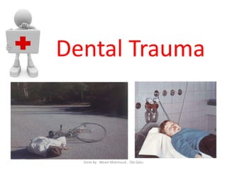
Dental Trauma Essential of trumatic injuries to the tooth by anderson Done BY : Weam faroun ,Ola Qatu

Recommended
More Related Content
What's hot
What's hot (20)
Similar to Dental Trauma
Similar to Dental Trauma (20)
More from Weam Faroun
More from Weam Faroun (11)
Recently uploaded
Recently uploaded (20)
Dental Trauma
- 1. Dental Trauma Done By : Weam Mahmoud .. Ola Qatu
- 2. Epidemiology • Most affected teeth are incisors • Peak ages 2 - 3 years 7 –- 11 years • Sex distribution m > f = 3:1 • Prevalence 50% of all children under 15 30% affecting deciduous teeth 20% affecting permanent teeth Done By : Weam Mahmoud .. Ola Qatu
- 3. • A patient presents to the clinic with a traumatic injury i.e. Fractured upper anterior tooth • Question: What should be done first? Done By : Weam Mahmoud .. Ola Qatu
- 4. Patient Examination • When patient is received for treatment of trauma, the oral region is usually heavily contaminated so the first step is to wash the patient’s face, in case of soft tissue wounds a mild detergent should be used Done By : Weam Mahmoud .. Ola Qatu
- 5. • Ask questions for diagnosis and treatment planning – Where/How/When did the injury occur? – Was there a period of unconsciousness? – Is there any disturbance in the bite? – Is there any reaction in the teeth to cold and/or heat exposure? • Ask about medical history (allergies/medical conditions ) Done By : Weam Mahmoud .. Ola Qatu
- 6. Clinical exam – Examine: face, lips and oral muscles for soft tissue lesions. – Palpate: facial skeleton for signs of fractures. – Inspect: dental trauma region for fractures or infarctions , tooth mobility, and abnormal response to percussion. – Pulp testing Percussion test Done By : Weam Mahmoud .. Ola Qatu
- 7. • Diagnose infarction by directing the light beam parallel to the labial surface of the injured tooth Done By : Weam Mahmoud .. Ola Qatu
- 8. • Mobility of group of teeth indication for alveolar fracture • Tenderness to percussion in axial direction indication for PDL damage Mobility test Done By : Weam Mahmoud .. Ola Qatu
- 9. Radiographic Examination Periapical Occlusal Panoramic Done By : Weam Mahmoud .. Ola Qatu
- 10. Radiographic examination • 1 occlusal & 3 periapiacal Occlusal give excellent view of : 1. Lateral luxation 2. Apical and mid root fracture 3. Alveolar fracture Periapical gives information 1.Cervical root fracture 2. Tooth displacement Done By : Weam Mahmoud .. Ola Qatu
- 11. Photographic registration • Used in treatment planning , legal claims or for clinical research Done By : Weam Mahmoud .. Ola Qatu
- 12. Done By : Weam Mahmoud .. Ola Qatu
- 13. Crown fracture • Crown fractures comprise the most frequent injuries in the permanent dentition- Apart from the loss of hard tissue; this injury can represent a hazard to the pulp • The closeness of the fracture to the pulp and the risk of bacteria or bacterial toxins penetrating dentin into the pulp are the primary sources of pulpal complications after crown fracture. Done By : Weam Mahmoud .. Ola Qatu
- 14. Crown fracture F/U: 6-8 wks and 1 yr TX: resin sealing TX: conservative treatment TX: pulp capping/partial pulpectomy/RCT Done By : Weam Mahmoud .. Ola Qatu
- 15. Treatment • some cases ---> selective grinding of the incisal edge is sufficient. Done By : Weam Mahmoud .. Ola Qatu
- 16. • In other cases, restoration with composite and the acid-etch technique is indicated. • fractures of enamel and dentin always require restoration in order to seal dentinal tubules and to restore esthetics Done By : Weam Mahmoud .. Ola Qatu
- 17. • In many situations, a temporary restoration may be indicated. These include pulpal involvement concomitant luxation injuries and lack of patient cooperation. • With respect to maintaining pulpal vitality, successful restoration of enamel-dentin crown fractures requires a hermetic seal of exposed dentinal tubules, this can be achieved by using glass ionomer cement, hard setting calcium hydroxide paste or a dentin bonding agent .While zinc oxide- eugenol cement has been found to be one of the best agents for producing a hermetic antibacterial seal (contraindicated with composite ) Done By : Weam Mahmoud .. Ola Qatu
- 18. Reattachment of crown fragment using a dentin bonding agent • Testing pulpal sensibility • The radiographic examination shows no displacement or root fracture. • Testing the fit of the fragment, the fragment fits exactly. • Temporary dentin coverage with calcium hydroxide, due to close proximity of the fracture surface to the pulp • Etching the enamel Done By : Weam Mahmoud .. Ola Qatu
- 19. • Storage of the crown fragment. The tooth is stored in physiologic saline for 1 month. The patient is given the fragment and is instructed to change the solution once a week to reduce contamination. • Bonding of the fragment after 1 month. • Preparation & Etching enamel • Removal of the etchant & Drying the fracture surfaces • Bonding the fragment & repositioning of the fragment, then light polymerization for composite • Finishing Done By : Weam Mahmoud .. Ola Qatu
- 20. Complicated Fracture involving enamel, dentin & pulp • Treatment option varies: • Level of root maturity (Pulpotomy or else) • Extent of damage restorability (Vital pulp therapy or RCT) • Time elapsed after fracture Crown. • Fracture with pulp involvement Done By : Weam Mahmoud .. Ola Qatu
- 21. R central without pulp exposure. L with pulp exposure Done By : Weam Mahmoud .. Ola Qatu
- 22. • Two treatment options exist: - Pulp capping - Partial pulpotomy. • the following conditions appear to favor pulpotomy rather than pulp capping: - Long exposure period after trauma (i.e. more than 24 h). - Large exposures (limit not established) - Reduced vascularity due to a concomitant luxation injury Done By : Weam Mahmoud .. Ola Qatu
- 23. • Pulp capping - Isolate the pulp exposure. - Cover the pulp with a calcium hydroxide material . - Restore the tooth either immediately with a bacteria-tight restoration, OR, after a 3- month period, where the exposure site is uncovered and the hard tissue barrier assessed. Thereafter, the hard tissue barrier is re-covered with hard-setting calcium hydroxide cement, glass ionomer cement or a composite resin retained with a dentin bonding agent, and thereafter restored. Done By : Weam Mahmoud .. Ola Qatu
- 24. • Pulpotomy - Isolate the pulp exposure. - Amputate the pulp to a level approximately 2 mm below the exposure site, or to where fresh bleeding is seen - If immediate restoration is desired, cover the exposure with hard-setting calcium hydroxide cement (e.g. - Dycal® ) Done By : Weam Mahmoud .. Ola Qatu
- 25. Crown root fracture • Most of these fractures occur as the result of a horizontal impact. • Crown-root fractures may or may not involve the pulp. Clinical diagnosis depends upon mobility of the coronal fragment. • Radiographic diagnosis, however, is uncertain as it is usually impossible to determine the oral extent of fracture. Done By : Weam Mahmoud .. Ola Qatu
- 26. treatment principles • Removal of the coronal fragment and supragingival restoration above gingival level (e.g., by bonding the original crown fragment after removing the sub gingival portion, with composite build-up or a crown) in order to permit sub gingival healing, presumably with a long functional epithelium. • Indication: This procedure should be limited to superficial fractures that do not involve the pulp. Done By : Weam Mahmoud .. Ola Qatu
- 27. • Removal of the coronal fragment supplemented by gingivectomy and/or osteotomy, in order to convert the sub gingival fracture surface to supragingival in situations where esthetics permit, thereafter, restoration (e.g. with a post-retained crown • Indication: Should only be used where the surgical technique does not compromise esthetic result Done By : Weam Mahmoud .. Ola Qatu
- 28. • Removal of the coronal fragment and surgical or orthodontic extrusion of the root, to move the fracture surface to a more optimal location for final restoration. • Indication: Should only be used where the root portion is long enough to accommodate a post-retained crown , ortho need more time Done By : Weam Mahmoud .. Ola Qatu
- 29. Root fracture • Root fractures are relatively uncommon injuries, but represent complex healing patterns due to concomitant injury to the pulp, periodontal ligament, dentin and cementum. • The fracture usually results from a horizontal impact. Fractures in the apical- and middle-thirds of root normally take an oblique course, being placed more apically on the labial aspect than on the palatal • Take radiographs with various angulations to diagnose fracture type and location. Done By : Weam Mahmoud .. Ola Qatu
- 30. Done By : Weam Mahmoud .. Ola Qatu
- 31. Done By : Weam Mahmoud .. Ola Qatu
- 32. Horizontal root fracture Vertical root fracture Treatment to facilitate pulpal and periodontal ligament healing, it is considered essential (although not proven) that a displaced coronal fragment be optimally repositioned. Furthermore, that splinting is maintained for a 3-month period in order to permit maximum stability of the hard tissue callus, repositioning and splinting of root fractures with different types of displacement (luxation) of the coronal fragment are demonstrated. Done By : Weam Mahmoud .. Ola Qatu
- 33. Concussion • Mechanism of concussion injury : A frontal impact leads to hemorrhage and edema in the periodontal ligament. • Least severe of Luxation injuries • Radiography: no signs of pathology Done By : Weam Mahmoud .. Ola Qatu
- 34. Done By : Weam Mahmoud .. Ola Qatu
- 35. Sub luxation • tooth tender to touch & slightly mobile (1+) but not displaced, possible hemorrhage from gingival crevice • No radiographic abnormalities • Mechanism of sub luxation injury: If the impact has greater force, periodontal ligament fibers may be torn resulting in loosening of the injured tooth. Done By : Weam Mahmoud .. Ola Qatu
- 36. Done By : Weam Mahmoud .. Ola Qatu
- 37. Treatment - Occlusal relief (e.g. by selective grinding of opposing teeth) and a soft diet. - Immobilization of the injured teeth may be appropriate for patient comfort. The fixation period is 2 weeks. Done By : Weam Mahmoud .. Ola Qatu
- 38. Extrusive luxation and lateral luxation - Extrusive luxation represents a rupture of the PDL and the pulp. - Lateral luxation represents a rupture of the PDL and the pulp as well as injury to the labial and/or palatal alveolar bone plate. - In both cases, healing includes both PDL repair and usually Pulpal revascularization. - Increase displacement ---> increase necrosisDone By : Weam Mahmoud .. Ola Qatu
- 39. • Treatment consists of: - Atraumatic repositioning and fixation. - In the case of lateral luxation, administration of local anesthetic is necessary prior to repositioning. - Radiographic examination after 2-3 weeks. *If radiographic examination reveals no sign of marginal breakdown, the splint can be removed. Otherwise further controls are necessary. *If radiographic examination reveals inflammatory resorption of the bone and root, immediate endodontic therapy is required. Done By : Weam Mahmoud .. Ola Qatu
- 40. Extrusive luxation • Pathogenesis of extrusive luxation : oblique forces displace the tooth out of socket. Only the gingival fibers palatally prevent the tooth from being avulsed. Both the PDL and the neurovascular supply to the pulp are severed. • Radiographically, a periapical bisecting angle exposure is more useful than an occlusal exposure Done By : Weam Mahmoud .. Ola Qatu
- 41. Clinical and radiographic feature of extrusive luxation The bisecting angle radiographic technique Is more useful than an occlusal exposure in revealing displacement. Done By : Weam Mahmoud .. Ola Qatu
- 42. Done By : Weam Mahmoud .. Ola Qatu
- 43. Lateral luxation • Pathogenesis of lateral luxation : Horizontal forces displace the crown palatally and the apex labially. Apart from severance of the PDL and the neurovascular supply to the pulp, compression of the PDL is found on the palatal aspect of the root. • Occlusally radiograph or eccentrically oriented exposure will tend to come between the root of the tooth and the empty socket, thus revealing the true nature of the injury. Done By : Weam Mahmoud .. Ola Qatu
- 44. Clinical and radiographic feature of lateral luxation The occlusal radiographic exposure or the eccentric bisecting angle exposures are more useful than an orthoradial bisecting angle in revealing displacement. Done By : Weam Mahmoud .. Ola Qatu
- 45. Done By : Weam Mahmoud .. Ola Qatu
- 46. Intrusion • Intrusion is the result of an axial, apical impact and results in extensive damage to the pulp and PDL. • Pathogenesis of intrusion: Axial Impact leads to extensive Injury to the pulp and periodontium Done By : Weam Mahmoud .. Ola Qatu
- 47. • ƒTreatment: - Immature root formation A wait spontaneous re-eruption, which usually takes 2-4 months. Monitor pulpal healing radiographically 3, 4 and 6 weeks after injury. - Mature root formation A wait spontaneous re-eruption or extrude orthodontically over a period of 2 - 3 weeks. Extirpate the pulp 2 weeks after injury, using calcium hydroxide paste as an interim dressing root till with a permanent gutta- percha filling once periodontal healing has been established radiographically. Done By : Weam Mahmoud .. Ola Qatu
- 48. Done By : Weam Mahmoud .. Ola Qatu
- 49. Done By : Weam Mahmoud .. Ola Qatu
- 50. Avulsion • Avulsion of permanent teeth is most common in the young dentition, where root development is still incomplete and the periodontium very resilient. Done By : Weam Mahmoud .. Ola Qatu
- 51. Done By : Weam Mahmoud .. Ola Qatu
- 52. • Replantation of avulsed teeth can result in successful healing if there has been only minimal damage to pulp and periodontal ligament. • The type of extra alveolar storage and length of storage period have an overwhelming effect upon later healing. • Replantation should be attempted only if the following conditions can he fulfilled : - Absence of gross caries and no major loss of periodontal support prior to injury. - Physiological storage of the tooth (in the case of an a vital PDL) Done By : Weam Mahmoud .. Ola Qatu
- 53. • Replantation procedure - Place the avulsed tooth in saline; saliva or milk - Examine the socket area - Rinse the periodontal ligament and apical foramen with saline - Flush the socket with saline - Replant the tooth with gentle finger pressure - Splint the tooth for 1 week with a semi-rigid splint - Begin antibiotic therapy as soon as possible after injury (e.g. penicillin) If the patient is not covered for tetanus ,tetanus vaccine should be administered Done By : Weam Mahmoud .. Ola Qatu
- 54. Done By : Weam Mahmoud .. Ola Qatu
- 55. Done By : Weam Mahmoud .. Ola Qatu
- 56. Alveolar Fracture -Verify the extent and position of the fracture clinically and radiographically, using a multiple radiographic exposure technique. - Place local anesthetic infiltration. - In case of an apical lock, the fragment must first be slightly extruded to free the apices. It is then possible to reposition the fragment. - Splint the fragment for 3 - 4 weeks, according to the age of the patient. - Monitor pulpal healing of the involved teeth. Done By : Weam Mahmoud .. Ola Qatu
- 57. Done By : Weam Mahmoud .. Ola Qatu
- 58. REACTION OF THE TOOTH TO TRAUMA 1. PULPAL HYPEREMIA Radiograph demonstrates almost complete obliteration of the pulp chambers and canals. Done By : Weam Mahmoud .. Ola Qatu
- 59. 2. Internal hemorrhage. 3. Calcific metamorphosis of dental pulp ( progressive canal calcification or dystrophic calcification ) 4. Internal resorption 5. Peripheral (external) root resorption 6. Pulpal necrosis 7. Ankylosis Done By : Weam Mahmoud .. Ola Qatu
- 60. Done By : Weam Mahmoud .. Ola Qatu
- 61. Done By : Weam Mahmoud .. Ola Qatu
- 62. Done By : Weam Mahmoud Faroun.21410298 Ola Abed Alrahman Qatu.21411059 Submitted to : DR Emad Qurrish Done By : Weam Mahmoud .. Ola Qatu
- 63. Reference • Essential of trumatic injuries to the tooth by anderson • McDONALD AND AVERY’S DENTISTRY FOR THE CHILD AND ADOLESCENT, TENTH EDITION Done By : Weam Mahmoud .. Ola Qatu