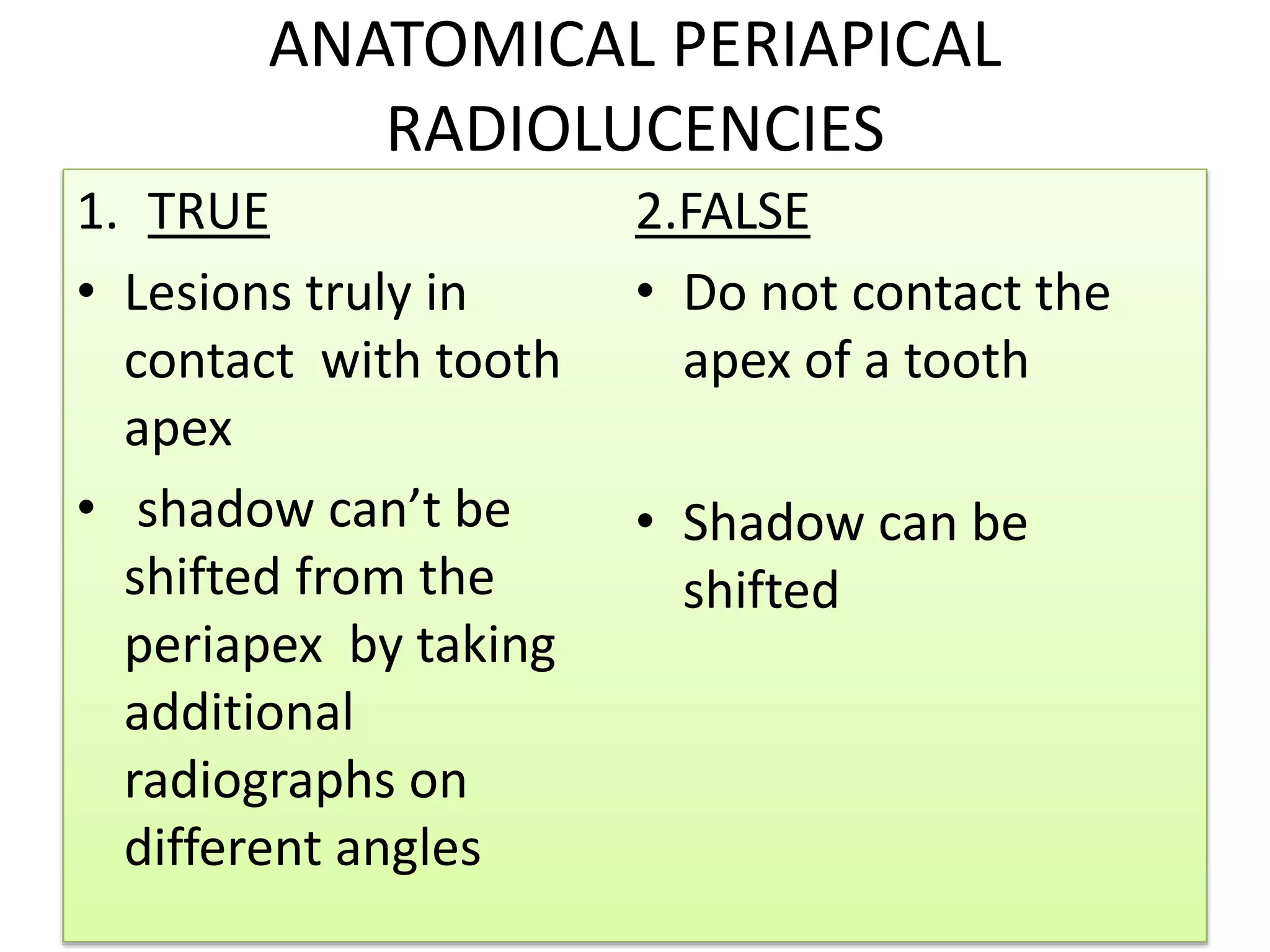
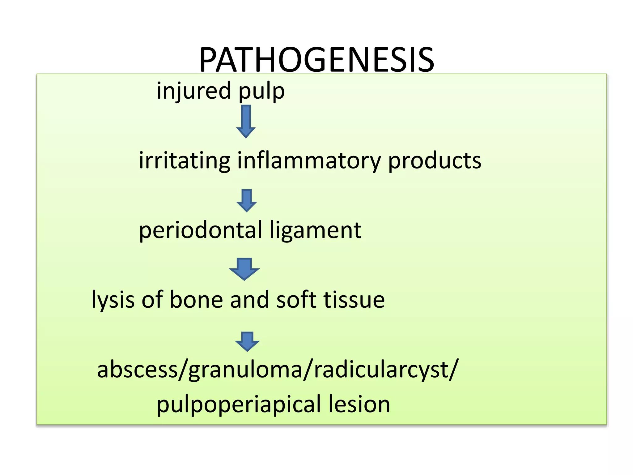
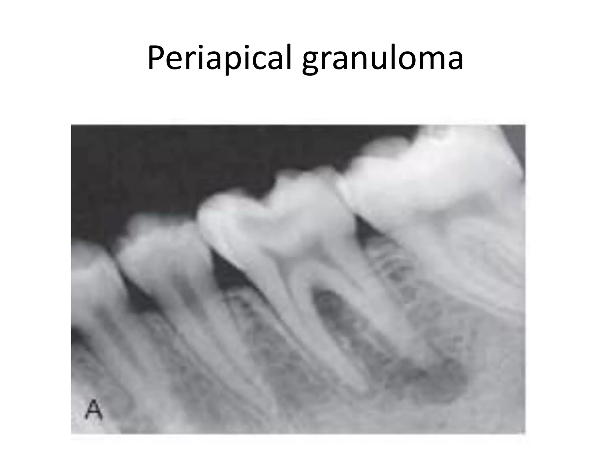
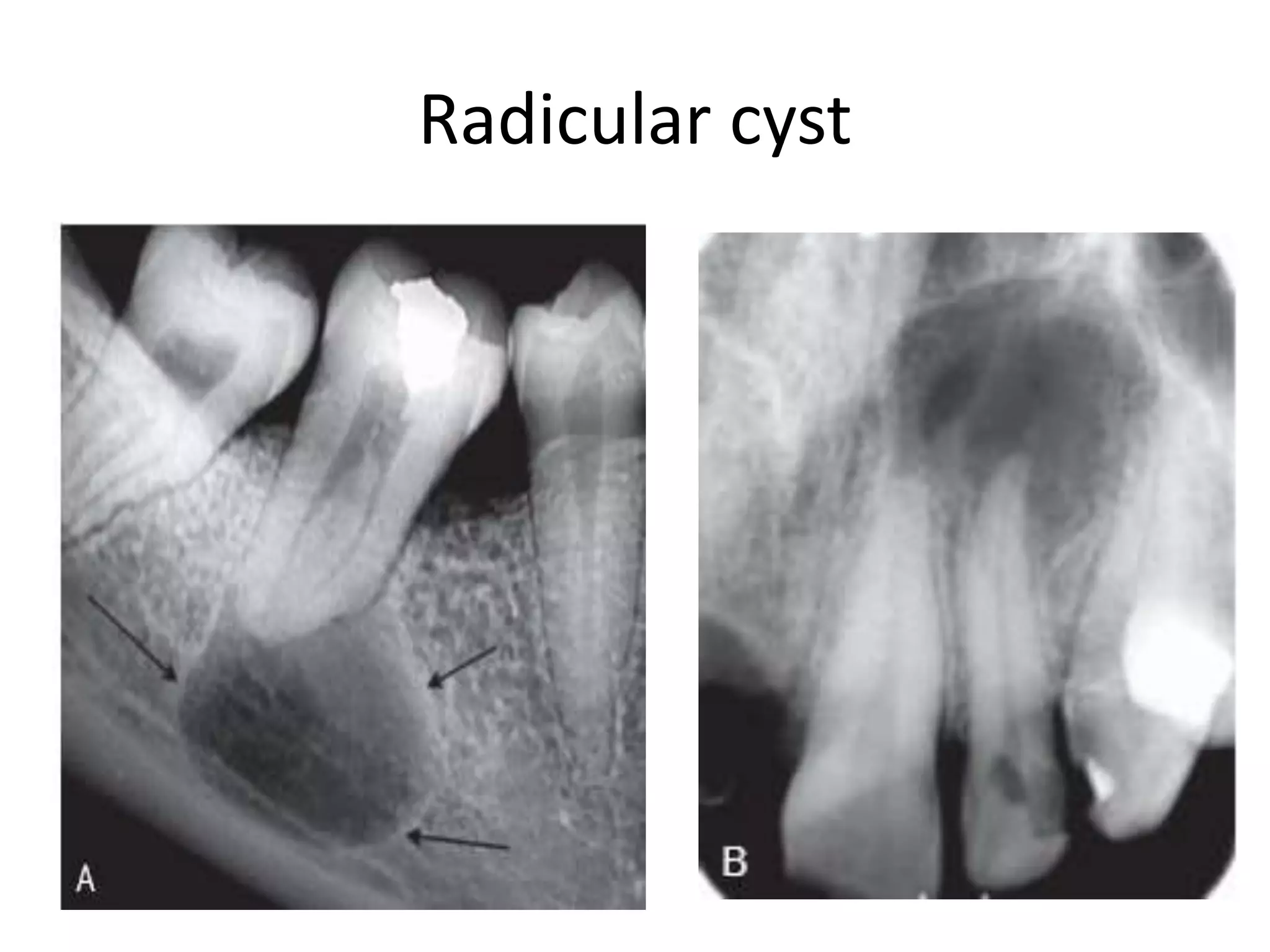
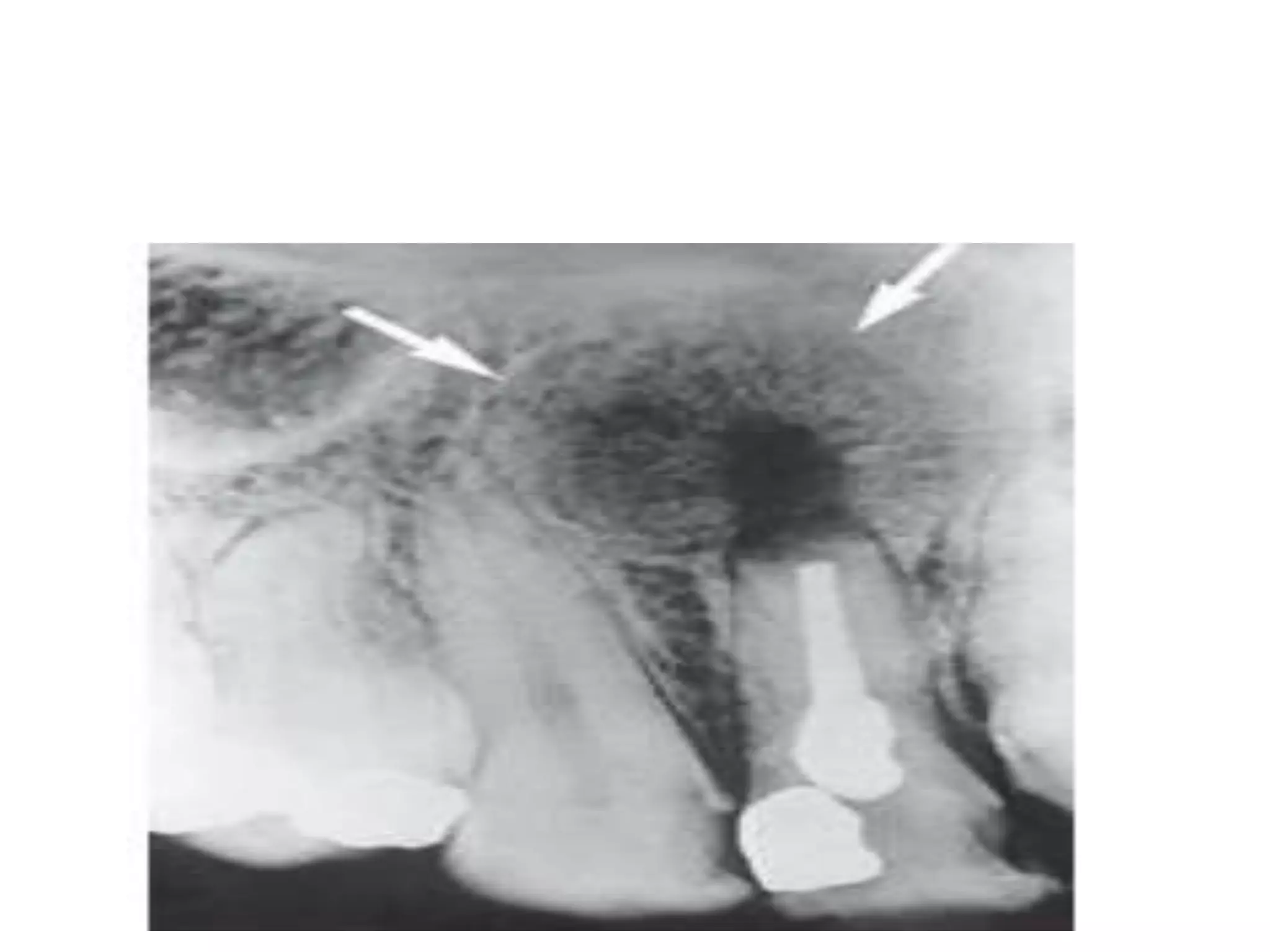
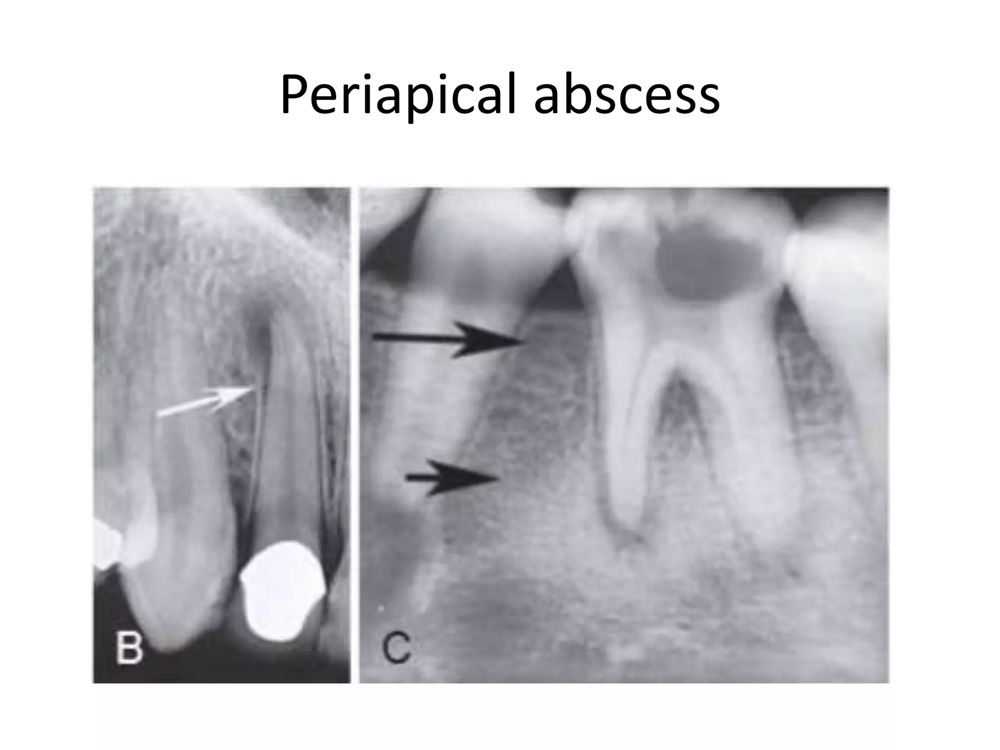
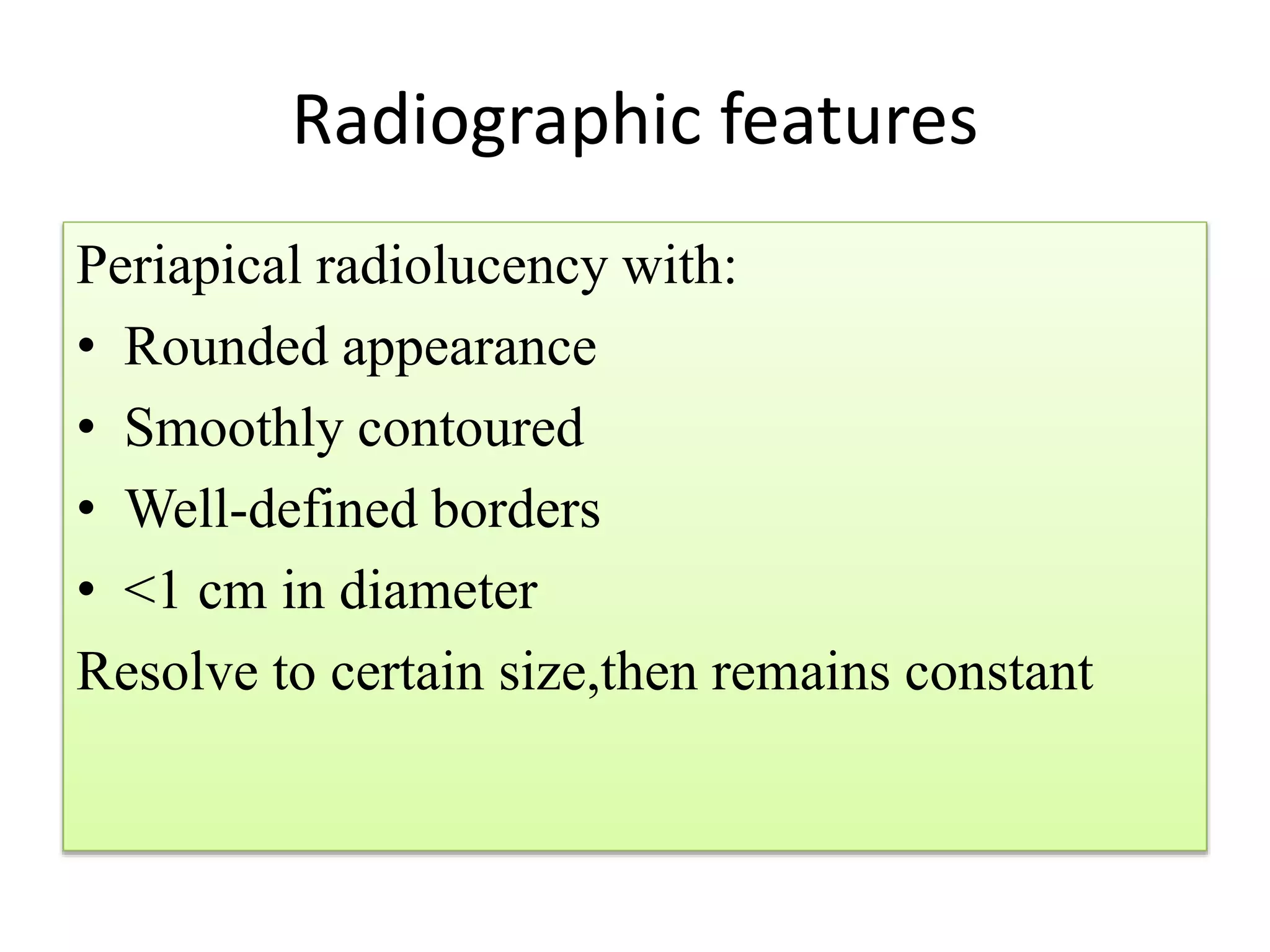
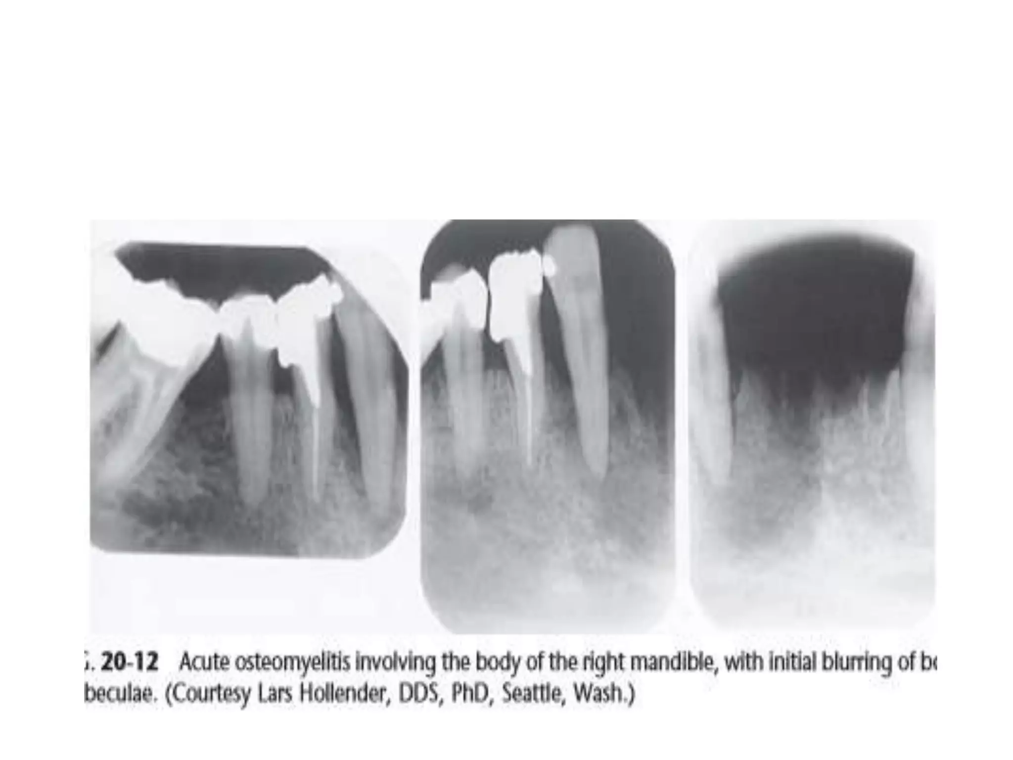
Periapical radiolucencies can have many causes, both benign and malignant. They are often classified as either anatomical pseudoperiapical radiolucencies, which do not contact the tooth apex, or true periapical radiolucent lesions, which do. Common true lesions include periapical granulomas, radicular cysts, and periapical abscesses. Periapical granulomas appear as well-defined radiolucencies, while radicular cysts can cause tooth displacement if left untreated. Management depends on the diagnosis and may involve root canal treatment, extraction, or surgery. Differential diagnosis considers conditions like osteomyelitis, dentigerous cysts,
3. INTRODUCTION
• Radiolucent shadows are cast over the
periapical regions of teeth in practically all oral
radiographic surveys of dentulous patients
• Some of this periapical radiolucencies
represent innocent anatomic variations,
• Others caused by benign condition
• Still others represent systemic disease
• Malignancies represent a small group of these
periapical shadows
5. ANATOMICAL PERIAPICAL
RADIOLUCENCIES
1. TRUE
• Lesions truly in
contact with tooth
apex
• shadow can’t be
shifted from the
periapex by taking
additional
radiographs on
different angles
2.FALSE
• Do not contact the
apex of a tooth
• Shadow can be
shifted
11. Clinical features
• Asymptomatic
• occasional severe toothache With/Without
facial swelling
• Fever
• Lymphadenopathy
• Non-vital pulp
• Swelling/expansion of cortical plates
12. Radiographic features
• Well-circumscribed radiolucency around the
apex of the tooth
• With thin radiopaque border
• Cannot be differentiated from radicular cyst
• Usually within a range of 2.5 cm
13. • LOCATION: epicenter at the apex of the
involved tooth
• PERIPHERY: ill-defined
• INTERNAL STRUCTURE: initially loss of bone
density,later,a mixture of sclerosis and
rarefraction,i.e ,periapical sclerosing osteitis
and periapical rarefying osteitis
14. • EFFECTS ON SURROUNDING STRUCTURES:
Loss of lamina dura,nearby cortical boundaries
can be destroyedfloor of nasal fossa ,buccal and
lingual plates of alveolar process
• Thin layer of new bone formed-halow shadow
17. 2.Radicular cyst
• 2 nd most common pulpoperiapical
• Most common of all odontogenic cyst
• Inflammatory cyst
Inflammatory products initiate the growth of
the epithelial component
• Odontogenic cyst
Origin in malassez rests,PDL cells,remnants of
epithelial root sheath
18. pre-existing periapical granuloma
proliferating epithelial cell nest
cell nest size increases
central cells degenerate and liquify
fluid filled cavity lined with epithelium
enlarging cyst on alveolar bone
osteoclastic activity
bone resorption
19. CLINICAL FEATURES
• Identical to periapical granuloma
• If periapical radiolucency >1.6 cm in dm
more likely radicular cyst
• if untreared,expand cortical plates
• Initially bony hard swelling to palpate
• Later, rubbery and fluctuant due to fluid
• Large cyst involves complete quadrant
• Occasionally tooth mobility,non-vital pulp
• If infected,painfull symptoms of an abscess
20. • Microscopic features:
Periphery is fibrous,
Inner region –granulation tissue where foci of
chronic inflammatory cells,foam cells, russell
bodies,cholesterol clefts
• Cholesterol crystal: shiny crystals from
aspirated noninfected cyst
21. Radiographic features
• Location: apex of the non-vital tooth,
Maxilla,around incisors and canine
Due to distal inclination of maxillary lateral
incisor,may invaginate the maxillary antrum
• Periphery and shape: well-defined cortical
border
• Internal structure:radiolucent
Dystrophic calcification in long standing cases
22. • Effects on internal structures:
Displacement and resorption of adjacent teeth
may occur
Invagination in to antrum
Outer cortical plates of maxilla and mandible
may expand in a curved or circular shape
Displace mandibular alveolar nerve canal in an
inferior direction
25. management
• Non-surgical endodontics
• If extraction ,curettage of periapical area should
be judiciously curetted
• Large radicular cyst:6 approaches
1. Surgical enucleation
2. Surgical enucleation and restoration with graft
3. Marsupialization
4. Decompression
5. Decompression with delayed enucleation
6. Creating common chamber with maxillary sinus
26. • Post surgical radiographs :
Average healing time for cyst of more than
10mm in diameter is approximately 2 and half
years .
27. 3.Periapical scar
• Composed of dense fibrous tissue
• In periapex of pulpless root canal filled teeth
• Represents previous periapical
granuloma/cyst/abscess,whose healing is
completed by dense scar formation
28. Clinical features
• Resemples periapical granuloma and cyst
• Causes well circumscribed radiolucency
• Asymptomatic
• Anterior region of maxilla
• Endodontically treated tooth
31. Chronic and acute dentoalveolar
abscess
Sudivided based on whether they are
radiolucent
1. Primary/neoteric abscess
2. Secondary/recrudescent abscess
32. Primary abscess
1. Normal on radiograph
2. Acute and exudative
3. Involving PDL tissues,at the tooth apex,
4. Necrotic pulp
5. Acute apical periodontitis
6. Very sensitive tooth
7. Alveolar swelling
8. Onset and course of infection are same
9. Sudden bone resorption
33. Secondary abscess
• Chronic/acute type,
• depending on organisms ,host,type and
timing of treatment
• Microflora is polymicrobial
• Majorly anerobes,co2 dependent
streptococci,and bacilli
34. Clinical features
• Periapical radiolucency in secondary abscess
• Depending on duration,margins of
radiolucency may vary
1.Well defined with hyperostotic border
2.Poorly defined in chronic abscess
• Root resorption
• Pain on percussion in acute abscess
• Non-vital
• Tooth mobility
• On progress,Space infection
39. Surgical defect
• Area where osseous tissue fail to fill after
surgery
• After root resection
• When labial and lingual plates are distroyed
• Clinically:
Mucosal scar from previous surgery
Palpated if large enough
40. Radiographic features
Periapical radiolucency with:
• Rounded appearance
• Smoothly contoured
• Well-defined borders
• <1 cm in diameter
Resolve to certain size,then remains constant
42. Osteomyelitis
• Defined as an infection of bone that involves:
1. Periosteum
2. Cortex
3. Marrow
Osteomyelitis –more aggressive diffuse
Osteitis-localized condition
2 major phases:acute and chronic
43. • Acute phase:
1. Synonyms:acute supportive osteomyelitis, garres
osteomyelitis,proliferative periostitis
2. caused by infection that has spread to bone
marrow
3. Medullary spaces of bone contain inflammatory
infiltrate consisting predominantly of neutrophils
4. Infection spread periosteally
elevating periosteum
stimulating bone formation
44. • Clinical features:
Affect any age
Strong male predilection
Mandible> maxilla (poor vascular supply)
Rapid onset,pain,swelling of adjacent soft
tissue,fever,lymphadenopathy,leukocytosis,
associated teeth may be mobile ,sensitive to
percussion,purulent drainage,paresthesia of
lower lip third division of 5 th cranial nerve is
not uncommon
45. • Radiological examination
Location: posterior body of the mandible
Periphery: ill defined periphery with a gradual
transition to normal trabeculae
Internal structure:slight decrease in density of
involved bone with loss of sharpness of existing
trabaculae
bone distruction,radiolucency in one focal
area/scattered regions throughout the involved
bone
sclerotic regions,sequestration formation
46. effects on surrounding structures
stimulate bone resorption/formation
cortical bone may be resorbed
inflammatory exudate lift the periosteum
stimulate bone formation
Radiographically,faint radiopaque line separated
from bone surface
onion skin appearance
,chronic phase
47.
48. Differential diagnosis
• Fibrous dysplasia
(in osteomyelitis, new bone formation is on
the periosteum,outer cortical plates get
thickened)
(But in fibrous dysplasia,new bone inside
mandible and outer cortex may be thinned)
• Malignant neoplasia (scc,osteosarcoma)
• Langerhans cells histiocytosis:rarely show a
sclerotic bone reaction
49. Chronic phase
• Synonyms
Chronic diffuse sclerosing osteomyelitis
Chronic non suppurative osteomyelitis
Chronic osteomyelitis with proliferative
periostitis
• A sequelae of inadequately treated acute
phase
• Symptoms are less severe and have longer
history
50. Radiographic features
• LOCATION:posterior mandible
• PERIPHERY :gradual transition is seen b/w
normal surrounding trabecular pattern and
dense granular pattern
• When disease is active,spread through
bone,periphery may be radiolucent and have
poorly defined borders
51. • INTERNAL STRUCTURE:
Lesion composed of more radiopaque
/sclerotic bone pattern
In older,internal density can be
radiopaque,equivalent to cortical bone.no
radiolucencies is evident.
Small regions of radiolucency may be
scattered through out the radiopaque bone
Island of sequestrum within the centre
52. • EFFECT ON SURROUNDING STRUCTURE:
Stimulate formation of periosteal new
bone,seen as single radiopaque line /series of
radiopaque line parallel to surface of cortical
bone/onion skin
53.
54. • Over time,radiolucent strips that separates new
bone from outer cortical bone.when this occurs,it
may not be possible to identify the original
cortex,which makes it difficult to determine
whether the new bone is formed is formed from
periosteum
• Roots of teeth may undergo external resorption
,lamina dura become less apparent as it blends
with surrounding granular sclerotic bone
• If tooth is non-vital ,the PDL space usually
enlarged in the apical region .in patients with
extensive chronic osteomyelitis,disease may
slowly spread to mandible condyle and in to the
joint ,resulting in a septic arthritis
• Spread involve inner ear and mastoid air cells
55. • Chronic lesions develop a draining
fistula,appear as a well defined break in the
outer cortex in the periosteal new bone .
58. Hyperplasia of maxillary sinus lining
• Pulpo periapical inflammation and
periodontal conditions frequantly produce
local inflammatory hyperplasia of adjacent
antral soft tissue floor.
• These appear as gray shadows that may be
dome shaped in the maxillary sinus floor or
gray radiopacity that appears as a cap over
adjacent troublesome root
59. • The free margin of soft tissue shadow is
usually smoothly contoured and distinct
although the soft tissue shadow may show
varying degree of grayness
• Soft tissue shadows disappear after treatment
61. Dentigerous cyst
• Follicular cyst
• Cyst forms the crown of an erupted tooth
• It begins when fluid accumulates in the layers
of reduced enamel epithelium /b/w the
epithelium and crown of an erupted tooth
• An eruption cyst is the soft tissue counterpart
of unerupted tooth
• An eruption cyst is the soft tissue counterpart
of a dentigerous cyst
62. Clinical features
• 2 nd most common type of cyst in jaw
• Develop around crown of an unerupted or
supernumerary tooth
• Clinical examination reveals a missing tooth
/teeth,possibly a hard swelling occasionally
resulting in facial asymmetry
• Patient has no pain /discomfort
63. Radiographic features
• Location
Just above the involved tooth
Mandibular/maxillary 3 rd molar/maxillary
canine
This cyst attaches to cementoenamel junction
Some are eccentric, developing from lateral
aspect of follicle so that they occupy an area
beside crown instead of above crown
64. • Cyst related to maxillary 3 rd molar grow to
maxillary antrum
• Mandibular molar-extend in to ramus
Periphery and shape
• Well-defined cortex with curved outline
• Infection if present ,cortex may be missing
Internal structure
Completely radiolucency exept for the crown of
involved tooth
65. • Effects on surrounding structures
Displace and resorb adjacent teeth
Floor of maxillary antrum may displace as the
cyst invaginate the antrum and the cyst may
displace the inferior alveolar nerve canal in an
inferior direction
Slow growing—expands the outer cortical
boundary of the involved jaw
66.
67. Differential diagnosis
• Hyperplastic follicle(cyst cause tooth
displacement and some degree of bone
expansion,if follicular space exeeds 5mm,a
dentigerous cyst is more likely)
• OKC
• Adenomatoid odantodenic tumour
68. Periapical cementosseous dysplasia
• Periapical cementoma
• most common fibrocementosseous lesion
• Reactive fibroosseous lesion
• Thought to arise from elements in the
versatile PDL,where mature osteoblasts,and
precursor cells reside
69. Radiographic features
• The early stage:osteolytic/osteoblastic
Radiolucent
Microstructure:cellular fibroblastic stroma
that may contain a few small foci of calcified
material
• Intermediate stage:radiolucent area
containingradiopaque foci
• The final stage:mature lesion
completely calcified,Welldefined,solid,
homogeneous radiopacity surounded by a thin
radiolucent border
70. • Calcified material in periapical lesions may be
entirely cementum,entirely
osseous/combination
• Well defined borders
• Non vital teeth
• Mandible predilection
• Periapical region of incisors
• Solitary/multiple exeeding 1cm
• Asymptomatic,cortical plate expansion
74. Periodontal disease
• Radiolucency caused by advanced periodontal
bone loss involving one teeth more severly
than the teeth immediately adjacent
• Appear to be floating in a radiolucency
• Apex is well defined radiolucencywhich in one
projection seems to surrounded by bone
• Pocket depth relative to root length of
associated tooth can be demonstrated by
placing gutta –percha points in the pockets to
their full depth.
75. • Teeth with advanced pdl distruction are
mobile
• Sensitive to percussion
• Vitality positive
76.
77. Traumatic bone cyst
• Hemorrhagic bone cyst
• Extravasation cyst
• Simple bone cyst
• Solitary bone cyst
• Progressive bony cyst
• Blood cyst
• False cyst of bone because it does not have an
epithelial lining
78. Clinical features
• Asymptomatic
• Mandible
• Commonly in premolar and molar regions
• Symphysis is frequently involved
• teeth : vital,intact lamina dura
• Usually in patient under 25 yrs of age
79. Radiographic examination
• Well-defined(cyst like) radiolucency above the
mandibular canal
• Location:Predominantly round to oval
positioned somewhat symmetrically about
the periapex of a root
o Ramus and posterior mandible
o Frequantly occurs in cemento-osseous and
fibrous dysplasia
80. • Periphery and shape:
Margin vary from well-defined to ill-defined
Often scallops blw the roots
• Internal structure:
Totally radiolucent
Multilocular although the lesion doesn’t
contain true septa
Pronounced scalloping of the endosteal
surface of either buccal and lingual plates
81. • Effects on surrounding structure:
Intact lamina dura,/partly disrupted
Sparing of cortical boundary of the crypt
around a developing tooth
Tendency to scallop endosteal surface of the
outer cortex of the mandible
Tendency to grow along the long axis of the
bone,causing minimal expansion
82.
83. Differential diagnosis
• Radicular cyst
• PCOD
Management:
Open the area surgically,remove the tissue
debris,curette the walls of the bony cavity to
induce bleeding ,close the soft tissue flap
securely
84. Non-radicular cysts
• May be projected over the apices of teeth
• Incisive canal,midpalatine cyst,median
mandibular cyst,primordial cyst
• Specific regions of the jawbone(expn for
primordial cyst)
• Teeth associated are vital
87. Malignant tumours
• Single periapical radiolucency
• Malignant tumours may be primary or secondary
• Mistaken as pulpoperiapical lesions
• Biopsy is mandatory for periapical lesions that do
not respond to endodontic treatment
• SCC,malignant tumour if minor salivary gland
,metastatic tumours ,osteolytic
sarcamo,chondrosarcoma,
melanoma,fibrosarcoma,reticulum cell sarcoma
and multiple myeloma
88. • Granted enlarging lesions with ragged,moth
eaten borders
• Mesenchymal malignant tumours and
metastatic tumours originating within bone
are more apt to produce a localized
radiolucency than a peripheral SCC which
originates in the surface and erodes through
alveolar bone to arrive at the apex
• SCC originating within a cyst could be seen as
localized periapical radiolucency
89. • They may produce
1. Well-defined periapical radiolucency
2. Poorly defined periapical radiolucency
3. A large ragged,well defined radiolucent
tumour that has destroyed a large segment
of surface bone and has widening of PDL
spacing
90. features
• More common in patients of middle and old
age
• Pain may be a feature,involved teeth may
retain their vitality
• If tumour is
advanced,migration,loosening,tipping,and
spreading of teeth .
• Gingival bleeding
• Paresthesia or anesthesia of soft tissues is
sometimes present
91. • Expansion of jaw in advanced lesions
Differential diagnosis
Early lesions mimic benign condns
Management
2 basic principles
First:tooth and area in question are observed
with periodic clinical and radiographic
examination,the lesion and tooth are treated
with conservative endodontic techniques
Second:if clinician chooses to perform a root
resection in addition to root canal filling