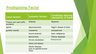
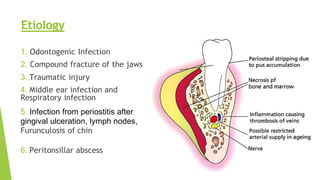
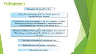
Osteomyelitis
Presented by : Ankit Thummar A.
Roll No.:- 92
Batch : H
• Osteomyelitis is defined as an inflammation of the
medullary portion of the bone. The process rarely is
confined to the endosteum and usually affects the cortical
bone and the periosteum.
• osteomyelitis may be defined as an inflammatory condition
of the bone that begins as an infection of the medullary
cavity, rapidly involves the Haversian system and quickly
extends to the periosteum
• Osteon = Bone, Myelos = Bone Marrow (in Greek word)
N.b. Don’t confused with Myositis and Myelitis
Definition
Local factors Systemic factors Conditions altering
vascularity of bone
Trauma and road traffic
accidents
Diabetes Radiation
splints Agranulocytosis Paget’s disease of bone
gunshot wounds Leukaemia osteopetrosis
Severe Anaemia bone malignancy
Malnutrition Fibrous Dysplasia
Chronic Alcoholism Osteoporosis
Sickle cell disease
febrile illnesses
such as typhoid,Scarlet
Fever
Predisposing Factor
1. Odontogenic Infection
2. Compound fracture of the jaws
3. Traumatic injury
4. Middle ear infection and
Respiratory infection
5. Infection from periostitis after
gingival ulceration, lymph nodes,
Furunculosis of chin
6. Peritonsillar abscess
Etiology
Pathogenesis
Sequelae of
Pulpitis
According to
anatomic
location of
infectious
process:
According to
duration and
severity:
Depending on
Suppuration:
Osteomyelitis
due
to specific
infection:
Intra-medullary Acute Suppurative Actinomycosis
Sub-periosteal Chronic Non - Suppurative Tuberculosis
Periosteal Syphilis
Classification
Suppurative
 Acute suppurative
osteomyelitis
 Chronic suppurative
osteomyelitis
1.Primary
2.Secondary
 Infantile osteomyelitis
Non Suppurative
 Chronic nonsuppurative
OML
1. Focal Sclerosing
2. Diffuse Sclerosing
 Radiation osteomyelitis
 Garre's sclerosing
osteomyelitis
Based on Suppuration
Acute Suppurative OML
It is a serious sequelae of periapical infection that often results in a diffuse spread of
infection throughout the medullary spaces with subsequent necrosis of a variable
amount of bone
 Clinical Feature :
Deep Intense Jaw Pain
Abscess Formation
High Intermittent Fever
Paresthesia of Lip
No fistula, Diffuse Swelling, Mobile teeth, Trismus
 Radiographic Feature:
- Moth Eaten Appearance
 Clinical Feature:
Paraesthesia of the lip may be seen, though not classically seen in
chronic suppurative osteomyelitis
It develop after acute phase of the disease has subsided (secondary), or
it may arise from a dental infection without a preceding acute stage
(primary)
 Radiographic Feature:
Irregular radiolucent areas superimposed on more sclerotic and
nontrabeculated zones
Chronic Suppurative OML
Chronic Focal Sclerosing OML
 Clinical Feature:
occurs in cases of extremely high tissue resistance or in cases of low-
grade medullary infection which causes endosteal or periosteal reactions.
Most common in younger individuals in the mandibular first molar region
with mild pain and decreased sensitivity in the tooth
 Radiographic Feature:
well circumscribed radiopaque mass of
sclerotic bone surrounding and extending
below the apex of one or both roots
Chronic Diffuse sclerosing OML
 Clinical Feature:
It Occur at any age
first symptom may be a fistula on the mucosal surface
patient may complain of vague pain and a bad taste.
chronic condition & represents a proliferative reaction of the bone to a low-grade
infection
 Radiographic Features
cotton wool appearance
 Clinical Feature:
It occurs commonly in children and young adults.
Mandible is affected more commonly than maxilla.
toothache or pain in the jaw and bony hard swelling in the outer surface of the jaw.
 Radiographic Feature:
Onion Skin Appearance
Chronic Osteomyelitis with Proliferative Periostitis Or
Garre’s Osteomyelitis Or Periostetitis Ossificans
Infantile osteomyelitis is believed to occur by haematogenous
route or from perinatal trauma that occurs few weeks after birth and usually
involves the maxilla.
 Clinical Feature:
Facial cellulitis centred about the orbit
Inner and outer canthal swelling
Palpebral oedema
Closure of the eye and proptosis
Purulent discharge from the nose and medial canthus
Generalised symptoms : fever, irritability, malaise, anorexia, dehydration,
convulsions and vomiting
Infantile
Osteomyelitis
Treatment of Osteomyelitis
Primary Treatment Measure Supportive Measures
Empirical Therapy of Antibiotics Sequesterectomy
Analgesics Saucerisation
Incision & Drainage Decortication
Removal of Offending teeth Resection & Reconstruction
Debridement Hyperbaric Oxygen Therapy
Bed Rest & Tx. of underlying disease
• Removal of sequestrum
• It may be carried out under proper cover of antibiotics
• Small Sequestrum → Under Local Anesthesia
• Bigger Sequestrum → Under General Anesthesia
• Involved site & Incision
Sequestrectomy
Involved Site Incision
For Lower border of mandible Risdon'sIncision
Ramus of Mandible Retromandiblur Incision
Coronoid Process Anterior broeder of ramus of
mandible
Consular Process Preauricular Incision
• After removal of the sequestrum the residual granulation tissue is
curetted till white shining bone appears.
• Bleeding is controlled by pressure pack
• When complete infection has been eradicated the wound can be closed
by primary closure
• When the elimination of the infection is doubtful, a glove drain or a
rubber drain is kept in place and is changed every 24 hours till no
discharge from the bone
• Sufficient bone is removed to convert the deep cavity into Shallow saucer
• It consists of elimination for bony cavity in the jaw bone to avoid collection of blood
and formation of large hematoma which is liable to get infected
• Saucerization is carried out by existing the wall of the bony cavity by means of
Rongeur bone-cutting forceps or burs and bone is smoothen by file
• The wound can be partially or completely closed depending upon the amount of
suppuration
Saucerization
Decortication:
First introduced in 1917 by Mowlem
Decortication involves removal of the chronically infected
cortex, usually the buccal and the inferior border are removed
1–2 cm beyond the affected area
Resection & Reconstruction
Excision of a part of the mandible is seldom necessary
Antibiotic therapy is started 24 h before operation
IMF is done, reconstruction plate contoured and screws. drilled
before resection in order to maintain preoperative occlusion
Partial resection involves
preservation of the condylar and coronoid processes
Total disarticulation of the mandible involves complete stripping of
the
coronoid and condylar processes
Thank you! 🦷

Osteomyelitis with Types and Treatment..

  • 1.
    Osteomyelitis Presented by :Ankit Thummar A. Roll No.:- 92 Batch : H
  • 2.
    • Osteomyelitis isdefined as an inflammation of the medullary portion of the bone. The process rarely is confined to the endosteum and usually affects the cortical bone and the periosteum. • osteomyelitis may be defined as an inflammatory condition of the bone that begins as an infection of the medullary cavity, rapidly involves the Haversian system and quickly extends to the periosteum • Osteon = Bone, Myelos = Bone Marrow (in Greek word) N.b. Don’t confused with Myositis and Myelitis Definition
  • 3.
    Local factors Systemicfactors Conditions altering vascularity of bone Trauma and road traffic accidents Diabetes Radiation splints Agranulocytosis Paget’s disease of bone gunshot wounds Leukaemia osteopetrosis Severe Anaemia bone malignancy Malnutrition Fibrous Dysplasia Chronic Alcoholism Osteoporosis Sickle cell disease febrile illnesses such as typhoid,Scarlet Fever Predisposing Factor
  • 4.
    1. Odontogenic Infection 2.Compound fracture of the jaws 3. Traumatic injury 4. Middle ear infection and Respiratory infection 5. Infection from periostitis after gingival ulceration, lymph nodes, Furunculosis of chin 6. Peritonsillar abscess Etiology
  • 5.
  • 7.
  • 8.
    According to anatomic location of infectious process: Accordingto duration and severity: Depending on Suppuration: Osteomyelitis due to specific infection: Intra-medullary Acute Suppurative Actinomycosis Sub-periosteal Chronic Non - Suppurative Tuberculosis Periosteal Syphilis Classification
  • 9.
    Suppurative  Acute suppurative osteomyelitis Chronic suppurative osteomyelitis 1.Primary 2.Secondary  Infantile osteomyelitis Non Suppurative  Chronic nonsuppurative OML 1. Focal Sclerosing 2. Diffuse Sclerosing  Radiation osteomyelitis  Garre's sclerosing osteomyelitis Based on Suppuration
  • 10.
    Acute Suppurative OML Itis a serious sequelae of periapical infection that often results in a diffuse spread of infection throughout the medullary spaces with subsequent necrosis of a variable amount of bone  Clinical Feature : Deep Intense Jaw Pain Abscess Formation High Intermittent Fever Paresthesia of Lip No fistula, Diffuse Swelling, Mobile teeth, Trismus  Radiographic Feature: - Moth Eaten Appearance
  • 11.
     Clinical Feature: Paraesthesiaof the lip may be seen, though not classically seen in chronic suppurative osteomyelitis It develop after acute phase of the disease has subsided (secondary), or it may arise from a dental infection without a preceding acute stage (primary)  Radiographic Feature: Irregular radiolucent areas superimposed on more sclerotic and nontrabeculated zones Chronic Suppurative OML
  • 12.
    Chronic Focal SclerosingOML  Clinical Feature: occurs in cases of extremely high tissue resistance or in cases of low- grade medullary infection which causes endosteal or periosteal reactions. Most common in younger individuals in the mandibular first molar region with mild pain and decreased sensitivity in the tooth  Radiographic Feature: well circumscribed radiopaque mass of sclerotic bone surrounding and extending below the apex of one or both roots
  • 13.
    Chronic Diffuse sclerosingOML  Clinical Feature: It Occur at any age first symptom may be a fistula on the mucosal surface patient may complain of vague pain and a bad taste. chronic condition & represents a proliferative reaction of the bone to a low-grade infection  Radiographic Features cotton wool appearance
  • 14.
     Clinical Feature: Itoccurs commonly in children and young adults. Mandible is affected more commonly than maxilla. toothache or pain in the jaw and bony hard swelling in the outer surface of the jaw.  Radiographic Feature: Onion Skin Appearance Chronic Osteomyelitis with Proliferative Periostitis Or Garre’s Osteomyelitis Or Periostetitis Ossificans
  • 15.
    Infantile osteomyelitis isbelieved to occur by haematogenous route or from perinatal trauma that occurs few weeks after birth and usually involves the maxilla.  Clinical Feature: Facial cellulitis centred about the orbit Inner and outer canthal swelling Palpebral oedema Closure of the eye and proptosis Purulent discharge from the nose and medial canthus Generalised symptoms : fever, irritability, malaise, anorexia, dehydration, convulsions and vomiting Infantile Osteomyelitis
  • 16.
    Treatment of Osteomyelitis PrimaryTreatment Measure Supportive Measures Empirical Therapy of Antibiotics Sequesterectomy Analgesics Saucerisation Incision & Drainage Decortication Removal of Offending teeth Resection & Reconstruction Debridement Hyperbaric Oxygen Therapy Bed Rest & Tx. of underlying disease
  • 17.
    • Removal ofsequestrum • It may be carried out under proper cover of antibiotics • Small Sequestrum → Under Local Anesthesia • Bigger Sequestrum → Under General Anesthesia • Involved site & Incision Sequestrectomy Involved Site Incision For Lower border of mandible Risdon'sIncision Ramus of Mandible Retromandiblur Incision Coronoid Process Anterior broeder of ramus of mandible Consular Process Preauricular Incision
  • 18.
    • After removalof the sequestrum the residual granulation tissue is curetted till white shining bone appears. • Bleeding is controlled by pressure pack • When complete infection has been eradicated the wound can be closed by primary closure • When the elimination of the infection is doubtful, a glove drain or a rubber drain is kept in place and is changed every 24 hours till no discharge from the bone
  • 19.
    • Sufficient boneis removed to convert the deep cavity into Shallow saucer • It consists of elimination for bony cavity in the jaw bone to avoid collection of blood and formation of large hematoma which is liable to get infected • Saucerization is carried out by existing the wall of the bony cavity by means of Rongeur bone-cutting forceps or burs and bone is smoothen by file • The wound can be partially or completely closed depending upon the amount of suppuration Saucerization
  • 20.
    Decortication: First introduced in1917 by Mowlem Decortication involves removal of the chronically infected cortex, usually the buccal and the inferior border are removed 1–2 cm beyond the affected area
  • 21.
    Resection & Reconstruction Excisionof a part of the mandible is seldom necessary Antibiotic therapy is started 24 h before operation IMF is done, reconstruction plate contoured and screws. drilled before resection in order to maintain preoperative occlusion Partial resection involves preservation of the condylar and coronoid processes Total disarticulation of the mandible involves complete stripping of the coronoid and condylar processes
  • 23.