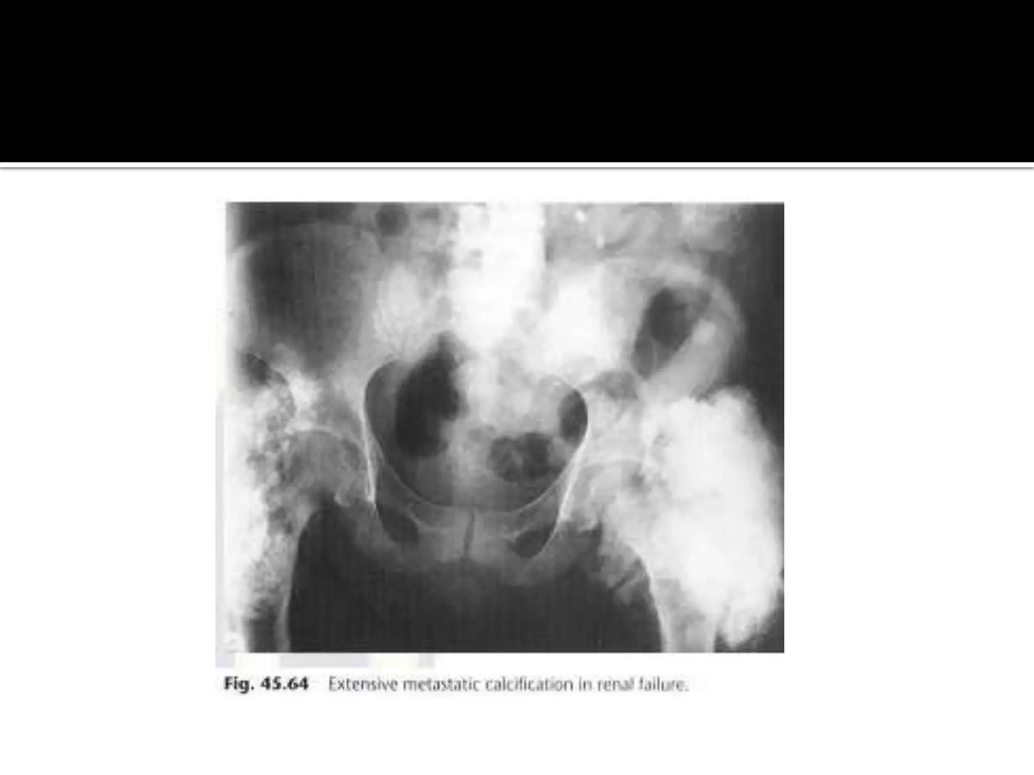
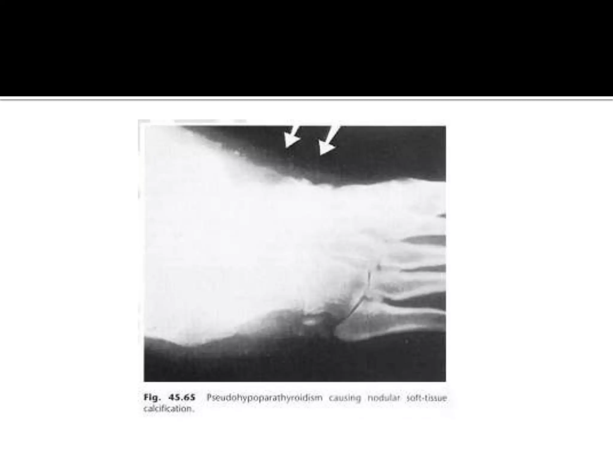
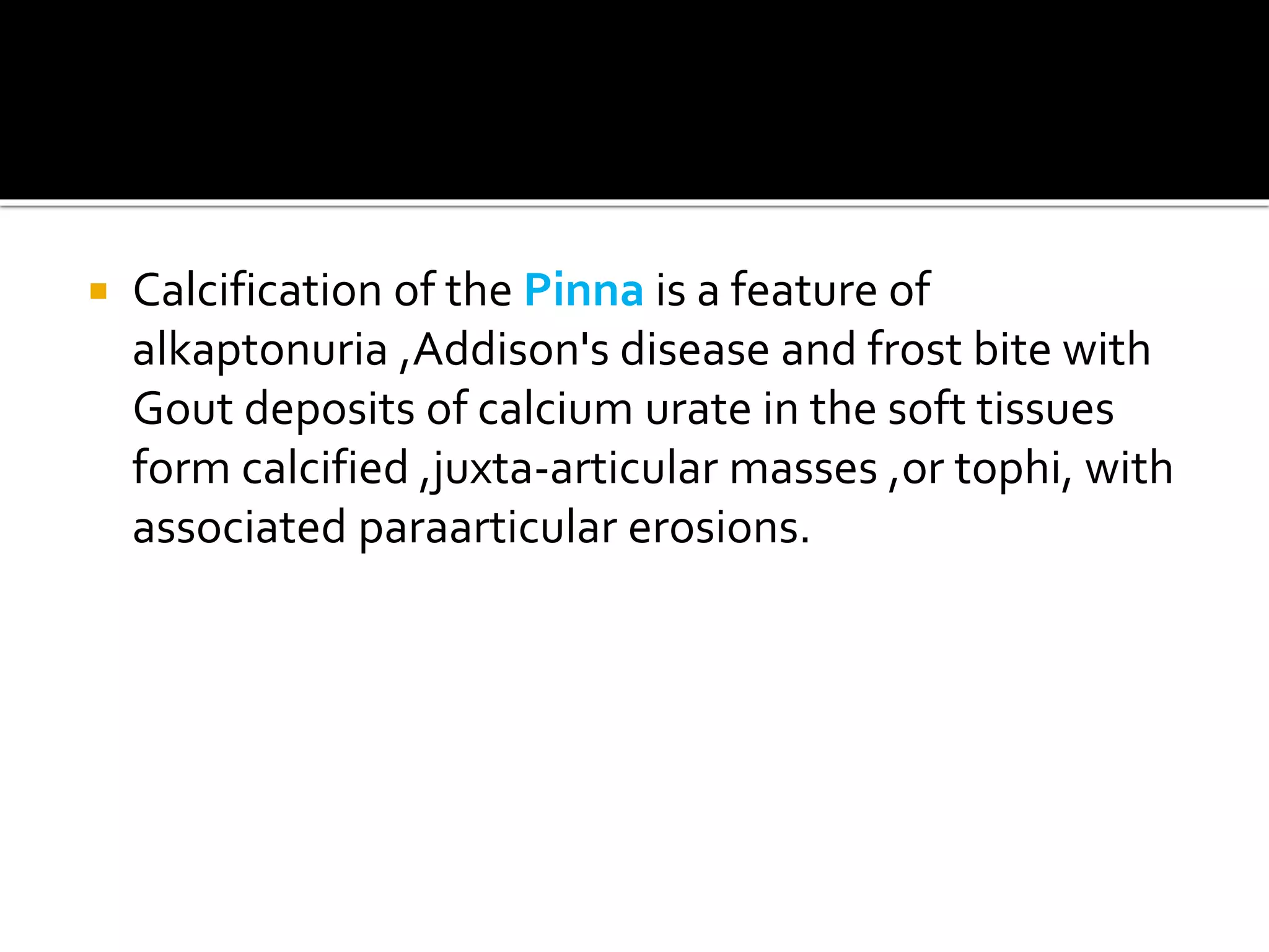
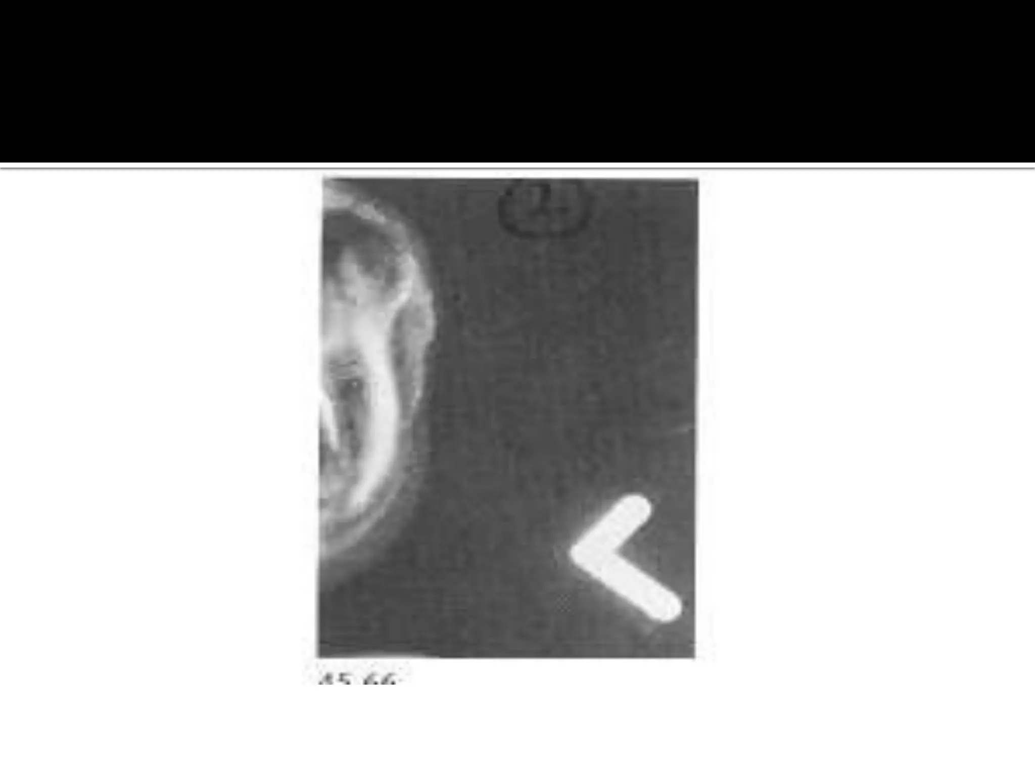
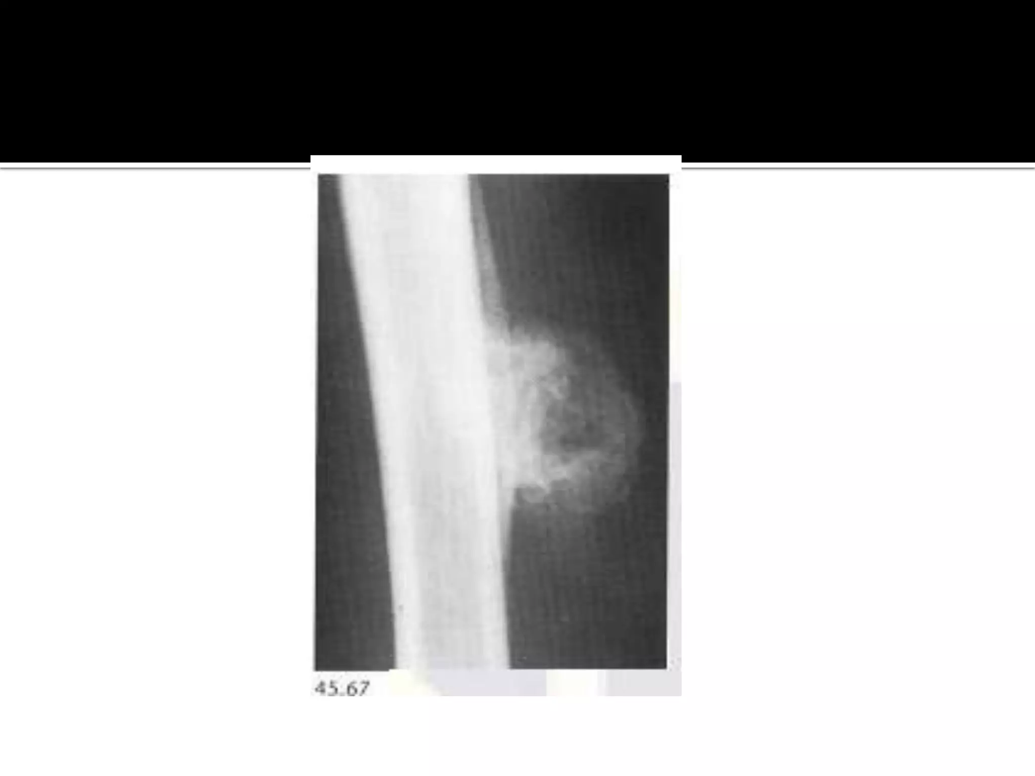
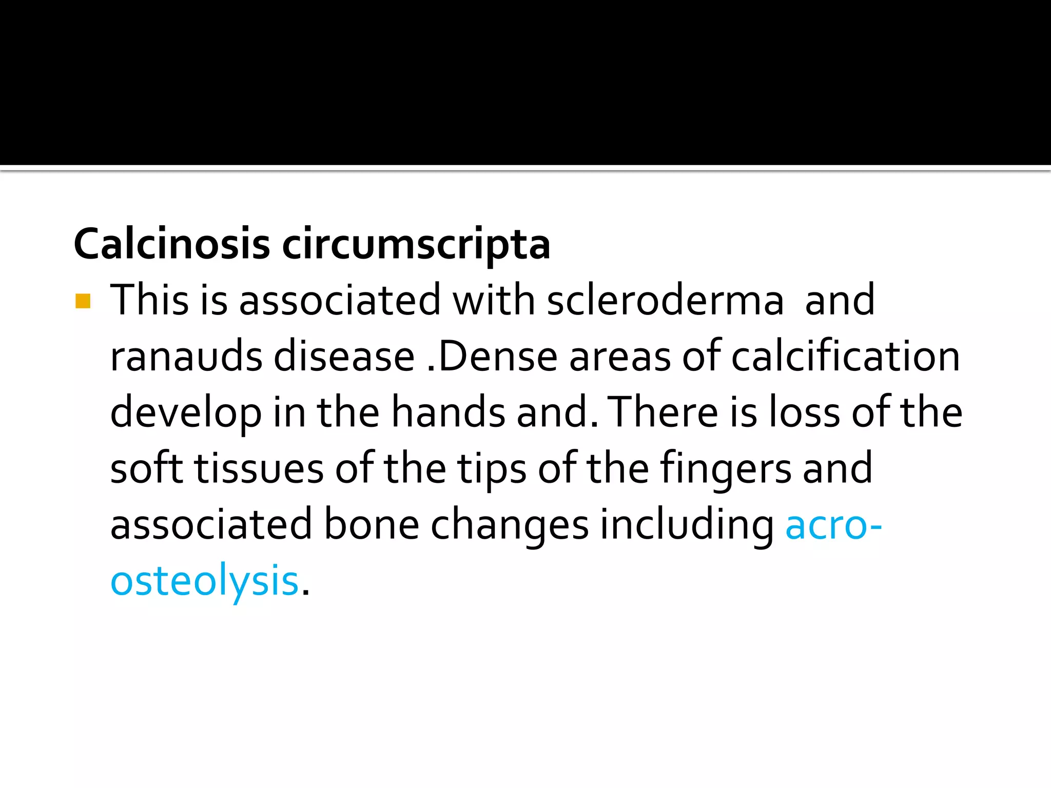
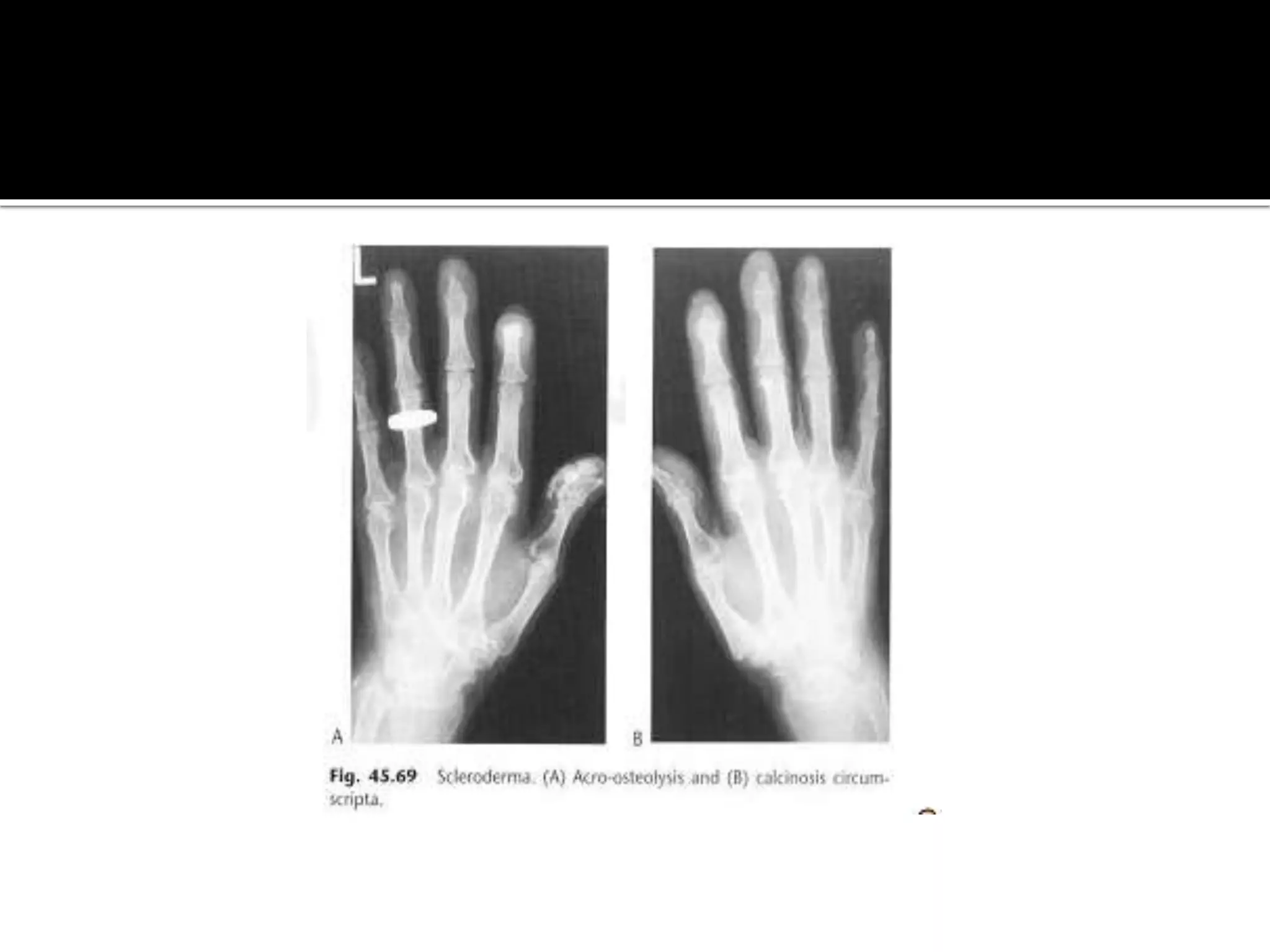
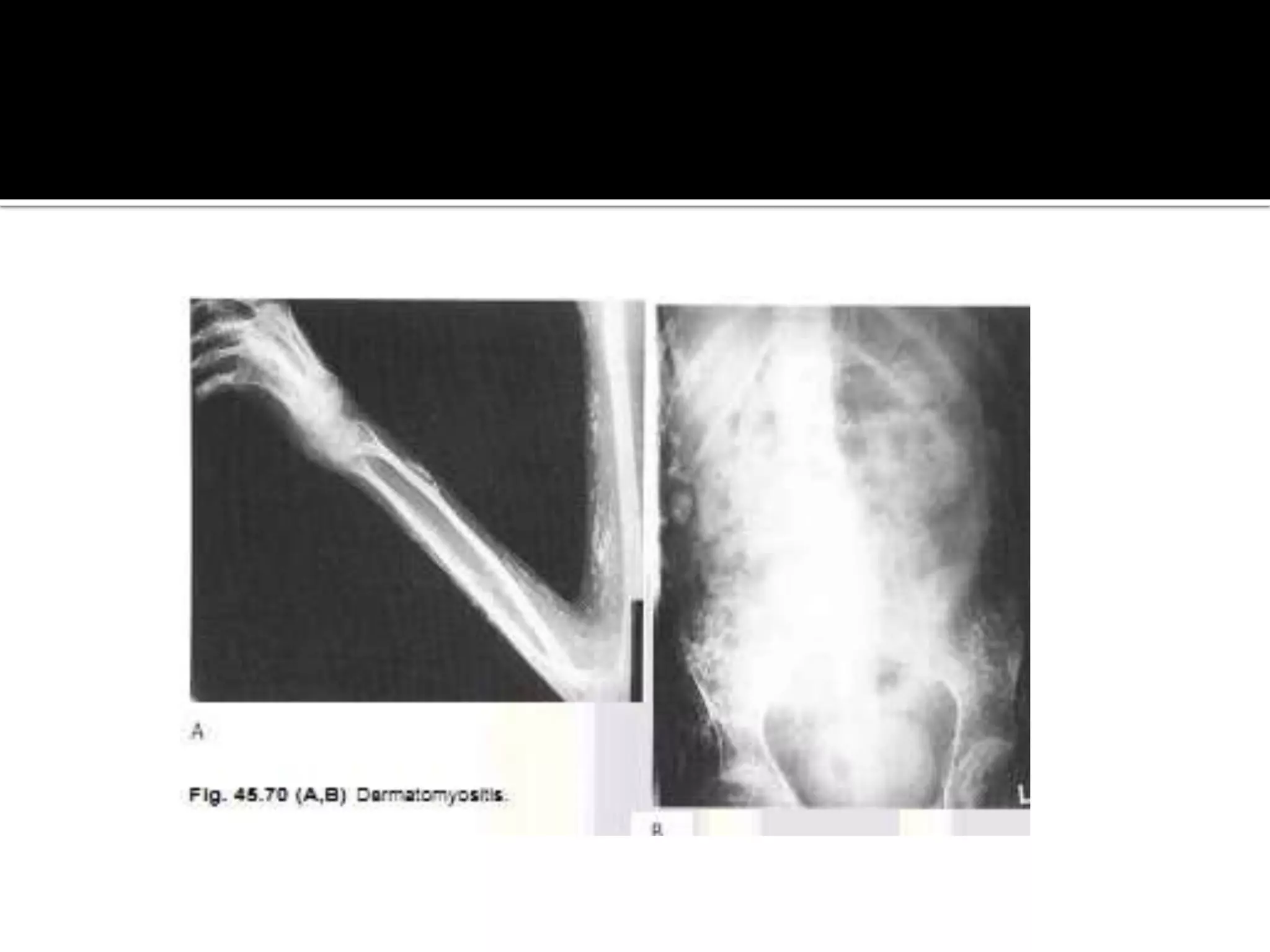
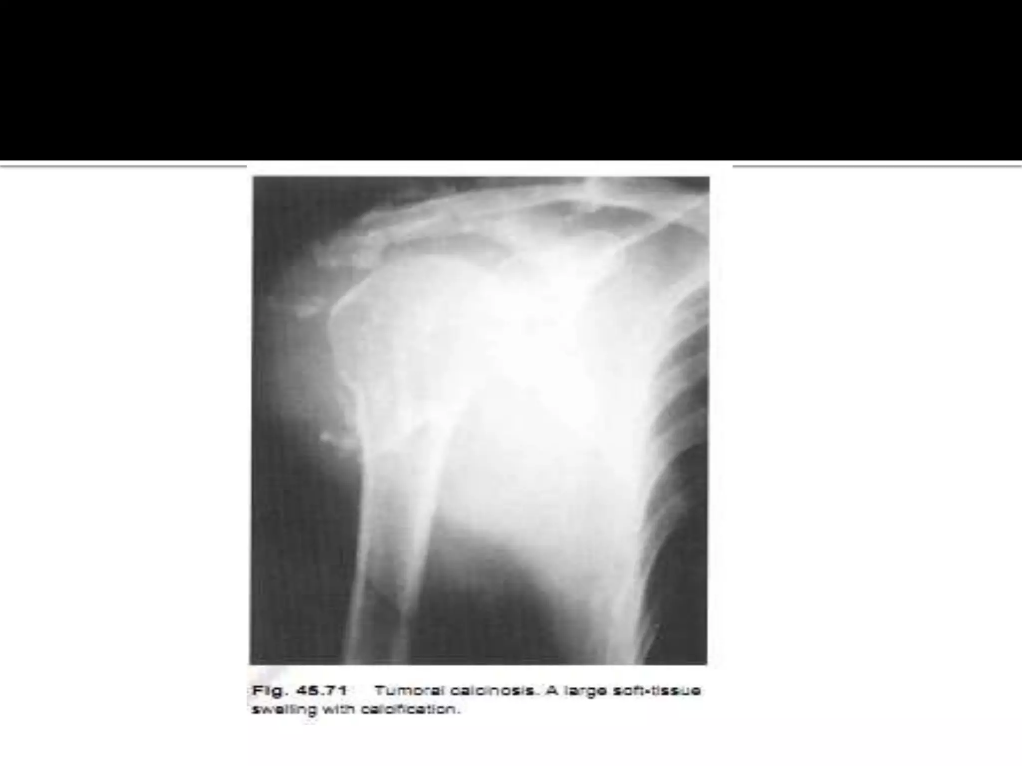
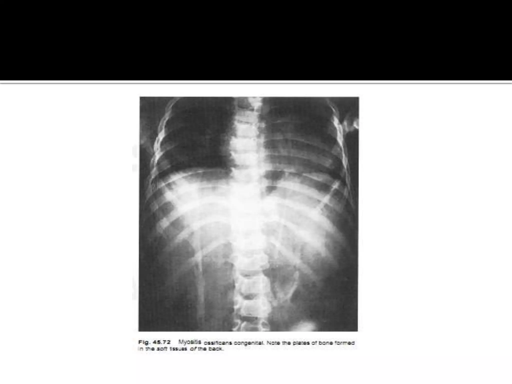
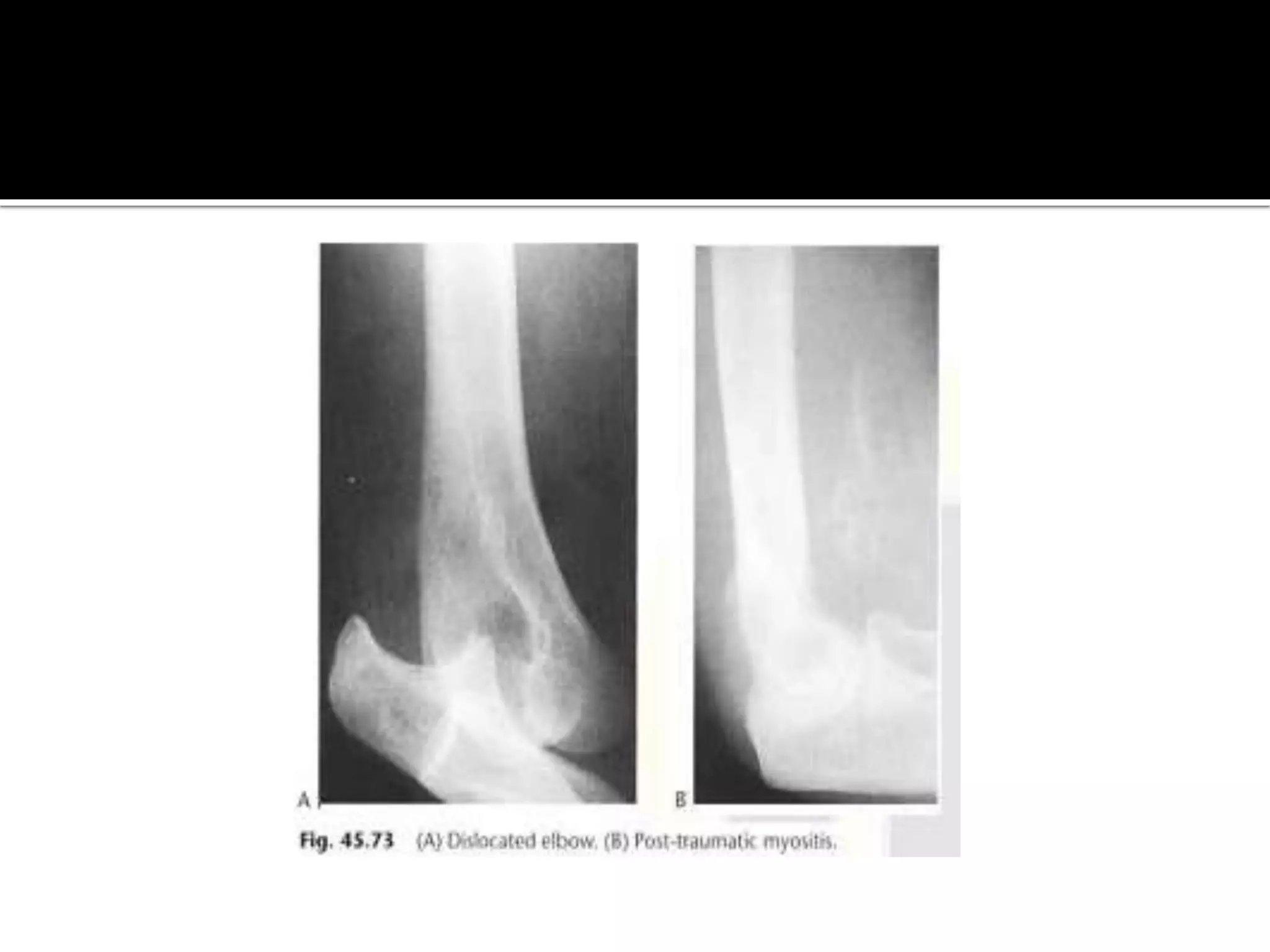
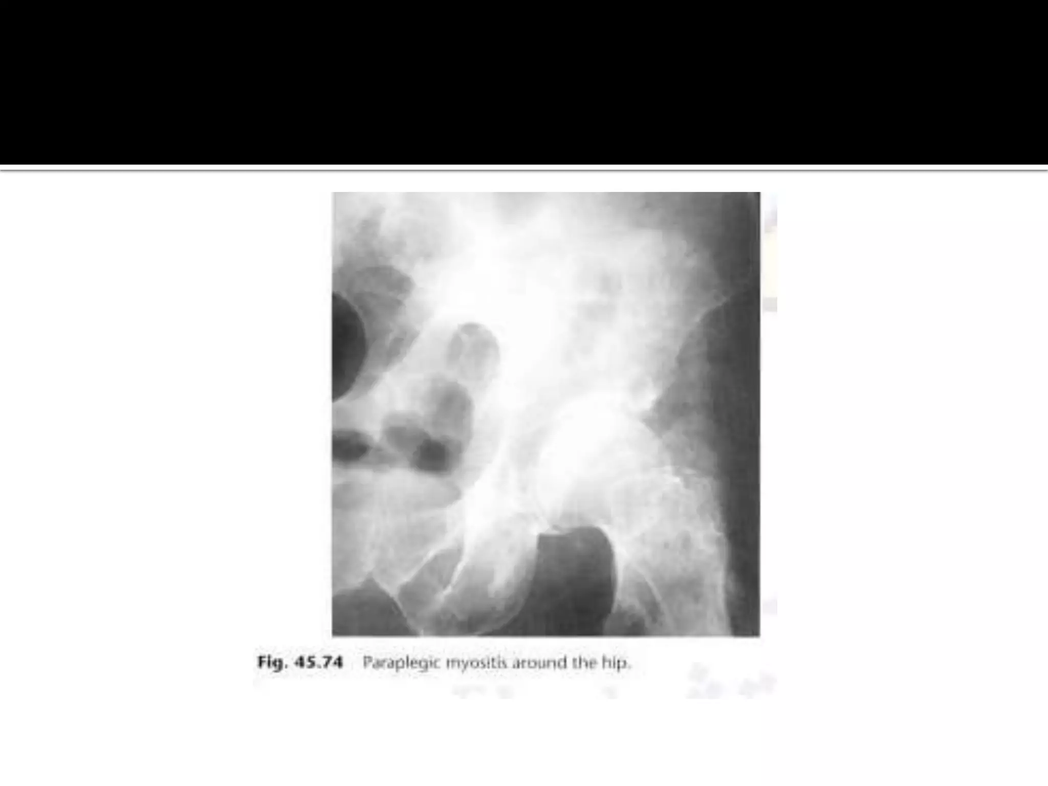
Types of calcification
 1.Metastatic calcification (abnormal calcium
metabolism)
 2.Calcinosis(normal calcium metabolism,
calcinosis circumscripta,calcinosis interstitialis
universalis)
 3.dystrophic calcification (related to tissue
damage)
 Arterial calcification
 Venous calcification
 Neural calcification
 Calcification in lymph nodes
 Calcification in tendons
 Calcification associated with parasites
 Calcification associated with metabolic
disorders.
 Calcification associated with Hematomas
 Calcification associated soft tissue necrosis
Ossification
 Types.
 1.Myocitis ossificans
 2.Post traumatic myocitis ossificans.
Calcification in Arteries
 Irregularly athermanous plaques(Aorta,illiac,femoral
arteries)
 Extent is not correlated to the severity of the disease.
 Curvilinear calcification in a soft tissue mass adjacent
to a vessel is typical for an aneurysm.
 Monkeberg,s medial atherosclerosis only media is
involved no narrowing of the lumen of the artery
appearance is a pipestem ring-like pattern.
Small vessels calcification DM,Hyperparthyroidism.
 Calcification of the vessels is not a feature of
Buerger,s disease.
Calcification in veins
 Phlebolith is a calcified thrombus in a vein ( 3-5 in
length and has a central lucency)
 Normal finding in uterine and prostatic veins in the
pelvis
 when a focal collection of phleboliths is seen
elsewhere is a characteristic
cavernous haemangioma.
 Chronic venous stasis may cause subcutaneous
calcification in a linear branching or plaque like
pattern.
 Ulceration, phleboliths and well- organized
periosteal reactions my also be present.
 Sometimes thrombus in a vein may calcify along its
length.
 Neural calcification. Rare but may occur in
Leprosy & neurofibromatosis.
Calcification in tendons,patella,Achilles
 Due to chronic trauma or inflammation
 Rotator cuff especially supraspinatus tendon most
commonly affected
 Calcification lies directly over the humeral head in
crescentic distribution. it may be present
symptomatic patents.
 Carpi radialis in violinists and medial
collateral ligament of the knee.(pellegrini
steida lesion )
Parasites
 Ova of the pork tape worm ,taenia solium are
ingested by eating under cooked infected meat with
the pig being the intermediate host.
 The embryo enters the blood stream and the
parasites ,cysticercosis cellulosa, becomes encysted
within the soft tissues ,brain and meninges.
 In muscles the parasites calcifies forming ellipsoids
masses 10-15 mm long often with a translucent
centre and which is lie in the direction of the muscle
fibers.
 The guinea worm Dracunculus medinensis dies in
the muscles and forms a spherical calcified mass or
a long string like calcified worm 10-12 cm in
length.(India,Africa,Middle East)
 Loa loa or micro filariasis ,is a small parasite which is
common in west Africa. it dies and calcifies
appearing as coiled threads in the hands and feet.
 Armillilifer arnillatus is a parasite which infest
snakes and infects human who eat them. it appears
as C-shaped calcifications less than 1 cm in length in
the serosa of the chest and abdomen.
 Hydatid cysts in the soft tissues is rare
Calcification in Metabolic disorders.
 Metabolic and metastatic calcification is the result
of abnormal calcium and or phosphate metabolism,
in particular any cause of hypercalcemia. commonly
occurs in secondary hyperparathyroidism
Periarticular calcinosis
 develops as well as metastatic calcification in the
soft tissues.This may be so extensive that layering
of the calcium is seen.With treatment of
hypercalcemia the soft tissue calcification may
resolves.
 Periarticular and interstitial calcinosis may be seen
with hypoparathyroidism.
Pseudohypoparathyroidism
 there may be punctate or nodular areas of soft
tissue calcification .
 In pseudo pesudo hypoparathyroidism calcification
of the skin ,sub cutaneous tissues ,fascia, planes and
tendons is present in 30 -50 % of cases. other
associations are calcification are the dentate
nucleus ,Basal ganglia and cataract.
 Calcification of the Pinna is a feature of
alkaptonuria ,Addison's disease and frost bite with
Gout deposits of calcium urate in the soft tissues
form calcified ,juxta-articular masses ,or tophi, with
associated paraarticular erosions.
Calcification associated with Hematomas
 initially there is an increase in soft tissue density at
the site of injury ,Although calcification is rare but
may appear within a few days in children .It usually
forms in the periphery of the hematoma ,at this
stage an isotope bone scan will show increased
uptake.
 an associated periosteal reaction ,the resulting
appearance can be difficult to differentiate from a
parotseal sarcoma on the plain film.
 In neonates a sub periosteal bleed in the parietal
bone results in a cephalhaematoma which later on
calcifies .This slowly absorbs and disappears with
time although an irregular bone density may
persists for years.
Calcification with soft tissues necrosis.
 Necrosis of the soft tissues following injections and
other forms trauma, frost bite and burns .Post
traumatic fat necrosis results in coarse spheroid
calcified masses .
 Frost bite typically affects the pinna .
 Burns occasionally calcify, particularly in children.
Calcinosis
 1.Calcinosis circumsripta.
 2.calcinosis interstitalis universalis or
dermatomyositis.
 3.Tumoural calcinosis.
Calcinosis circumscripta
 This is associated with scleroderma and
ranauds disease .Dense areas of calcification
develop in the hands and.There is loss of the
soft tissues of the tips of the fingers and
associated bone changes including acro-
osteolysis.
Calcinosis interstitials universalis or
dermatomyositis.
there is degeneration of the collagen tissue resulting
in diffuse subcutaneous plaques or nodules of
calcium or reticular calcification often with
overlying ulceration . In addition with progression
,calcified masses or sheets of calcium form in the
deeper tissues and lie in the direction of the limbs.
 There is normal calcium and phosphate metabolism
 those affected have pain and muscle weakness
 if progressive the disease may be fatal. In adults
there is an association between dermatomyositis
and malignancy.
 Ehler,s Danlos syndrome is hereditary disorder
with findings including lax joints ,fragile and elastic
skin and blood vessels ,acro osteolysis and
subcutaneous nodules
Tumoral Calcinosis
 Tumoral calcinosis typically presents in young
african men with swelling ,which may be painful,
around the large joints and predominantly affecting
the extensors surfaces.
 The hips are the most frequently involved ,followed
by the elbows and the shoulders .
Ossification.
 Myocitis ossificans.
 1.Myocitis osificans progressiva.
 2.post traumatic myocitis osificans
 3.paraplegic myocitis ossificans.
Myocitis osificans progressiva.
 the progressive form myocitis ossificans is a
hereditary disease commoner in boys.
 it presents with ossification of the perimuscular
fascia within one year and is associated with many
congenital bone lesions such as phalangeal and
metacarpalhypoplasia of the thumbs and big toes .
 the condition is usually fatal.
 in the limbs the appearance needs to be
distinguished from parosteal sarcoma.
Post-traumatic myositis
 it results in a soft tissue mass which ossifies with a lacy
pattern by 4-8 weeks.
 common sites are the elbow and knee.The ossification
may resolves.
Paraplegic myocitis ossificans.
 it develops in paraplegics within 3-4 weeks below the
level of the paralysis.There is hetrotopic periarticular
calcification and ossification around the joints
particularly the hip with irregular plaques of bone
forming.
Thank you

Ossification & calcification

  • 2.
    Types of calcification 1.Metastatic calcification (abnormal calcium metabolism)  2.Calcinosis(normal calcium metabolism, calcinosis circumscripta,calcinosis interstitialis universalis)  3.dystrophic calcification (related to tissue damage)
  • 3.
     Arterial calcification Venous calcification  Neural calcification  Calcification in lymph nodes  Calcification in tendons  Calcification associated with parasites  Calcification associated with metabolic disorders.
  • 4.
     Calcification associatedwith Hematomas  Calcification associated soft tissue necrosis
  • 5.
    Ossification  Types.  1.Myocitisossificans  2.Post traumatic myocitis ossificans.
  • 6.
    Calcification in Arteries Irregularly athermanous plaques(Aorta,illiac,femoral arteries)  Extent is not correlated to the severity of the disease.  Curvilinear calcification in a soft tissue mass adjacent to a vessel is typical for an aneurysm.  Monkeberg,s medial atherosclerosis only media is involved no narrowing of the lumen of the artery appearance is a pipestem ring-like pattern.
  • 7.
    Small vessels calcificationDM,Hyperparthyroidism.  Calcification of the vessels is not a feature of Buerger,s disease. Calcification in veins  Phlebolith is a calcified thrombus in a vein ( 3-5 in length and has a central lucency)  Normal finding in uterine and prostatic veins in the pelvis  when a focal collection of phleboliths is seen elsewhere is a characteristic
  • 8.
    cavernous haemangioma.  Chronicvenous stasis may cause subcutaneous calcification in a linear branching or plaque like pattern.  Ulceration, phleboliths and well- organized periosteal reactions my also be present.  Sometimes thrombus in a vein may calcify along its length.
  • 9.
     Neural calcification.Rare but may occur in Leprosy & neurofibromatosis.
  • 11.
    Calcification in tendons,patella,Achilles Due to chronic trauma or inflammation  Rotator cuff especially supraspinatus tendon most commonly affected  Calcification lies directly over the humeral head in crescentic distribution. it may be present symptomatic patents.
  • 14.
     Carpi radialisin violinists and medial collateral ligament of the knee.(pellegrini steida lesion )
  • 16.
    Parasites  Ova ofthe pork tape worm ,taenia solium are ingested by eating under cooked infected meat with the pig being the intermediate host.  The embryo enters the blood stream and the parasites ,cysticercosis cellulosa, becomes encysted within the soft tissues ,brain and meninges.
  • 17.
     In musclesthe parasites calcifies forming ellipsoids masses 10-15 mm long often with a translucent centre and which is lie in the direction of the muscle fibers.  The guinea worm Dracunculus medinensis dies in the muscles and forms a spherical calcified mass or a long string like calcified worm 10-12 cm in length.(India,Africa,Middle East)
  • 18.
     Loa loaor micro filariasis ,is a small parasite which is common in west Africa. it dies and calcifies appearing as coiled threads in the hands and feet.  Armillilifer arnillatus is a parasite which infest snakes and infects human who eat them. it appears as C-shaped calcifications less than 1 cm in length in the serosa of the chest and abdomen.
  • 21.
     Hydatid cystsin the soft tissues is rare Calcification in Metabolic disorders.  Metabolic and metastatic calcification is the result of abnormal calcium and or phosphate metabolism, in particular any cause of hypercalcemia. commonly occurs in secondary hyperparathyroidism
  • 23.
    Periarticular calcinosis  developsas well as metastatic calcification in the soft tissues.This may be so extensive that layering of the calcium is seen.With treatment of hypercalcemia the soft tissue calcification may resolves.  Periarticular and interstitial calcinosis may be seen with hypoparathyroidism.
  • 24.
    Pseudohypoparathyroidism  there maybe punctate or nodular areas of soft tissue calcification .  In pseudo pesudo hypoparathyroidism calcification of the skin ,sub cutaneous tissues ,fascia, planes and tendons is present in 30 -50 % of cases. other associations are calcification are the dentate nucleus ,Basal ganglia and cataract.
  • 26.
     Calcification ofthe Pinna is a feature of alkaptonuria ,Addison's disease and frost bite with Gout deposits of calcium urate in the soft tissues form calcified ,juxta-articular masses ,or tophi, with associated paraarticular erosions.
  • 28.
    Calcification associated withHematomas  initially there is an increase in soft tissue density at the site of injury ,Although calcification is rare but may appear within a few days in children .It usually forms in the periphery of the hematoma ,at this stage an isotope bone scan will show increased uptake.
  • 30.
     an associatedperiosteal reaction ,the resulting appearance can be difficult to differentiate from a parotseal sarcoma on the plain film.  In neonates a sub periosteal bleed in the parietal bone results in a cephalhaematoma which later on calcifies .This slowly absorbs and disappears with time although an irregular bone density may persists for years.
  • 32.
    Calcification with softtissues necrosis.  Necrosis of the soft tissues following injections and other forms trauma, frost bite and burns .Post traumatic fat necrosis results in coarse spheroid calcified masses .  Frost bite typically affects the pinna .  Burns occasionally calcify, particularly in children.
  • 33.
    Calcinosis  1.Calcinosis circumsripta. 2.calcinosis interstitalis universalis or dermatomyositis.  3.Tumoural calcinosis.
  • 34.
    Calcinosis circumscripta  Thisis associated with scleroderma and ranauds disease .Dense areas of calcification develop in the hands and.There is loss of the soft tissues of the tips of the fingers and associated bone changes including acro- osteolysis.
  • 36.
    Calcinosis interstitials universalisor dermatomyositis. there is degeneration of the collagen tissue resulting in diffuse subcutaneous plaques or nodules of calcium or reticular calcification often with overlying ulceration . In addition with progression ,calcified masses or sheets of calcium form in the deeper tissues and lie in the direction of the limbs.
  • 38.
     There isnormal calcium and phosphate metabolism  those affected have pain and muscle weakness  if progressive the disease may be fatal. In adults there is an association between dermatomyositis and malignancy.  Ehler,s Danlos syndrome is hereditary disorder with findings including lax joints ,fragile and elastic skin and blood vessels ,acro osteolysis and subcutaneous nodules
  • 39.
    Tumoral Calcinosis  Tumoralcalcinosis typically presents in young african men with swelling ,which may be painful, around the large joints and predominantly affecting the extensors surfaces.  The hips are the most frequently involved ,followed by the elbows and the shoulders .
  • 41.
    Ossification.  Myocitis ossificans. 1.Myocitis osificans progressiva.  2.post traumatic myocitis osificans  3.paraplegic myocitis ossificans.
  • 42.
    Myocitis osificans progressiva. the progressive form myocitis ossificans is a hereditary disease commoner in boys.  it presents with ossification of the perimuscular fascia within one year and is associated with many congenital bone lesions such as phalangeal and metacarpalhypoplasia of the thumbs and big toes .
  • 44.
     the conditionis usually fatal.  in the limbs the appearance needs to be distinguished from parosteal sarcoma.
  • 45.
    Post-traumatic myositis  itresults in a soft tissue mass which ossifies with a lacy pattern by 4-8 weeks.  common sites are the elbow and knee.The ossification may resolves. Paraplegic myocitis ossificans.  it develops in paraplegics within 3-4 weeks below the level of the paralysis.There is hetrotopic periarticular calcification and ossification around the joints particularly the hip with irregular plaques of bone forming.
  • 48.