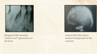
This document discusses several diseases that can affect bone density and structure, including hyperparathyroidism, osteoporosis, osteomalacia, leukemia, Langerhans cell disease, Paget's disease, and multiple myeloma. It provides details on the clinical features, radiographic manifestations, differential diagnosis, and management of each condition. The document contains radiographic images showing examples of bone changes associated with some of the diseases.
The Indian Dental Academy is the Leader in continuing dental education , training dentists in all aspects of dentistry and
offering a wide range of dental certified courses in different formats.
Solitary cyst like radiolucencies not contacting teeth/ dental coursesIndian dental academy
The Indian Dental Academy is the Leader in continuing dental education , training dentists in all aspects of dentistry and
offering a wide range of dental certified courses in different formats.
Systemic Diseases Manifested in the Jawsvahid199212
Systemic Diseases Manifestation the Jaws based on chapter25
Oral Radiology
P R I N C I P L E S
a n d I N T E R P R E T A T I O N Sixth Edition
White and pharoah
ORN is an inflammatory condition of bone that occurs after the bone has been exposed to therapeutic doses of radiation usually given for a malignancies.
The Indian Dental Academy is the Leader in continuing dental education , training dentists in all aspects of dentistry and
offering a wide range of dental certified courses in different formats.
Solitary cyst like radiolucencies not contacting teeth/ dental coursesIndian dental academy
The Indian Dental Academy is the Leader in continuing dental education , training dentists in all aspects of dentistry and
offering a wide range of dental certified courses in different formats.
Systemic Diseases Manifested in the Jawsvahid199212
Systemic Diseases Manifestation the Jaws based on chapter25
Oral Radiology
P R I N C I P L E S
a n d I N T E R P R E T A T I O N Sixth Edition
White and pharoah
ORN is an inflammatory condition of bone that occurs after the bone has been exposed to therapeutic doses of radiation usually given for a malignancies.
Cancer of the oral cavity accounts for approximately 3% of all malignancies diagnosed annually in 270,000 patients world-wide. Oral cancer is the 12th most common cancer in women and the 6th in men. Many oral squamous cell carcinomas develop from potentially malignant disorders (PMDs). Lack of awareness about the signs and symptoms of oral PMDs in the general population and even healthcare providers is believed to be responsible for the diagnostic delay of these entities.
WHO has defined it as an intraosseous lesion consisting of cellular and fibrous tissue that contains multiple foci of hemorrhage, aggregation of multinucleated giant cells and occasionally trabeculae of woven bone.
JAFFE (1953): considered this lesion to be a local reparative reaction of bone, possibly to intramedullary hemorrhage or trauma, hence the term reparative giant cell granuloma was accepted.
Charles A Waldron & W G Shafer (1966) suggested trauma to be an important etiological factor in the initiation of the CGCG of the jaws.
Thoma K H (1986) suggested that, the lesion may be due to capillary injury caused by defective wall due to some type of trauma
J V Soames and J C southam (1997) suggested that it could be a reaction to some form of hemodynamic disturbance in bone marrow, perhaps associated with trauma and hemorrhage REGEZI AND SCIUBBA(1999) :
Suggested that
Response to previous traumatic or inflammatory episodes.
This lesion is characterised by proliferation of fibroblasts and multinucleated giant cells, in a densely packed stroma
The Indian Dental Academy is the Leader in continuing dental education , training dentists in all aspects of dentistry and
offering a wide range of dental certified courses in different formats.for more details please visit
www.indiandentalacademy.com
Odontogenic keratocyst (OKC) is the cyst arising from the cell rests of dental lamina. It can occur anywhere in the jaw, but commonly seen in the posterior part of the mandible. Radiographically, most OKCs are unilocular when presented at the periapex and can be mistaken for radicular or lateral periodontal cyst.
The Indian Dental Academy is the Leader in continuing dental education , training dentists in all aspects of dentistry and offering a wide range of dental certified courses in different formats.
Indian dental academy provides dental crown & Bridge,rotary endodontics,fixed orthodontics,
Dental implants courses.for details pls visit www.indiandentalacademy.com ,or call
0091-9248678078
The Indian Dental Academy is the Leader in continuing dental education , training dentists in all aspects of dentistry and
offering a wide range of dental certified courses in different formats.
The Indian Dental Academy is the Leader in continuing dental education , training dentists in all aspects of dentistry and
offering a wide range of dental certified courses in different formats.for more details please visit
www.indiandentalacademy.com
New Drug Discovery and Development .....NEHA GUPTA
The "New Drug Discovery and Development" process involves the identification, design, testing, and manufacturing of novel pharmaceutical compounds with the aim of introducing new and improved treatments for various medical conditions. This comprehensive endeavor encompasses various stages, including target identification, preclinical studies, clinical trials, regulatory approval, and post-market surveillance. It involves multidisciplinary collaboration among scientists, researchers, clinicians, regulatory experts, and pharmaceutical companies to bring innovative therapies to market and address unmet medical needs.
Acute scrotum is a general term referring to an emergency condition affecting the contents or the wall of the scrotum.
There are a number of conditions that present acutely, predominantly with pain and/or swelling
A careful and detailed history and examination, and in some cases, investigations allow differentiation between these diagnoses. A prompt diagnosis is essential as the patient may require urgent surgical intervention
Testicular torsion refers to twisting of the spermatic cord, causing ischaemia of the testicle.
Testicular torsion results from inadequate fixation of the testis to the tunica vaginalis producing ischemia from reduced arterial inflow and venous outflow obstruction.
The prevalence of testicular torsion in adult patients hospitalized with acute scrotal pain is approximately 25 to 50 percent
Knee anatomy and clinical tests 2024.pdfvimalpl1234
This includes all relevant anatomy and clinical tests compiled from standard textbooks, Campbell,netter etc..It is comprehensive and best suited for orthopaedicians and orthopaedic residents.
micro teaching on communication m.sc nursing.pdfAnurag Sharma
Microteaching is a unique model of practice teaching. It is a viable instrument for the. desired change in the teaching behavior or the behavior potential which, in specified types of real. classroom situations, tends to facilitate the achievement of specified types of objectives.
Title: Sense of Smell
Presenter: Dr. Faiza, Assistant Professor of Physiology
Qualifications:
MBBS (Best Graduate, AIMC Lahore)
FCPS Physiology
ICMT, CHPE, DHPE (STMU)
MPH (GC University, Faisalabad)
MBA (Virtual University of Pakistan)
Learning Objectives:
Describe the primary categories of smells and the concept of odor blindness.
Explain the structure and location of the olfactory membrane and mucosa, including the types and roles of cells involved in olfaction.
Describe the pathway and mechanisms of olfactory signal transmission from the olfactory receptors to the brain.
Illustrate the biochemical cascade triggered by odorant binding to olfactory receptors, including the role of G-proteins and second messengers in generating an action potential.
Identify different types of olfactory disorders such as anosmia, hyposmia, hyperosmia, and dysosmia, including their potential causes.
Key Topics:
Olfactory Genes:
3% of the human genome accounts for olfactory genes.
400 genes for odorant receptors.
Olfactory Membrane:
Located in the superior part of the nasal cavity.
Medially: Folds downward along the superior septum.
Laterally: Folds over the superior turbinate and upper surface of the middle turbinate.
Total surface area: 5-10 square centimeters.
Olfactory Mucosa:
Olfactory Cells: Bipolar nerve cells derived from the CNS (100 million), with 4-25 olfactory cilia per cell.
Sustentacular Cells: Produce mucus and maintain ionic and molecular environment.
Basal Cells: Replace worn-out olfactory cells with an average lifespan of 1-2 months.
Bowman’s Gland: Secretes mucus.
Stimulation of Olfactory Cells:
Odorant dissolves in mucus and attaches to receptors on olfactory cilia.
Involves a cascade effect through G-proteins and second messengers, leading to depolarization and action potential generation in the olfactory nerve.
Quality of a Good Odorant:
Small (3-20 Carbon atoms), volatile, water-soluble, and lipid-soluble.
Facilitated by odorant-binding proteins in mucus.
Membrane Potential and Action Potential:
Resting membrane potential: -55mV.
Action potential frequency in the olfactory nerve increases with odorant strength.
Adaptation Towards the Sense of Smell:
Rapid adaptation within the first second, with further slow adaptation.
Psychological adaptation greater than receptor adaptation, involving feedback inhibition from the central nervous system.
Primary Sensations of Smell:
Camphoraceous, Musky, Floral, Pepperminty, Ethereal, Pungent, Putrid.
Odor Detection Threshold:
Examples: Hydrogen sulfide (0.0005 ppm), Methyl-mercaptan (0.002 ppm).
Some toxic substances are odorless at lethal concentrations.
Characteristics of Smell:
Odor blindness for single substances due to lack of appropriate receptor protein.
Behavioral and emotional influences of smell.
Transmission of Olfactory Signals:
From olfactory cells to glomeruli in the olfactory bulb, involving lateral inhibition.
Primitive, less old, and new olfactory systems with different path
Ozempic: Preoperative Management of Patients on GLP-1 Receptor Agonists Saeid Safari
Preoperative Management of Patients on GLP-1 Receptor Agonists like Ozempic and Semiglutide
ASA GUIDELINE
NYSORA Guideline
2 Case Reports of Gastric Ultrasound
Recomendações da OMS sobre cuidados maternos e neonatais para uma experiência pós-natal positiva.
Em consonância com os ODS – Objetivos do Desenvolvimento Sustentável e a Estratégia Global para a Saúde das Mulheres, Crianças e Adolescentes, e aplicando uma abordagem baseada nos direitos humanos, os esforços de cuidados pós-natais devem expandir-se para além da cobertura e da simples sobrevivência, de modo a incluir cuidados de qualidade.
Estas diretrizes visam melhorar a qualidade dos cuidados pós-natais essenciais e de rotina prestados às mulheres e aos recém-nascidos, com o objetivo final de melhorar a saúde e o bem-estar materno e neonatal.
Uma “experiência pós-natal positiva” é um resultado importante para todas as mulheres que dão à luz e para os seus recém-nascidos, estabelecendo as bases para a melhoria da saúde e do bem-estar a curto e longo prazo. Uma experiência pós-natal positiva é definida como aquela em que as mulheres, pessoas que gestam, os recém-nascidos, os casais, os pais, os cuidadores e as famílias recebem informação consistente, garantia e apoio de profissionais de saúde motivados; e onde um sistema de saúde flexível e com recursos reconheça as necessidades das mulheres e dos bebês e respeite o seu contexto cultural.
Estas diretrizes consolidadas apresentam algumas recomendações novas e já bem fundamentadas sobre cuidados pós-natais de rotina para mulheres e neonatos que recebem cuidados no pós-parto em unidades de saúde ou na comunidade, independentemente dos recursos disponíveis.
É fornecido um conjunto abrangente de recomendações para cuidados durante o período puerperal, com ênfase nos cuidados essenciais que todas as mulheres e recém-nascidos devem receber, e com a devida atenção à qualidade dos cuidados; isto é, a entrega e a experiência do cuidado recebido. Estas diretrizes atualizam e ampliam as recomendações da OMS de 2014 sobre cuidados pós-natais da mãe e do recém-nascido e complementam as atuais diretrizes da OMS sobre a gestão de complicações pós-natais.
O estabelecimento da amamentação e o manejo das principais intercorrências é contemplada.
Recomendamos muito.
Vamos discutir essas recomendações no nosso curso de pós-graduação em Aleitamento no Instituto Ciclos.
Esta publicação só está disponível em inglês até o momento.
Prof. Marcus Renato de Carvalho
www.agostodourado.com
The prostate is an exocrine gland of the male mammalian reproductive system
It is a walnut-sized gland that forms part of the male reproductive system and is located in front of the rectum and just below the urinary bladder
Function is to store and secrete a clear, slightly alkaline fluid that constitutes 10-30% of the volume of the seminal fluid that along with the spermatozoa, constitutes semen
A healthy human prostate measures (4cm-vertical, by 3cm-horizontal, 2cm ant-post ).
It surrounds the urethra just below the urinary bladder. It has anterior, median, posterior and two lateral lobes
It’s work is regulated by androgens which are responsible for male sex characteristics
Generalised disease of the prostate due to hormonal derangement which leads to non malignant enlargement of the gland (increase in the number of epithelial cells and stromal tissue)to cause compression of the urethra leading to symptoms (LUTS
Prix Galien International 2024 Forum ProgramLevi Shapiro
June 20, 2024, Prix Galien International and Jerusalem Ethics Forum in ROME. Detailed agenda including panels:
- ADVANCES IN CARDIOLOGY: A NEW PARADIGM IS COMING
- WOMEN’S HEALTH: FERTILITY PRESERVATION
- WHAT’S NEW IN THE TREATMENT OF INFECTIOUS,
ONCOLOGICAL AND INFLAMMATORY SKIN DISEASES?
- ARTIFICIAL INTELLIGENCE AND ETHICS
- GENE THERAPY
- BEYOND BORDERS: GLOBAL INITIATIVES FOR DEMOCRATIZING LIFE SCIENCE TECHNOLOGIES AND PROMOTING ACCESS TO HEALTHCARE
- ETHICAL CHALLENGES IN LIFE SCIENCES
- Prix Galien International Awards Ceremony
Flu Vaccine Alert in Bangalore Karnatakaaddon Scans
As flu season approaches, health officials in Bangalore, Karnataka, are urging residents to get their flu vaccinations. The seasonal flu, while common, can lead to severe health complications, particularly for vulnerable populations such as young children, the elderly, and those with underlying health conditions.
Dr. Vidisha Kumari, a leading epidemiologist in Bangalore, emphasizes the importance of getting vaccinated. "The flu vaccine is our best defense against the influenza virus. It not only protects individuals but also helps prevent the spread of the virus in our communities," he says.
This year, the flu season is expected to coincide with a potential increase in other respiratory illnesses. The Karnataka Health Department has launched an awareness campaign highlighting the significance of flu vaccinations. They have set up multiple vaccination centers across Bangalore, making it convenient for residents to receive their shots.
To encourage widespread vaccination, the government is also collaborating with local schools, workplaces, and community centers to facilitate vaccination drives. Special attention is being given to ensuring that the vaccine is accessible to all, including marginalized communities who may have limited access to healthcare.
Residents are reminded that the flu vaccine is safe and effective. Common side effects are mild and may include soreness at the injection site, mild fever, or muscle aches. These side effects are generally short-lived and far less severe than the flu itself.
Healthcare providers are also stressing the importance of continuing COVID-19 precautions. Wearing masks, practicing good hand hygiene, and maintaining social distancing are still crucial, especially in crowded places.
Protect yourself and your loved ones by getting vaccinated. Together, we can help keep Bangalore healthy and safe this flu season. For more information on vaccination centers and schedules, residents can visit the Karnataka Health Department’s official website or follow their social media pages.
Stay informed, stay safe, and get your flu shot today!
4. • Maintenance of normal bone density or mass requires normal
function and the interaction of a number of intricate and delicate
systems:osseous endocrine hematopoetic ,renal
gastrointestinal,nutritional and neurocirculatory.
• Radiographs reveal a considerable variation in the density of
normal jaw bones.
• All the rarefying diseases included here represent a disruption of
bone homeostasis that may result from an imbalance among the
factors noted or a direct influence of a disese process on the bone
itself.
INTRODUCTION
5. HYPERPARATHYROIDISM
Hyperparathyroidism is an endocrine abnormality in which there is
an excess of circulating parathyroid hormone(PTH).
TYPES
• Primary hyperparathyroidism
Results from hyperplasia or benign tumour of one of the four
parathyroid glands , resulting in production of excess PTH.
• Secondary hyperparathyroidism
This results from compensatory increase in the output of PTH in
response to hypocalcaemia.
6. Tertiary Hyperparathyroidism
Occasionally,the parathyroid tumour after long standing secondary
hyperparathyroidism may develop a condition called tertiary
hyperparathyroidism.
Ectopic hyperparathyroidism
Caused due to excessive parathyroid hormone synthesis in a patient
with malignant disease.
7. CLINICAL FEATURES
More common in females
Occurs mainly in 30-60 years of age
Classis triad and signs of hyperparathyroidism are described as
having “stones , bones, and abdominal groans”
Stones refers to the development of renal calculi (kidney
stones,nephrolithiasis) because of the elevated serum calcium levels
Metastatic calcifications involving blood vessel walls,subcutaneous
soft tissues, sclera, dura and the regions around the jaws
8. Bones refers to variety of osseous changes that may occur in conjunction
with hyperparathyroidism.
Bone deformities such as bending of long bones,occasional pathologic
fractures,collapse of vertebrae and pigeon chest
Loosening and drifting of teeth leading to malocclusion with diastema
formation.
Abdominal groans refers to the tendency
for the development of duodenal ulcers.
Palatal enlargement associated with
secondary hyperparathyroidism.
9. RADIOGRAPHIC FEATURES
Radigraphically observable changes are seen in 1 out of 5 patients
Earliest changes are subtle erosions of the bone from the periosteal
surfaces of the phalanges from hand
Overall demineralization of the skeleton resulting in overall grayness
and lack of normal contrast on the radiograph
Generalized osteoporosis
In prominent hyperparathyroidism ,the entire calvarium has a
granular appearance of the bone ,caused by loss of central trabeculae
and thinning of cortical tables
10. Osteitis fibrosa cystica or osteitis fibrosa generalisata are localized regions
of bone loss produced by osteoclastic activity,resulting in loss of all
apparent bone structure. These appear cyst like on radiograph
Brown tumor occur late in the disease .
A change in normal trabecular pattern gives ground glass appearance of
numerous ,small , randomly oriented
trabeculae.
Periapical radiographs reveal loss of lamina
dura.
13. DIFFERENTIAL DIAGNOSIS
UNILOCULAR
Post extraction socket
Primordial bone cyst
MULTILOCULAR
Pagets disease
Ameloblastoma
Central giant cell granuloma
Cherubism
Aneurysmal bone cyst
Osteomalacia
Fibrous dysplasia
Multiple myeloma
14. MANAGEMENT
• After successful surgical removal of the causative factor,almost all
radiographic changes revert to normal,except for brown tumours
which heals with bone that is radigraphically more sclerotic than
normal
15. OSTEOPOROSIS
Osteoporosis is defined as “ a disease characterized by low bone mass
and micro architectural deterioration of bone tissue leading to
enhanced bone fragility and an increase in fracture risk.
There are 2 types:
1. primary
• Associated with aging process
2. Secondary
• Results from nutritional deficiencies,hormonal
imbalance,inactivity or corticosteroid or heparin therapy.
16. CLINICAL FEATURES
• Post menopausal women are at risk
• Bones are fragile and succeptible to fractures
• Congenital form is inherited as an autosomal recessive disorder which is
fatal in early life due to massive haemorrhage, anemia and rampant
bone infections occurring due to progressive loss of bone marrow and
their cellular products
• They may notice a gradual loss of height due to shortening of trunk,
characterized by attacks of severe pain, aggrevated by movements
17. • Percussion over the affected vertebrae is painful
• Bone becomes succeptible to osteomyelitis
• Fracture of the jaws is common during tooth extraction with common
occurrence of osteomyelitis
• Teeth may show delayed eruption, early loss of teeth, arrested root
formation, enamel hypoplasia and are more prone to caries due to poor
calcification
• Patients have anemia due to replacement of hemolytic marrow by bone.
(RBC count below 1,000,000 per cumm)
18. RADIOGRAPHIC FEATURES
• Results in overall reduction in bone density,which may be observed in
the jaws by using the unaltered density of the teeth in comparison.
• Thinning of the cortical boundaries of the inferior mandibular cortex
• Reduction in the volume of cancellous bone
• Lamina dura appear thinner than normal
• Anatomical shadows such as the nasal fossa and maxillary sinus are
less distinct
• Wedge shaped appearance of the effected vertebrae
19. Wedge shaped fracture
of vertebrae
Panoramic view showing
generalized granular appearance of
the trabeculae involving the entire
maxilla and mandible.
20. DIFFERENTIAL DIAGNOSIS
Infantile cortical hyperostosis
MANAGEMENT
• Estrogen , calcium and vitaminD supplement after
menopause
• Weight-bearing exercise programs are also effective
21. OSTEOMALACIA
• “Rickets” in children and “osteomalacia” in adults embrace a
group of disorders characterized by an accumulation of osteoid
in place of mineralized bone.
• They are caused by deficiency of calcium.
• It may be a result of 1 or a combination of the following:
• Vitamin D Deficiency
• Calcium malabsorption
• Liver and renal disorders
• Prolonged anticonvulsive drug therapy
• Hypophosphatemic rickets
22. CLINICAL FEATURES
RICKETS
• In the 1st 6 months of life,tetany and convulsions are the
clinical problems resulting due to hypocalcaemia
• This may be followed with swelling of the wrist and ankles
• Craniotabes .a softening of the posterior of the parietal
bones may be the initial sign of the disease
• Short stature and deformity of the extremities are common
• Development of dentition is delayed ,
23. OSTEOMALACIA
• Patient will have bone pain and muscle weakness
• Pelvic deformities are common in females
• They have a peculiar waddling or penguin gait ,tetany and green
stick(incomplete) bone fractures.
• Dental abnormalities are rarely seen ,there may be incidence of
periodontitis.
24. RADIOGRAPHIC FEATURES
General radiographic features
• Widening and fraying of the epiphyses of the long
bones
• Bowing of the weight bearing bones
• Bone cortex may be thin
• Pseudofractures occurs most commonly in the ribs,pelvis and weight
bearing bones.
25. Radiographic features of the jaws
• The jaws cortical structures , such as the inferior mandibular border ,
follicular walls of the developing teeth and lamina dura are thinned or
missing
• Within the cancellous portion of the jaw,the trabeculae become reduced
in density ,number and thickness
Radiographic features associated with teeth
• Teeth may be poorly formed,with thin enamel caps and large pulp
chamber and root canals.
• Periapical and periodontal abscess occur frequently
26. Thinning or decreased mineralization of
the enamel seen in bitewing view.
Large pulp chamber
28. LEUKEMIA
Definition
Leukemia is a malignant tumour of hematopoietic stem cells.
These malignant cells displace normal bone marrow constituents and spill
out into the peripheral blood.
31. CLINICAL FEATURES
ACUTE LEUKEMIA
• More common in young and adult males
• Onset is usually abrupt,with pyrexia and enlargement of spleen
• Patient may complaint weakness ,fever ,headache ,vomiting ,generalized
swelling of lymph nodes ,petechae or hemorrhage in the skin and mucous
membrane
• Recurrent infection of the lungs,urinary tract,skin mouth rectum and upper
respiratory tract.
32. • Localized tumours consisting of leukemic cells called chloroma,whose
surface turns green when exposed to light are seen.
• Oral cavity shows pallor,ulceration with necrosis petechiae and
bleeding
• Gingiva appears boggy,hypertrophic with cyanotic discoloration
• Paresthesia of the lower lip and chin with toothache due to leukemic
cell infiltration of the dental pulp.
• There may be associated normochromic anemia ,thrombocytopenia
and decrease in the normal functioning of the neutrophils.
33. CHRONIC LEUKEMIA
• Usually occurs in the older age group
• No presenting signs or complaints
• Patient may complaint of acute left upper abdominal pain ,weakness ,
fatigue and dyspnea on exertion ,petechiae , ecchymosis and hemorrhage
on the skin and mucous membrane
• Oral findings are-hypertrophy of the gingiva, with ulceration , necrosis
and dark gangrenous degeneration. Swollen tongue.,loosening of the
teeth,regional lymphadenopathy
• Shows presence of normocytic and normochromic anemia.
34. Extensive hemorrhagic enlargement of
the maxillary and mandibular gingiva.
The ulcerated soft tissue nodule of the
hard palate represents leukemc cells that
have proliferated in this area
35. RADIOGRAPHIC FEATURES
• The lesions are seen as patchy areas of radiolucency which may later coalesce
to form generalized areas of ill-defined radiolucency of the bone
• The lesions are usually bilaterally present
• Foci of leukemic cells may be present as a mass (chloroma) which may
behave like a localized malignant tumor. These are rare in the jaws
• Developing teeth in their crypts and teeth undergoing eruption may be
displaced in the occlusal direction or into the oral cavity before root
development resulting in the premature loss of teeth
• If the lesion involves periodontal structures, the crestal bone may also be lost
36. Periapical radiographs of left mandible illustrating multi focal areas of bone destruction
and widening of portions of periodontal ligament space.
38. DIFFERENTIAL DIAGNOSIS
• Metabolic disorders : these cause generalized rarefaction of the bone.
May be differentiated on the basis of the blood picture
• Lymphoma, neuroblastoma : the clinical presentation is different and it
may be differentiated on the basis of the blood picture
• Rarefying osteitis : careful clinical and radiological examination will
show no apparent cause for the same in case of leukemia
39. MANAGEMENT
Leukemia is primarily treated with a combination of chemotherapy
with or without allogenic or autologous bone marrow
transplantation
40. LANGERHAN’S CELL DISEASE
Definition
Disorders included in this category are abnormalities that result from the
abnormal proliferation of langerhan’s cells or their precursers.
These are classified into 3 distinct clinical forms :
1. Eosinophilic granuloma (solitary)
2. Hand-Schuller-Christian disease (chronic disseminated)
3. Letterer-Siwe disease (acute disseminated)
The acute disseminated form ; the most aggressive and lethal can produce a
generalized rarefactions of the bones
41. CLINICAL FEATURES
• Often occurs in infants below 3 years of age
• Initial onset is manifested as skin rash involving the trunk,scalp and extremities.
• The rash may be erythematous,purpuric and ecchymotic and sometimes it may
be ulcerative.
• Soft tissue and bony granulomatous lesions are disseminated through out the
body.
• Oral cavity is affected with multiple ulcers and enlargement of gingival tissues.
• Diffuse destruction of maxilla and mandible leading to loosening and
premature loss of teeth.
42. RADIOGRAPHIC FEATURES
• Rarified appearance of jaw bones closely mimic leukemic changes.
• Bone destruction in Langerhans cell disease commences at alveolar crest .
• Extrusion of teeth , thinning of the cortices, and lamina dura and crypt
and tooth involvement may be present in Langerhans cell disease.
• New bone formation in the form of proliferative periostitis is commonly
seen in juvenile cases.
43. Marked bone loss resulting in
“floating-in-air “ appearance
Severe bone loss that resemble advanced
periodontitis.
44. DIFFERENTIAL DIAGNOSIS
Periodontal disease
Traumatic bone cyst
Cherubism
leukemia
Multiple myloma
MANAGEMENT
Surgical curettage and limited radiation therapy.disseminated disease is
treated with chemotherapy.
45. PAGETS DISEASE
DEFINITION
This is a condition of resorption and apposition of osseous tissue in one or
more than one bone simultaneously
CLINICAL FEATURES
• Disease of later middle and old age, more in males
• It affects the maxilla more than the mandible and the disease is bilateral
• The affected bone is usually enlarged and commonly deformed resulting in
bowing of the legs ,curvature of the spine and enlargement of skull.
46. • When affected the jaws also enlarge,with increase in alveolar width. Mouth
remain open as lips are too small to cover the jaws.
• Movement and migration of the affected teeth,malocclusion
and in edentulous cases poor fit of the denture
• Bone pain,more often in the weight bearing bones
• Extraction sites heal slowly.
• Associated complications are osteogenic sarcoma ,osteomylelitis ,giant cell
tumour,carcinoma of overlying mucosa and maxillary antrum,pathological
fractures and facial paralysis.
• Elevated levels of serum alkaline phosphatase.
47.
48. RADIOGRAPHIC FEATURES
It has 3 radiographic stages.
An early radiolucent resorptive stage
• In the mandible,the inferior cortex appears osteoporotic and has a
laminated structure
• Trabeculae may be altered in shape and decreased in number ,they
may be long and align themselves in a horizontal linear pattern.
• Root resorption may be seen
• In the skull the early lytic lesion may be seen as discrete radiolucent
area termed as osteoporosis circumscripta.
49. A granular or ground glass second stage:
• The trabeculae become short,with random orientation and have a
granular pattern
• There are rounded patches of abnormal bone of greater density ,within the
radiolucent bone.
A denser more radiopaque appositional stage:
• The trabeculae may be more organized into rounded radiopaque patches
of abnormal bone ,creating a cotton wool appearance.
• Bone is enlarged and nearly 4 times its normal thickness on the lateral
radiographs.
51. • Irregular enlargement of the alveolar process,which become prominent
and bulge.
• Pagets disease in the maxilla may encroach the maxillary sinus
• Hypercementosis is seen on the involved teeth and the lamina dura and
periodontal ligament space is obliterated around the hypercementosed
and normal roots resulting in ankylosis.
• Development of osteogenic sarcoma may produce frank dissolution or
destruction of the bone
53. • Chondroma and chondrosarcoma
• Metabolic disease
Advanced stage(purely radiopaque appearance)
• Florid osseous dysplasia
• Tori
• Osteoma
54. MANAGEMENT
• managed medically by using either calcitonin or sodium etidronate.
• Surgery may be required to correct the deformities of the bone
• Complications of the disease treated with radiotherapy
55. MULTIPLE MYELOMA
DEFINITION
It is a malignant neoplasm of plasma cells of the bone marrow with
widespread involvement of the skeletal system including the skull and jaws.
56. CLINICAL FEATURES
• Common in men between the age of 35 and 70 years.
• Mandible is more commonly involved than the maxilla
• Patient will complain of-fatigue,weight loss,fever,bone pain(lower back
pain) and anemia. Hypercalcemia is also seen.
• Intraoral swelling is usually tender and elicite egg shell cracking,tends to be
ulcerated and bluish red.
• Intraoral hemorrhage and susceptibility to infection may also be present.
• Oral amyloidosis is a complication of this disease.
57. RADIOGRAPHIC FEATURES
• Lesions are bilateral,well-defined but not corticated radiolucencies,with an
oval or a cystic shape –punched out appearance. It may appear ragged and
even infiltrative.
• The inferior alveolar canal looses its cortical boundary. The mandibular
lesions cause thinning of lower border of mandible or endosteal scalloping.
58. Multiple circular radiolucent
lesions in the skull
Panoramic image depicting
multiple areas of well-defined
bone destruction lacking any
cortical boundary.
Cropped panoramic image
shows a solitary lesion in
the condylar neck region
and a pathologic fracture
59. DIFFERENTIAL DIAGNOSIS
Metastatic carcinoma
Simple bone cyst
Hyperparathyroidism
MANAGEMENT
• Chemotherapy with or without autologous or allogenic bone
marrow transplantation.
• Radiation therapy may be used.
60. RARITIES
• Acromegaly
• Agranulocytosis
• Burkitts lymphoma
• Central cavernous hemangioma
• Congenital heart disease
• Cyclic neutropenia
• Diabetes
• Down syndrome
• Erythroblastosis foetalis
• Gauchers disease
• Hypoparathyroidism
• Hypophosphatemia
• Hypovitaminosis C
These diseases may produce generalized rarefactions of the skeleton . Either these diseases
occur rarely or they seldom produce rarefactions of the jaws or bones
61. Differential diagnosis of oral and maxillofacial lesions-5th edition -
NORMAN K WOOD & PAUL W GOAZ
Oral radiology-principles and interpretation
STUART C WHITE & MICHAEL J PHAROAH
Burket’s oral medicine – MICHAEL GLICK
Essentials of oral and maxillofacial radiology -FRENY R KARJODKAR
Oral and maxillofacial pathology-Neville , damm, allen , bouquot
REFERENCE