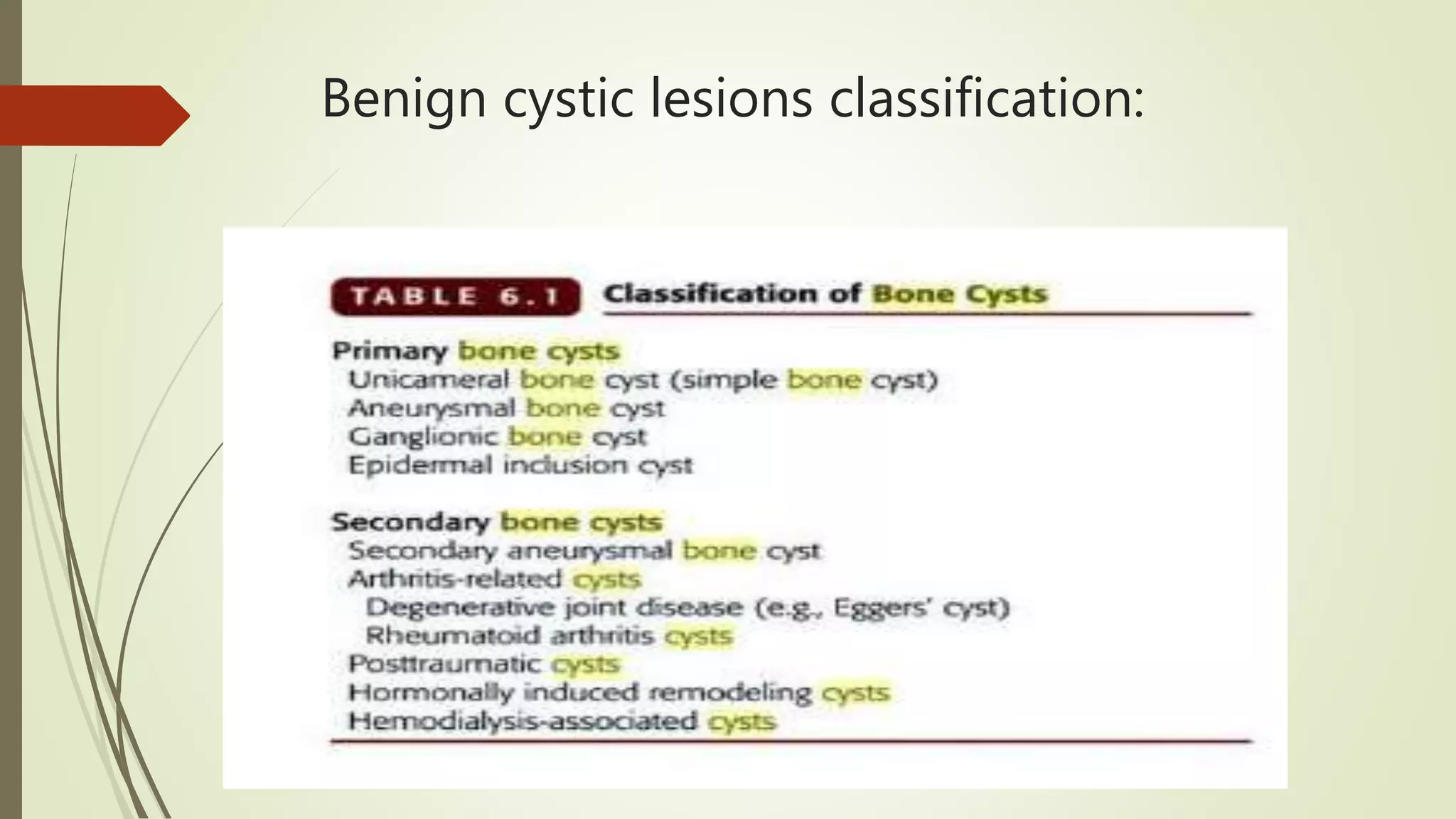
SlideShare a Scribd company logo
APPROACH TO CYSTIC
LESIONS OF BONE
PRESENTER:DR.ARGHA BARUAH
MODERATOR:DR.TN SURESH
DR.SIDDARAM.N PATIL
Outline
 1.Definition
 2.Classification
 3.Approach to cystic lesions
 4.Cystic lesions(individually)
 5.Cases
 6.Summary
Case1 :20 year female presented with progressive pain in the
clavicular region since 4 months
Anastomosing trabeculae of osteoid.Rimmed by
single layer of benign activated
osteoblasts.Numerous osteoclasts
CASE 2: 20 YEAR F PRESENTED WITH PAIN AND SWELLING IN THE KNEE
JOINT SINCE 2 MONTHS.
well-defined, expansile osteolytic lesion with thin peripheral bone shell
What is a cystic lesion of bone?
 Cysts are closed capsule or sac-like structures, typically filled with liquid, semisolid
or gaseous material .
 Cysts lesions of bone develop when one or several factors that affect bone
remodelling cause a focally increased rate of resorption relative to the formation
Numerous influences may produce this net
effect:
 1)tumor
 2)vascular disturbances(ABC)
 3)intraosseous enzymatic effect(Unicameral bone cyst)
 4)hormonal influences such as parathormone linked osteoclastic resorption
 4)external factors-trauma, infection (osteomyelitis),inflammation(rheumatoid
arthritis),degenerative joint disease
Benign cystic lesions classification:
Malignant cystic lesions of bone:
 Giant cell tumor
 Osteosarcoma
 Chondrosarcoma
 Multiple myeloma
 Ewings sarcoma
 Metastasis-lung,renal ,thyroid
Osteolytic lesion of bone:
A L T - M C P S
Age
Location
Transitional zone
Matrix
Cortex
Periosteal reaction
Soft tissue swelling
According to the age:
According to location:
Epiphysis:
 In young patients it is likely to be either a chondroblastoma or an infection.
 In patients over 20, a giant cell tumor has to be included in the differential
diagnosis.
 In older patients a geode, i.e. degenerative subchondral bone cyst must be added
to the differential diagnosis.
Metaphysis:
 NOF, SBC, Osteosarcoma, Chondrosarcoma, Enchondroma and infections.
Diaphysis:
 Ewing's sarcoma, SBC, ABC, Enchondroma, Fibrous dysplasia
Location:
Centric in long bone:
SBC, eosinophilic granuloma, fibrous dysplasia, ABC and enchondroma are lesions that
are located centrally within long bones.
Eccentric in long bone:
Osteosarcoma, NOF, chondroblastoma,, GCT and osteoblastoma are located
eccentrically in long bones.
Zone of transition:
In order to classify osteolytic lesions as well-defined or ill-defined, we need to look at
the zone of transition between the lesion and the adjacent normal bone.
Wide zone of transition: An ill-defined border with a broad zone of transition is a
sign of aggressive growth
Small zone of transition:A small zone of transition results in a sharp, well-defined
border and is a sign of slow growth.
Matrix
Ground Glass – Fib.Dys.
Calcification -
Chondrosarcoma
Matrix
“Cloud” like Ossific densities in Bone
= Osteosarcoma
Wide Zone of
Transition and
Broken Cortex
could be signs of
Aggressiveness /
Malignancy
Cortex:
Intact or
Broken
Soft Tissue
Enormous soft tissue seen in Ewing’s Sarcoma
According to symptoms(if no pain):
 Fibrous Dysplasia
 Enchondroma
 NOF
 Solitary Bone Cyst
Multiple Lesions????
 Mnemonic: FEEMHI
 Fibrous dysplasia
 EG
 Enchondroma
 Mets, Myeloma
 Hyperparathyroidism
 Infection
ANEURYSMAL BONE CYST
Solitary expansile Blood filled reactive lesion of bone .
Locally destructive.
Age :- usually in 1st ,2nd & 3rd decade of life .
Sex :- slight female predominance.
Location :- any bone may be involved.
-Most common is proximal humerus
,distal femur,proximal tibia & spine.
Types :-
Primary :apears de novo following intraosseous
A-V fistula.
Secondary :-results of cystic changes in GCT
,OSTEOBLASTOMA etc.
ANEURYSMAL BONE CYST
Clinical feature :-
-Pt complain of mild to moderate pain.
-Swelling
-Limitation of joint movements.
-Neurological deficit or radicular pain.
-Rapid growth may occur which clinically
presents as a malignancy.
X ray findings :-
Affected bone is expanded, cystic & ballooned
outward.
Eccentrically placed.
Cortex is breached.
Surrounded by a faint outline representing new
periosteal bone.
ANEURYSMAL BONE CYST
Other investigation:-
Bone scan :- shows diffuse or peripheral tracer uptake
with decreased uptake in center.
Ct scan :- helpful in delineating the cyst in complex
area ex. Spine ,pelvis .
MRI :- shows multi loculated cavities & fluid level.
ANEURYSMAL BONE CYST
PATHOLOGY :-
GROSS :-
-Large mass is attached by a broad base to the shaft of
long bone .
-Growing outwards & displacing the soft tissue.
-Thin shell of bone enclosing the blood filled spaces.
Spongy honeycombed blood filled mass with cystic
spaces
Microscopic :-
-Bone & marrow are replaced by the large pool of blood
enclosed in fibro osseous septae.
-Connective tissue bordering the vascular space contain
multinucleated giant cells, new bone formation &
calcium deposits.
Treatment :-
 Curettage & bone grafting.
 Marginal resection Is indicated in expendable bone
 Low dose radiation has been used effectively ,associated with rapid ossification.
SOLITARY BONE CYST
Is a common lesion
Age :- most commonly in 5-15 yrs
rarely after 20 yrs
Sex :- male predominant (2:1)
Location :- long tubular bones particularly humerus & femur
-metaphyseal region is the preferred site.
2 types :-
Active cystic lesion :- develops under 10yrs age . Within 1cm of epiphysis
Benign latent cyst :- develops after 20 yrs age. Towards diaphysis
- seperated from epiphysis
SOLITARY BONE CYST
Physical findings :-
- Usually asymptomatic.
-Cyst lying adjacent to growth plate can
lead to growth disturbances.
X ray findings :- centrally located,lytic lesion.
-Expansion of the bone.
-Well marginated outline.
-Thinning of the cortex( scalloping).
- Fallen fragment sign
--loculated appearance is due to presence of
ridges over inner surface of cyst.
SOLITARY BONE CYST
Pathology :-
Gross :-
- Bone displays an area of fusiform
expansion.
- Underlying bone is egg-shell thin.
- Cavity contain yellow fluid.
-
Microscopic :-
- Connective tissue composed of fibroblast
contaning multinucleated giant cells , foam
cell.
- Cortical wall consist of loosely trabeculated
osseous tissue & many thin walled vessels.
Unicameral bone cyst is filled with straw colored fluid rich in
prostaglandins, GAGs, mucopolysaccharides and lysosomal enzymes. This
fluid contributes to bone resorption.
SOLITARY BONE CYST
Treatment :-
Curettage
Aspiration
Steroid injection
FIBROUS DYSPLASIA
-Non inherited, sporadic developmental anomaly.
-Replacement of bony tissue with fibrous tissue.
Age :- begins in childhood(<10 yrs).
Sex :- M:F=1:1
Location :- base of the skull & long bones.
Clinical features:-
-Pain
-Limp
-Deformity
-Asymmetry of face
-Endocrine disorder
-Limb length discrepancy.
-Sexual precocity
It can be
 Monostotic form (85%)
 Polyostotic form (15%)
 Craniofacial form- Leontiasis Ossea
 Cherubism
 McCune McCune-Albright Syndrome (10%)
Polyostotic , café-au au-lait , endocrine dysfunction
(precocious female puberty (20%), hyperthyroidism)
 MAZABRAUD’s syndrome: ( polyostotic F.D + intramus. Myxomas)
 RIBS>FEMUR>TIBIA>MAXILLA>MANDIBLE>SKULL>HUMREUS .
INTRAMEDULLARY ,ECENTRIC OR CENTRAL LOCATION.
FIBROUS DYSPLASIA
Radiological findings :-
-intramedullary & predominantly diaphyseal.
-central or eccentric in location.
-Ground glass appearance.
-lesion is well defined with sclerotic margins
-Cortex thinning & expansion is seen.
-Pathological fractures .
-Bowing of bones.
-HARRISON’S GROOVE.
FIBROUS DYSPLASIA
Pathology :-
Gross :-
-Bone is irregular in shape & bent.
-Grey tough fibrous tissue that cut with a gritty resistance “SANDPAPER”
-Well circumscribed,
-Tan-white-yellow, gritty
-Cortical bone often thin and expanded
Micro :-
-composed of dense,mature collagenous tissue.
-fibroblast are oriented in linear /whorl pattern.
-Curvilinear trabeculae (Chinese letters) of metaplastic woven bone
in hypocellular, fibroblastic stroma
TREATMENT
- Conservative management includes bracing & modification of
activities.
Surgical management :-
-Pathological fracture usually heals but develops deformity.
-Fracture of long bone should be treated preferably with I.M
nails along with bone grafts.
Large lesion which jeopardize the integrity of bone should be
treated with curettage & bone grafting
Arthritis related cyst:
 Subchondral cyst: multiple,vary in size,occur in one or both sides of joint,central
location,more in weight bearing bones.
 Pyriform in shape and multilocular.
 Contiguous with the joint or may have fine bone plate.End of the bone shows
degenerative changes
INTRAOSSEOUS GANGLION CYST
-Lesion of uncertain pathogenesis.
Age :- can occur at any age.
-Most commonly seen in 20-60yrs age .
Location :- subchondral region of bone ,
acetabulum & carpel bones.
Clinical feature :-are usually clinically silent.
-Chronic mild pain
-Swelling if associated with soft tissue ganglion.
-Usually solitary
INTRAOSSEOUS GANGLION CYST
Radiographic findings :-
-Well demarcated ,sharply circumscribed
osteolytic lesion.
-Sclerotic margin is evident.
-In para articular location gas may be
evident.
Treatment :-
- Local excision of overlying soft tissue &
curettage of involved bone.
- Recurrence is rare.
EPIDERMOID CYST
 Uncommon.
 M >F
 Age :- usually in 2nd ,3rd & 4th decade of life.
 Location :- skull & phalanges of hand are commonly affected.
Clinically :-
 Pain & swelling
Microscopically :-
 They resemble epidermal cyst of skin.
 Cysts are filled with keratinous material & lined by squamous epithelium.
EPIDERMOID CYST
RADIOGRAPHIC :-
Well defined osteolytic lesion with
sclerotic margins .
CT & MRI are rarely required.
Treatment :-
Excision
Giant cell reparative granuloma:
CHONDROMA
•Benign lesions of hyaline cartilage.
•It can arise within a bone ( ENCHONDROMA)
or on the surface of bone ( PERIOSTEAL
/JUXTACORTICAL CHONDROMA).
ENCHONDROMA
Age :- usually all age group are affected .
More commonly young individual in 2nd -4th decade.
Sex :- equally (M:F=1:1)
Location:- any bone can be affected but commonly phalanges of
hand & feet.
-solitary enchondromas involve humerus , tibia & femur.
Clinical findings :-
-Swelling
-Tenderness
-Pain
ENCHONDROMA
X RAYS FINDINGS :-
-well-defined lytic lesion in the hand is almost always
an enchondroma
-small loculated area with well defined margins.
-Cortex is thinned & expanded.
-Interlesional calcification :- calcification is irregular
Stippled ,punctate or popcorn.
-no reactive bone formation
CT scan :-
Evaluate endosteal erosion to rule out
chondrosarcoma.
ENCHONDROMA
Pathology :-
Gross :-
-Tumour is surrounded by a fibrous capsule.
-The neoplastic tissue is composed of bluish
white translucent cartilage
- Areas of calcification.
Microscopically :-
-Tumour shows stages of cartilage formation.
-Mesenchymal tissue is seen at periphery.
-Most mature cartilage is seen at center.
 Ollier’s Disease= multiple enchondromas which usually develop in childhood. The
growth of these enchondromas usually stops after skeletal maturation.The affected
extremity is shortened (asymmetric dwarfism) and sometimes bowed due to
epiphyseal fusion anomalies. Persons with Ollier disease are prone to breaking
bones and normally have swollen, aching limbs.
 Maffucci’s Syndrome =characterized by enchondromas and soft tissue
hemangiomas
TREATMENT
 Treatment of solitary enchondroma consist of observation with serial radiographs.
 If the lesion appears to be radiographically stable & asymptomatic then no further
investigation is required.
 If the lesion appears to be unstable , growing then extended curettage is done.
CHONDROMYXOID FIBROMA
-Least common cartilage tumour.
-Not known to metastasize.
-2% of all benign tumour.
-Seen in patients <30yrs specially in 2nd
& 3rd decade
-Tumour is more common in male
(M:F=2:1).
-Observed in long tubular bones
specially in lower extremity.
CHONDROMYXOID FIBROMA
Clinical features:-
-Pain near the joint without h/o trauma
-Occasionally swelling
-Pathological fractures ( rare)
-May presents with soft tissue swelling.
Radiographic findings :-
-Translucent mass located eccentrically.
-Fusiform expansion in small bones.
-Cortex is expanded & thinned.
-Margin’s are scalloped & sclerosis +.
-Periosteal reaction is uncommon.
CHONDROMYXOID FIBROMA
Pathology :-
Gross :-
-Cut surface reveals solid tumour mass
containing small cavities with mucoid tissue.
-Calcified areas are unusual.
-Surface is sharply demarcated ,lobulated &
surrounded by thin scalloped border of dense
bone.
Microscopic :-
-Composed of lobulated areas of stellate cells
with indistinct cytoplasmic borders.
-At the periphery ,the appearance is more
cellular & collagenous.
TREATMENT
-local excision & filling the cavity with autogenous bone.
-Curettage is not sufficient as tumour may recure especially
childrens.
-Wide en- bloc excision gives high rate of cure.
-If tumour recure even after en bloc excision then studies has to
be done to rule out the malignant transformation.
FIBROUS CORTICAL DEFECT
 Benign well circumscribed fibrous growth with in a small area of long bone.
 A/k/a nonossifying fibroma,fibroxanthoma
 Localised form of fibrous dysplasia.
 Age :- <30yrs
 Location :- metaphysis of long bone.-usually lower limb.
 Sex :-M:F=1:1
Clinical feature :-
 -usually asymptomatic
 -occasionally presents with pathological fracture.
FIBROUS CORTICAL DEFECT
X ray findings :-
-well defined lobulated lesion.
-Ecentrically placed.
-Cortex thinning & expansion
-Multilocular appearance or ridges in bony
wall.
-No periosteal reaction.
Treatment :-
Excision & curettage
FIBROUS CORTICAL DEFECT
Pathology :-
Gross :- thin cortex enclose soft /tough rubbery gray –yellow or reddish brown tissue.
Microscopic :-
 Spindle shaped cell distributed in a whorled pattern.
 Fibroblastic proliferation with high cellularity.
 Giant & foam cells are present.
BROWN TUMOR OF HYPERPARATHYROIDISM
-Hyperparathyroidism results in disorder of bone & mineral
metabolism.
-Diffuse & focal lesion may arise in multiple bones.
-These lesion are K/as brown tumours due to the presence of
hemorrhage in the lesion.
Location :-
Any bone can be affected.
Mainly diaphysis of long bone.
Clinical features :-
Stones
Bones
Groans
BROWN TUMOR OF HYPERPARATHYROIDISM
Radiological features :-
Salt & pepper skull
Multiple osteolytic lesions
Laboratory findings :-
↑ed levels of PTH
Hypercalcemia
↑ed serum phosphate & urate.
TREATMENT :- treatment of the primary cause
will cause the healing of lesion.
Eosinophilic granuloma:
 EG is like osteomyelitis a great mimicker of benign and malignant bone tumors.
 Must be included in the differential diagnosis of almost any ill-defined osteolytic
bone lesion in patients under the age of 30.
 May also present as a well-defined lesion.
 EG can be excluded in patients > 30 years.
 Either solitary bone involvement, multiple bone involvement (variable skin
involvement) or multiple organ involvement (bone, liver, spleen, other sites)
Microscopy:
 Infiltration by Langerhans cells (polygonal cells with eosinophilic cytoplasm,
nuclei with longitudinal grooves resembling coffee beans)
 Eosinophils, giant cells, neutrophils, foam cells, lymphocytes, plasma cells,
fibrosis, necrosis; may have typical and atypical mitotic figures
Giant cell tumor:
 Giant cell tumor is a benign lesion
 Most commonly seen in age 20-40 years.
 A well-defined margin is associated with a locally less aggressive biologic behavior.
 In many cases the margin is ill-defined
 These tumors often thin the cortex and may expand into the soft tissues
surrounding the bone.
Stromal cells are mononuclear, resemble macrophages
Giant cells are large, multinucleated (10 - 50 nuclei) with similar nuclei as stromal cells
Treatment
Surgical curettage (34% recur) or en bloc excision (7% recur); may implant into
adjacent soft tissue
Radiation therapy only if surgical excision impossible since it may promote
transformation
Osteosarcoma
 Most common primary bone tumor after myeloma
 60% male; usually ages 10 - 25 years or ages 40+ with other diseases
 Sites: metaphysis of long bones (distal femur, proximal tibia, proximal humerus;
sites of peak mitotic activity for bone cells); occasionally diaphysis, rarely epiphysis;
less common in flat bones or short bones; usually arises within medullary cavity
and extends to cortex
Osteosarcoma:
 Hallmark of osteosarcoma is the production of bony matrix, which is reflected by the sclerosis
 Usually typical malignant features including permeative-motheaten pattern of destruction, irregular
cortical destruction and aggressive (interrupted) periosteal reaction.
 Radio: Large, destructive, lytic or blastic mass with permeative margins.May break through cortex
and elevate periosteum.Sunburst pattern due to new bone formation in soft tissue
 Occasionally an osteosarcoma may present as a lytic or mixed lytic-sclerotic lesion.
 Completely lytic osteosarcoma may represent telangiectatic subtype, composed of multiple
cavities filled with blood (may resemble aneurysmal bone cyst!).
mixed osteolytic and sclerotic lesion in the proximal humerus with irregular cortical
destruction.
There is an aggressive periosteal reaction and a soft tissue mass.
Gross:
 Big, bulky, gritty, hemorrhagic with cystic degeneration
 Spreads within medullary cavity, destroys cortical bone, elevates periosteum and
invades soft tissue
 May form satellite nodules (“skip metastases”)
 Usually has well defined proximal and distal margins
Chondrosarcoma:
 Third most common bone malignancy after myeloma and osteosarcoma
 Usually ages 30 - 60 years, 75% males
 Most commonly presents as a well-defined lesion, but uncommonly it can be
encountered as an ill-defined lytic lesion.
Partially ill-defined osteolytic lesion
There are cloud-like calcifications
indicating a chondroid matrix.
Pearly white or light blue, often with focal calcification
May have small cysts or myxoid change
Micro:
 Tumor cells produce cartilaginous matrix
 Either well, moderate or poorly differentiated
 May have only minor or focal atypia, but consider malignant if malignant radiologic
features
 Intracytoplasmic hyaline globules common in low grade tumors
 Grading: based on cellularity and nuclear changes in chondrocytes; well, moderate
or poorly differentiated correspond to grades 1 - 3; grade 4 is spindled tumor
representing either chondroblastic osteosarcoma or dedifferentiated
chondrosarcoma
Mutiple myeloma:
 Multiple myeloma must be included in the differential diagnosis of any lytic
bone lesion, either well-defined or ill-defined in age > 40.
 Most common presentation: multiple lytic 'punched out' lesions. A solitary
presentation is referred to as plasmacytoma.
 Usually no increased uptake on bone scan.
 The most common location is in the axial skeleton (spine, skull, pelvis and ribs)
and in the diaphysis of long bones (femur and humerus).
Round lesions filled with a soft reddish material are indicative of foci of
myeloma in this section of vertebral bone. Larger lesions may weaken the
bone to cause fracture with back pain.
Micro:
Ewings sarcoma
 Most common presentation: ill-defined osteolytic lesion with multiple small holes
in the diaphysis of a long bone in a child with a large soft tissue mass.
 Presentation with pain, mass, fever, anemia and leukocytosis.
 Most common location: femur, iliac bone, fibula, rib, tibia.
 Frequently aggressive type of periosteal reaction, but never a benign type.
Gross description
 White, fleshy, ill defined tumor with extensive involvement of medulla and cortex
with periosteal elevation
 May be necrotic or resemble pus
 Excised specimen show fibrosis, hemorrhage and necrosis
 Necrosis can be used as a predictor of long term outcome
Micro:
At lower power, is undifferentiated and densely cellular with "light" cell and "dark" cell appearance.Sheets of small,
round, uniform cells with scant clear cytoplasm, divided into irregular lobules by fibrous strands.Indistinct cell
membranes
Minimal amounts of stroma
Metastasis :
 Must be considered in the differential diagnosis of any bone lesion in a patient > 40
years.
 May present as well-defined osteolytic, ill-defined osteolytic and also as sclerotic bone
lesion.
 Majority of osteolytic metastases originate from lung, kidney, colon, melanoma.
 Cancer most commonly spreads to these sites in the skeleton:
Vertebra,Pelvis, Ribs, Skull,Upper arm and Long bones of the leg
Case3 :
30 YEAR F PRESENTED WITH PAIN AND SWELLING IN THE KNEE JOINT SINCE 1 WEEK
.PATIENT ALSO HAS FEVER SINCE 1 WEEK
Osteomyelitis :
 Osteomyelitis is the great mimicker of bone tumors.
 It has a broad spectrum of radiographic features and occurs at any age and has no
typical location.
 In the chronic stage it can mimic a benign bone tumor (Brodies abscess).
 In the acute stage it can mimic a malignant bone tumor with ill-defined margins,
cortical destruction and an aggressive type of periostitis.
 Only when there is a thick solid periosteal reaction we can recognize the non-malignant
underlying process
Case4:
Spotter:
“If I were you, I will run
to the Radiology
department
and get the x-ray films
and make them
available before the
Histology slides are
studied”
Prof. Peter G Bullough
Professor of Orthopedic Pathology
Hospital for Special Surgery,
Cornell University,
NEW YORK
Diagnosis of Cystic Tumours
Final Diagnosis
CLINICAL
PATHOLOGY
IMAGEOLOGY
(Radiology)
Take home message:
Following points to be kept in mind while reporting :
 History
 Age
 Location
 Multiple/single
 Radiological findings
References:
 1.Orthopaedic pathology Vincent J Vigorita
 2.Dahlin’s bone tumors
 3. The Radiology Assistant : Bone tumor - well-defined osteolytic tumors and
tumor-like lesions
 4.https://radiopaedia.org
 5.www.pathologyoutlines.com
Cystic diseases of bone

More Related Content

What's hot

Radiology of Bone Tumours
Radiology of Bone TumoursRadiology of Bone Tumours
Radiology of Bone Tumours
Muhammad Eimaduddin
 
Giant Cell Tumour
Giant Cell TumourGiant Cell Tumour
Giant Cell Tumour
BipulBorthakur
 
Bone forming tumors
Bone forming tumorsBone forming tumors
Bone forming tumorsKemUnited
 
Bone tumours
Bone tumoursBone tumours
Bone tumours
Usman Shams
 
EWINGS SARCOMA
EWINGS SARCOMAEWINGS SARCOMA
EWINGS SARCOMA
Prashanth Kumar
 
Tuberculosis of bones and joints
Tuberculosis of bones and jointsTuberculosis of bones and joints
Tuberculosis of bones and jointsVishal Sankpal
 
Malignant bone tumours
Malignant bone tumoursMalignant bone tumours
Malignant bone tumours
Archana Koshy
 
Chondroblastoma
Chondroblastoma   Chondroblastoma
Chondroblastoma orthoprince
 
Bone tumors
Bone tumorsBone tumors
Bone tumors
Dhruv Taneja
 
Osteolytic lesions of Bone
Osteolytic lesions of BoneOsteolytic lesions of Bone
Osteolytic lesions of Bone
Vineel Bezawada
 
Round cell tumours
Round cell tumoursRound cell tumours
Round cell tumours
kanwalpreet15
 
Osteomyelitis
OsteomyelitisOsteomyelitis
Osteomyelitis
shahnawaz65
 
An approach to malignant bone tumors
An approach to malignant bone tumors An approach to malignant bone tumors
An approach to malignant bone tumors
Dr.Suhas Basavaiah
 
Giant cell tumor
Giant cell tumorGiant cell tumor
Giant cell tumor
Sudheer Kumar
 
Bone Tumor
Bone TumorBone Tumor
Pigmented villonodular synovitis31may16
Pigmented villonodular synovitis31may16Pigmented villonodular synovitis31may16
Pigmented villonodular synovitis31may16
Suprashant Kumar
 
Benign bone tumor.
Benign bone tumor.Benign bone tumor.
Benign bone tumor.
Dr-ATF
 
Ewings sarcoma- BONE TUMORS
Ewings sarcoma- BONE TUMORS Ewings sarcoma- BONE TUMORS
Ewings sarcoma- BONE TUMORS
Dr.Nikhil. S.U
 

What's hot (20)

Radiology of Bone Tumours
Radiology of Bone TumoursRadiology of Bone Tumours
Radiology of Bone Tumours
 
Giant Cell Tumour
Giant Cell TumourGiant Cell Tumour
Giant Cell Tumour
 
Soft tissue tumor
Soft tissue tumorSoft tissue tumor
Soft tissue tumor
 
Bone forming tumors
Bone forming tumorsBone forming tumors
Bone forming tumors
 
Bone tumours
Bone tumoursBone tumours
Bone tumours
 
EWINGS SARCOMA
EWINGS SARCOMAEWINGS SARCOMA
EWINGS SARCOMA
 
Tuberculosis of bones and joints
Tuberculosis of bones and jointsTuberculosis of bones and joints
Tuberculosis of bones and joints
 
Malignant bone tumours
Malignant bone tumoursMalignant bone tumours
Malignant bone tumours
 
Chondroblastoma
Chondroblastoma   Chondroblastoma
Chondroblastoma
 
Bone tumors
Bone tumorsBone tumors
Bone tumors
 
Osteolytic lesions of Bone
Osteolytic lesions of BoneOsteolytic lesions of Bone
Osteolytic lesions of Bone
 
Round cell tumours
Round cell tumoursRound cell tumours
Round cell tumours
 
Osteomyelitis
OsteomyelitisOsteomyelitis
Osteomyelitis
 
An approach to malignant bone tumors
An approach to malignant bone tumors An approach to malignant bone tumors
An approach to malignant bone tumors
 
Giant cell tumor
Giant cell tumorGiant cell tumor
Giant cell tumor
 
Bone Tumor
Bone TumorBone Tumor
Bone Tumor
 
Pigmented villonodular synovitis31may16
Pigmented villonodular synovitis31may16Pigmented villonodular synovitis31may16
Pigmented villonodular synovitis31may16
 
Benign bone tumor.
Benign bone tumor.Benign bone tumor.
Benign bone tumor.
 
Osteoid+Osteoma
Osteoid+OsteomaOsteoid+Osteoma
Osteoid+Osteoma
 
Ewings sarcoma- BONE TUMORS
Ewings sarcoma- BONE TUMORS Ewings sarcoma- BONE TUMORS
Ewings sarcoma- BONE TUMORS
 

Similar to Cystic diseases of bone

osteogenic bone tumors & fibrous dysplasia
osteogenic bone tumors & fibrous dysplasiaosteogenic bone tumors & fibrous dysplasia
osteogenic bone tumors & fibrous dysplasia
Dr Arun Haldia
 
SKELETAL SYSTEM pathology nursing ppt.pptx
SKELETAL SYSTEM pathology nursing ppt.pptxSKELETAL SYSTEM pathology nursing ppt.pptx
SKELETAL SYSTEM pathology nursing ppt.pptx
AkilanN5
 
Simple and aneurysmal Bone cyst - Definition, Classfication, Investigations, ...
Simple and aneurysmal Bone cyst - Definition, Classfication, Investigations, ...Simple and aneurysmal Bone cyst - Definition, Classfication, Investigations, ...
Simple and aneurysmal Bone cyst - Definition, Classfication, Investigations, ...
orthoslides
 
Aneurysmal bone cyst.pptxavhndbjamnxivsbxhbd
Aneurysmal bone cyst.pptxavhndbjamnxivsbxhbdAneurysmal bone cyst.pptxavhndbjamnxivsbxhbd
Aneurysmal bone cyst.pptxavhndbjamnxivsbxhbd
sunnysam4072
 
Benign bone tumor ppt
Benign bone tumor pptBenign bone tumor ppt
Benign bone tumor ppt
Saurabh Chahar
 
benign bone tumors contd...
benign bone tumors contd...benign bone tumors contd...
benign bone tumors contd...
Anand Dev
 
FOLs
FOLs FOLs
Systemic approach to bone tumor radiology
Systemic approach to bone tumor radiologySystemic approach to bone tumor radiology
Systemic approach to bone tumor radiology
Ramin Sadeghi
 
anandbenignbonetumors-150803083037-lva1-app6892.pptx
anandbenignbonetumors-150803083037-lva1-app6892.pptxanandbenignbonetumors-150803083037-lva1-app6892.pptx
anandbenignbonetumors-150803083037-lva1-app6892.pptx
asdgja
 
Osteomyelitis of jaw--department of oral medicine and radiology
Osteomyelitis of jaw--department of oral medicine and radiologyOsteomyelitis of jaw--department of oral medicine and radiology
Osteomyelitis of jaw--department of oral medicine and radiology
Government Dental College and Hospital, Shimla
 
tumorlikelesionsofbone-130620140455-phpapp02.pdf
tumorlikelesionsofbone-130620140455-phpapp02.pdftumorlikelesionsofbone-130620140455-phpapp02.pdf
tumorlikelesionsofbone-130620140455-phpapp02.pdf
Dishan Mandania
 
Dr.salah.radiology.radiological approach to bone diseases
Dr.salah.radiology.radiological approach to bone diseasesDr.salah.radiology.radiological approach to bone diseases
Dr.salah.radiology.radiological approach to bone diseasesabas_lb
 
Bone forming tumors rabins
Bone forming tumors rabinsBone forming tumors rabins
Bone forming tumors rabins
Robins Shah
 
Benign bone lesion 1
Benign bone lesion 1Benign bone lesion 1
Benign bone lesion 1
sabir khadka
 
Radiology 5th year, 1st lecture (Dr. Salah Mohammad Fatih)
Radiology 5th year, 1st lecture (Dr. Salah Mohammad Fatih)Radiology 5th year, 1st lecture (Dr. Salah Mohammad Fatih)
Radiology 5th year, 1st lecture (Dr. Salah Mohammad Fatih)
College of Medicine, Sulaymaniyah
 
Giant osteoid osteoma of tibial shaft: A rare case report
Giant osteoid osteoma of tibial shaft: A rare case reportGiant osteoid osteoma of tibial shaft: A rare case report
Giant osteoid osteoma of tibial shaft: A rare case report
Apollo Hospitals
 
Bone Tumors
Bone TumorsBone Tumors
Bone Tumors
Muhammad Eimaduddin
 
Presentation1.pptx, radiological imaging of benign bone tumour.
Presentation1.pptx, radiological imaging of benign bone tumour.Presentation1.pptx, radiological imaging of benign bone tumour.
Presentation1.pptx, radiological imaging of benign bone tumour.
Abdellah Nazeer
 

Similar to Cystic diseases of bone (20)

osteogenic bone tumors & fibrous dysplasia
osteogenic bone tumors & fibrous dysplasiaosteogenic bone tumors & fibrous dysplasia
osteogenic bone tumors & fibrous dysplasia
 
SKELETAL SYSTEM pathology nursing ppt.pptx
SKELETAL SYSTEM pathology nursing ppt.pptxSKELETAL SYSTEM pathology nursing ppt.pptx
SKELETAL SYSTEM pathology nursing ppt.pptx
 
Simple and aneurysmal Bone cyst - Definition, Classfication, Investigations, ...
Simple and aneurysmal Bone cyst - Definition, Classfication, Investigations, ...Simple and aneurysmal Bone cyst - Definition, Classfication, Investigations, ...
Simple and aneurysmal Bone cyst - Definition, Classfication, Investigations, ...
 
Aneurysmal bone cyst.pptxavhndbjamnxivsbxhbd
Aneurysmal bone cyst.pptxavhndbjamnxivsbxhbdAneurysmal bone cyst.pptxavhndbjamnxivsbxhbd
Aneurysmal bone cyst.pptxavhndbjamnxivsbxhbd
 
Benign bone tumor ppt
Benign bone tumor pptBenign bone tumor ppt
Benign bone tumor ppt
 
benign bone tumors contd...
benign bone tumors contd...benign bone tumors contd...
benign bone tumors contd...
 
FOLs
FOLs FOLs
FOLs
 
Systemic approach to bone tumor radiology
Systemic approach to bone tumor radiologySystemic approach to bone tumor radiology
Systemic approach to bone tumor radiology
 
anandbenignbonetumors-150803083037-lva1-app6892.pptx
anandbenignbonetumors-150803083037-lva1-app6892.pptxanandbenignbonetumors-150803083037-lva1-app6892.pptx
anandbenignbonetumors-150803083037-lva1-app6892.pptx
 
Osteomyelitis of jaw--department of oral medicine and radiology
Osteomyelitis of jaw--department of oral medicine and radiologyOsteomyelitis of jaw--department of oral medicine and radiology
Osteomyelitis of jaw--department of oral medicine and radiology
 
tumorlikelesionsofbone-130620140455-phpapp02.pdf
tumorlikelesionsofbone-130620140455-phpapp02.pdftumorlikelesionsofbone-130620140455-phpapp02.pdf
tumorlikelesionsofbone-130620140455-phpapp02.pdf
 
Dr.salah.radiology.radiological approach to bone diseases
Dr.salah.radiology.radiological approach to bone diseasesDr.salah.radiology.radiological approach to bone diseases
Dr.salah.radiology.radiological approach to bone diseases
 
Bone forming tumors rabins
Bone forming tumors rabinsBone forming tumors rabins
Bone forming tumors rabins
 
Benign bone lesion 1
Benign bone lesion 1Benign bone lesion 1
Benign bone lesion 1
 
Radiology 5th year, 1st lecture (Dr. Salah Mohammad Fatih)
Radiology 5th year, 1st lecture (Dr. Salah Mohammad Fatih)Radiology 5th year, 1st lecture (Dr. Salah Mohammad Fatih)
Radiology 5th year, 1st lecture (Dr. Salah Mohammad Fatih)
 
Bones And Muscles
Bones And MusclesBones And Muscles
Bones And Muscles
 
Bones And Muscles
Bones And MusclesBones And Muscles
Bones And Muscles
 
Giant osteoid osteoma of tibial shaft: A rare case report
Giant osteoid osteoma of tibial shaft: A rare case reportGiant osteoid osteoma of tibial shaft: A rare case report
Giant osteoid osteoma of tibial shaft: A rare case report
 
Bone Tumors
Bone TumorsBone Tumors
Bone Tumors
 
Presentation1.pptx, radiological imaging of benign bone tumour.
Presentation1.pptx, radiological imaging of benign bone tumour.Presentation1.pptx, radiological imaging of benign bone tumour.
Presentation1.pptx, radiological imaging of benign bone tumour.
 

More from Argha Baruah

Molecular Genetics and Recent updates of Soft tissue tumours Dr.Argha Baruah
Molecular Genetics and Recent updates of Soft tissue tumours Dr.Argha BaruahMolecular Genetics and Recent updates of Soft tissue tumours Dr.Argha Baruah
Molecular Genetics and Recent updates of Soft tissue tumours Dr.Argha Baruah
Argha Baruah
 
Recent diagnostic and Prognostic indices of Endometrial Cancers Dr.Argha Baruah
Recent diagnostic and Prognostic indices of Endometrial Cancers Dr.Argha BaruahRecent diagnostic and Prognostic indices of Endometrial Cancers Dr.Argha Baruah
Recent diagnostic and Prognostic indices of Endometrial Cancers Dr.Argha Baruah
Argha Baruah
 
Radiological and pathological correlation of bone tumours Dr.Argha Baruah
Radiological and pathological correlation of bone tumours  Dr.Argha BaruahRadiological and pathological correlation of bone tumours  Dr.Argha Baruah
Radiological and pathological correlation of bone tumours Dr.Argha Baruah
Argha Baruah
 
Thymic epithelial tumours and recent updates
Thymic epithelial tumours and recent updatesThymic epithelial tumours and recent updates
Thymic epithelial tumours and recent updates
Argha Baruah
 
Unusual type and site of Lymphoma(Extranodal Lymphoma) Dr.Argha Baruah
Unusual type and site of Lymphoma(Extranodal Lymphoma) Dr.Argha BaruahUnusual type and site of Lymphoma(Extranodal Lymphoma) Dr.Argha Baruah
Unusual type and site of Lymphoma(Extranodal Lymphoma) Dr.Argha Baruah
Argha Baruah
 
Recent updates and reporting of testicular tumors Dr.Argha Baruah
Recent updates and reporting  of testicular tumors  Dr.Argha BaruahRecent updates and reporting  of testicular tumors  Dr.Argha Baruah
Recent updates and reporting of testicular tumors Dr.Argha Baruah
Argha Baruah
 
WHO GIT RECENT UPDATES
WHO GIT RECENT UPDATESWHO GIT RECENT UPDATES
WHO GIT RECENT UPDATES
Argha Baruah
 
SPECIAL STAINS USED IN DIAGNOSING LIVER PATHOLOGY
SPECIAL STAINS USED IN DIAGNOSING LIVER PATHOLOGYSPECIAL STAINS USED IN DIAGNOSING LIVER PATHOLOGY
SPECIAL STAINS USED IN DIAGNOSING LIVER PATHOLOGY
Argha Baruah
 
What is new in Genitourinary pathology
What is new in Genitourinary pathology What is new in Genitourinary pathology
What is new in Genitourinary pathology
Argha Baruah
 
Mediastinal cyst
Mediastinal cyst Mediastinal cyst
Mediastinal cyst
Argha Baruah
 
CNS papillary neoplasm
CNS papillary neoplasm CNS papillary neoplasm
CNS papillary neoplasm
Argha Baruah
 
MALToma
MALTomaMALToma
MALToma
Argha Baruah
 
Milan cytology reporting
Milan cytology reportingMilan cytology reporting
Milan cytology reporting
Argha Baruah
 

More from Argha Baruah (13)

Molecular Genetics and Recent updates of Soft tissue tumours Dr.Argha Baruah
Molecular Genetics and Recent updates of Soft tissue tumours Dr.Argha BaruahMolecular Genetics and Recent updates of Soft tissue tumours Dr.Argha Baruah
Molecular Genetics and Recent updates of Soft tissue tumours Dr.Argha Baruah
 
Recent diagnostic and Prognostic indices of Endometrial Cancers Dr.Argha Baruah
Recent diagnostic and Prognostic indices of Endometrial Cancers Dr.Argha BaruahRecent diagnostic and Prognostic indices of Endometrial Cancers Dr.Argha Baruah
Recent diagnostic and Prognostic indices of Endometrial Cancers Dr.Argha Baruah
 
Radiological and pathological correlation of bone tumours Dr.Argha Baruah
Radiological and pathological correlation of bone tumours  Dr.Argha BaruahRadiological and pathological correlation of bone tumours  Dr.Argha Baruah
Radiological and pathological correlation of bone tumours Dr.Argha Baruah
 
Thymic epithelial tumours and recent updates
Thymic epithelial tumours and recent updatesThymic epithelial tumours and recent updates
Thymic epithelial tumours and recent updates
 
Unusual type and site of Lymphoma(Extranodal Lymphoma) Dr.Argha Baruah
Unusual type and site of Lymphoma(Extranodal Lymphoma) Dr.Argha BaruahUnusual type and site of Lymphoma(Extranodal Lymphoma) Dr.Argha Baruah
Unusual type and site of Lymphoma(Extranodal Lymphoma) Dr.Argha Baruah
 
Recent updates and reporting of testicular tumors Dr.Argha Baruah
Recent updates and reporting  of testicular tumors  Dr.Argha BaruahRecent updates and reporting  of testicular tumors  Dr.Argha Baruah
Recent updates and reporting of testicular tumors Dr.Argha Baruah
 
WHO GIT RECENT UPDATES
WHO GIT RECENT UPDATESWHO GIT RECENT UPDATES
WHO GIT RECENT UPDATES
 
SPECIAL STAINS USED IN DIAGNOSING LIVER PATHOLOGY
SPECIAL STAINS USED IN DIAGNOSING LIVER PATHOLOGYSPECIAL STAINS USED IN DIAGNOSING LIVER PATHOLOGY
SPECIAL STAINS USED IN DIAGNOSING LIVER PATHOLOGY
 
What is new in Genitourinary pathology
What is new in Genitourinary pathology What is new in Genitourinary pathology
What is new in Genitourinary pathology
 
Mediastinal cyst
Mediastinal cyst Mediastinal cyst
Mediastinal cyst
 
CNS papillary neoplasm
CNS papillary neoplasm CNS papillary neoplasm
CNS papillary neoplasm
 
MALToma
MALTomaMALToma
MALToma
 
Milan cytology reporting
Milan cytology reportingMilan cytology reporting
Milan cytology reporting
 

Recently uploaded

CDSCO and Phamacovigilance {Regulatory body in India}
CDSCO and Phamacovigilance {Regulatory body in India}CDSCO and Phamacovigilance {Regulatory body in India}
CDSCO and Phamacovigilance {Regulatory body in India}
NEHA GUPTA
 
Aortic Association CBL Pilot April 19 – 20 Bern
Aortic Association CBL Pilot April 19 – 20 BernAortic Association CBL Pilot April 19 – 20 Bern
Aortic Association CBL Pilot April 19 – 20 Bern
suvadeepdas911
 
Vision-1.pptx, Eye structure, basics of optics
Vision-1.pptx, Eye structure, basics of opticsVision-1.pptx, Eye structure, basics of optics
Vision-1.pptx, Eye structure, basics of optics
Sai Sailesh Kumar Goothy
 
Ophthalmology Clinical Tests for OSCE exam
Ophthalmology Clinical Tests for OSCE examOphthalmology Clinical Tests for OSCE exam
Ophthalmology Clinical Tests for OSCE exam
KafrELShiekh University
 
Pharma Pcd Franchise in Jharkhand - Yodley Lifesciences
Pharma Pcd Franchise in Jharkhand - Yodley LifesciencesPharma Pcd Franchise in Jharkhand - Yodley Lifesciences
Pharma Pcd Franchise in Jharkhand - Yodley Lifesciences
Yodley Lifesciences
 
Local Advanced Lung Cancer: Artificial Intelligence, Synergetics, Complex Sys...
Local Advanced Lung Cancer: Artificial Intelligence, Synergetics, Complex Sys...Local Advanced Lung Cancer: Artificial Intelligence, Synergetics, Complex Sys...
Local Advanced Lung Cancer: Artificial Intelligence, Synergetics, Complex Sys...
Oleg Kshivets
 
Knee anatomy and clinical tests 2024.pdf
Knee anatomy and clinical tests 2024.pdfKnee anatomy and clinical tests 2024.pdf
Knee anatomy and clinical tests 2024.pdf
vimalpl1234
 
How STIs Influence the Development of Pelvic Inflammatory Disease.pptx
How STIs Influence the Development of Pelvic Inflammatory Disease.pptxHow STIs Influence the Development of Pelvic Inflammatory Disease.pptx
How STIs Influence the Development of Pelvic Inflammatory Disease.pptx
FFragrant
 
Pharynx and Clinical Correlations BY Dr.Rabia Inam Gandapore.pptx
Pharynx and Clinical Correlations BY Dr.Rabia Inam Gandapore.pptxPharynx and Clinical Correlations BY Dr.Rabia Inam Gandapore.pptx
Pharynx and Clinical Correlations BY Dr.Rabia Inam Gandapore.pptx
Dr. Rabia Inam Gandapore
 
Role of Mukta Pishti in the Management of Hyperthyroidism
Role of Mukta Pishti in the Management of HyperthyroidismRole of Mukta Pishti in the Management of Hyperthyroidism
Role of Mukta Pishti in the Management of Hyperthyroidism
Dr. Jyothirmai Paindla
 
New Drug Discovery and Development .....
New Drug Discovery and Development .....New Drug Discovery and Development .....
New Drug Discovery and Development .....
NEHA GUPTA
 
Light House Retreats: Plant Medicine Retreat Europe
Light House Retreats: Plant Medicine Retreat EuropeLight House Retreats: Plant Medicine Retreat Europe
Light House Retreats: Plant Medicine Retreat Europe
Lighthouse Retreat
 
Superficial & Deep Fascia of the NECK.pptx
Superficial & Deep Fascia of the NECK.pptxSuperficial & Deep Fascia of the NECK.pptx
Superficial & Deep Fascia of the NECK.pptx
Dr. Rabia Inam Gandapore
 
micro teaching on communication m.sc nursing.pdf
micro teaching on communication m.sc nursing.pdfmicro teaching on communication m.sc nursing.pdf
micro teaching on communication m.sc nursing.pdf
Anurag Sharma
 
Top-Vitamin-Supplement-Brands-in-India.pptx
Top-Vitamin-Supplement-Brands-in-India.pptxTop-Vitamin-Supplement-Brands-in-India.pptx
Top-Vitamin-Supplement-Brands-in-India.pptx
SwisschemDerma
 
ARTHROLOGY PPT NCISM SYLLABUS AYURVEDA STUDENTS
ARTHROLOGY PPT NCISM SYLLABUS AYURVEDA STUDENTSARTHROLOGY PPT NCISM SYLLABUS AYURVEDA STUDENTS
ARTHROLOGY PPT NCISM SYLLABUS AYURVEDA STUDENTS
Dr. Vinay Pareek
 
Non-respiratory Functions of the Lungs.pdf
Non-respiratory Functions of the Lungs.pdfNon-respiratory Functions of the Lungs.pdf
Non-respiratory Functions of the Lungs.pdf
MedicoseAcademics
 
Flu Vaccine Alert in Bangalore Karnataka
Flu Vaccine Alert in Bangalore KarnatakaFlu Vaccine Alert in Bangalore Karnataka
Flu Vaccine Alert in Bangalore Karnataka
addon Scans
 
Triangles of Neck and Clinical Correlation by Dr. RIG.pptx
Triangles of Neck and Clinical Correlation by Dr. RIG.pptxTriangles of Neck and Clinical Correlation by Dr. RIG.pptx
Triangles of Neck and Clinical Correlation by Dr. RIG.pptx
Dr. Rabia Inam Gandapore
 
Ocular injury ppt Upendra pal optometrist upums saifai etawah
Ocular injury  ppt  Upendra pal  optometrist upums saifai etawahOcular injury  ppt  Upendra pal  optometrist upums saifai etawah
Ocular injury ppt Upendra pal optometrist upums saifai etawah
pal078100
 

Recently uploaded (20)

CDSCO and Phamacovigilance {Regulatory body in India}
CDSCO and Phamacovigilance {Regulatory body in India}CDSCO and Phamacovigilance {Regulatory body in India}
CDSCO and Phamacovigilance {Regulatory body in India}
 
Aortic Association CBL Pilot April 19 – 20 Bern
Aortic Association CBL Pilot April 19 – 20 BernAortic Association CBL Pilot April 19 – 20 Bern
Aortic Association CBL Pilot April 19 – 20 Bern
 
Vision-1.pptx, Eye structure, basics of optics
Vision-1.pptx, Eye structure, basics of opticsVision-1.pptx, Eye structure, basics of optics
Vision-1.pptx, Eye structure, basics of optics
 
Ophthalmology Clinical Tests for OSCE exam
Ophthalmology Clinical Tests for OSCE examOphthalmology Clinical Tests for OSCE exam
Ophthalmology Clinical Tests for OSCE exam
 
Pharma Pcd Franchise in Jharkhand - Yodley Lifesciences
Pharma Pcd Franchise in Jharkhand - Yodley LifesciencesPharma Pcd Franchise in Jharkhand - Yodley Lifesciences
Pharma Pcd Franchise in Jharkhand - Yodley Lifesciences
 
Local Advanced Lung Cancer: Artificial Intelligence, Synergetics, Complex Sys...
Local Advanced Lung Cancer: Artificial Intelligence, Synergetics, Complex Sys...Local Advanced Lung Cancer: Artificial Intelligence, Synergetics, Complex Sys...
Local Advanced Lung Cancer: Artificial Intelligence, Synergetics, Complex Sys...
 
Knee anatomy and clinical tests 2024.pdf
Knee anatomy and clinical tests 2024.pdfKnee anatomy and clinical tests 2024.pdf
Knee anatomy and clinical tests 2024.pdf
 
How STIs Influence the Development of Pelvic Inflammatory Disease.pptx
How STIs Influence the Development of Pelvic Inflammatory Disease.pptxHow STIs Influence the Development of Pelvic Inflammatory Disease.pptx
How STIs Influence the Development of Pelvic Inflammatory Disease.pptx
 
Pharynx and Clinical Correlations BY Dr.Rabia Inam Gandapore.pptx
Pharynx and Clinical Correlations BY Dr.Rabia Inam Gandapore.pptxPharynx and Clinical Correlations BY Dr.Rabia Inam Gandapore.pptx
Pharynx and Clinical Correlations BY Dr.Rabia Inam Gandapore.pptx
 
Role of Mukta Pishti in the Management of Hyperthyroidism
Role of Mukta Pishti in the Management of HyperthyroidismRole of Mukta Pishti in the Management of Hyperthyroidism
Role of Mukta Pishti in the Management of Hyperthyroidism
 
New Drug Discovery and Development .....
New Drug Discovery and Development .....New Drug Discovery and Development .....
New Drug Discovery and Development .....
 
Light House Retreats: Plant Medicine Retreat Europe
Light House Retreats: Plant Medicine Retreat EuropeLight House Retreats: Plant Medicine Retreat Europe
Light House Retreats: Plant Medicine Retreat Europe
 
Superficial & Deep Fascia of the NECK.pptx
Superficial & Deep Fascia of the NECK.pptxSuperficial & Deep Fascia of the NECK.pptx
Superficial & Deep Fascia of the NECK.pptx
 
micro teaching on communication m.sc nursing.pdf
micro teaching on communication m.sc nursing.pdfmicro teaching on communication m.sc nursing.pdf
micro teaching on communication m.sc nursing.pdf
 
Top-Vitamin-Supplement-Brands-in-India.pptx
Top-Vitamin-Supplement-Brands-in-India.pptxTop-Vitamin-Supplement-Brands-in-India.pptx
Top-Vitamin-Supplement-Brands-in-India.pptx
 
ARTHROLOGY PPT NCISM SYLLABUS AYURVEDA STUDENTS
ARTHROLOGY PPT NCISM SYLLABUS AYURVEDA STUDENTSARTHROLOGY PPT NCISM SYLLABUS AYURVEDA STUDENTS
ARTHROLOGY PPT NCISM SYLLABUS AYURVEDA STUDENTS
 
Non-respiratory Functions of the Lungs.pdf
Non-respiratory Functions of the Lungs.pdfNon-respiratory Functions of the Lungs.pdf
Non-respiratory Functions of the Lungs.pdf
 
Flu Vaccine Alert in Bangalore Karnataka
Flu Vaccine Alert in Bangalore KarnatakaFlu Vaccine Alert in Bangalore Karnataka
Flu Vaccine Alert in Bangalore Karnataka
 
Triangles of Neck and Clinical Correlation by Dr. RIG.pptx
Triangles of Neck and Clinical Correlation by Dr. RIG.pptxTriangles of Neck and Clinical Correlation by Dr. RIG.pptx
Triangles of Neck and Clinical Correlation by Dr. RIG.pptx
 
Ocular injury ppt Upendra pal optometrist upums saifai etawah
Ocular injury  ppt  Upendra pal  optometrist upums saifai etawahOcular injury  ppt  Upendra pal  optometrist upums saifai etawah
Ocular injury ppt Upendra pal optometrist upums saifai etawah
 

Cystic diseases of bone

  • 1. APPROACH TO CYSTIC LESIONS OF BONE PRESENTER:DR.ARGHA BARUAH MODERATOR:DR.TN SURESH DR.SIDDARAM.N PATIL
  • 2. Outline  1.Definition  2.Classification  3.Approach to cystic lesions  4.Cystic lesions(individually)  5.Cases  6.Summary
  • 3. Case1 :20 year female presented with progressive pain in the clavicular region since 4 months Anastomosing trabeculae of osteoid.Rimmed by single layer of benign activated osteoblasts.Numerous osteoclasts
  • 4. CASE 2: 20 YEAR F PRESENTED WITH PAIN AND SWELLING IN THE KNEE JOINT SINCE 2 MONTHS. well-defined, expansile osteolytic lesion with thin peripheral bone shell
  • 5.
  • 6. What is a cystic lesion of bone?  Cysts are closed capsule or sac-like structures, typically filled with liquid, semisolid or gaseous material .  Cysts lesions of bone develop when one or several factors that affect bone remodelling cause a focally increased rate of resorption relative to the formation
  • 7. Numerous influences may produce this net effect:  1)tumor  2)vascular disturbances(ABC)  3)intraosseous enzymatic effect(Unicameral bone cyst)  4)hormonal influences such as parathormone linked osteoclastic resorption  4)external factors-trauma, infection (osteomyelitis),inflammation(rheumatoid arthritis),degenerative joint disease
  • 8. Benign cystic lesions classification:
  • 9. Malignant cystic lesions of bone:  Giant cell tumor  Osteosarcoma  Chondrosarcoma  Multiple myeloma  Ewings sarcoma  Metastasis-lung,renal ,thyroid
  • 11.
  • 12. A L T - M C P S Age Location Transitional zone Matrix Cortex Periosteal reaction Soft tissue swelling
  • 14.
  • 15.
  • 16. According to location: Epiphysis:  In young patients it is likely to be either a chondroblastoma or an infection.  In patients over 20, a giant cell tumor has to be included in the differential diagnosis.  In older patients a geode, i.e. degenerative subchondral bone cyst must be added to the differential diagnosis. Metaphysis:  NOF, SBC, Osteosarcoma, Chondrosarcoma, Enchondroma and infections. Diaphysis:  Ewing's sarcoma, SBC, ABC, Enchondroma, Fibrous dysplasia
  • 17. Location: Centric in long bone: SBC, eosinophilic granuloma, fibrous dysplasia, ABC and enchondroma are lesions that are located centrally within long bones. Eccentric in long bone: Osteosarcoma, NOF, chondroblastoma,, GCT and osteoblastoma are located eccentrically in long bones.
  • 18. Zone of transition: In order to classify osteolytic lesions as well-defined or ill-defined, we need to look at the zone of transition between the lesion and the adjacent normal bone. Wide zone of transition: An ill-defined border with a broad zone of transition is a sign of aggressive growth Small zone of transition:A small zone of transition results in a sharp, well-defined border and is a sign of slow growth.
  • 19.
  • 20. Matrix Ground Glass – Fib.Dys. Calcification - Chondrosarcoma
  • 21. Matrix “Cloud” like Ossific densities in Bone = Osteosarcoma
  • 22. Wide Zone of Transition and Broken Cortex could be signs of Aggressiveness / Malignancy Cortex: Intact or Broken
  • 23.
  • 24. Soft Tissue Enormous soft tissue seen in Ewing’s Sarcoma
  • 25. According to symptoms(if no pain):  Fibrous Dysplasia  Enchondroma  NOF  Solitary Bone Cyst
  • 26. Multiple Lesions????  Mnemonic: FEEMHI  Fibrous dysplasia  EG  Enchondroma  Mets, Myeloma  Hyperparathyroidism  Infection
  • 27. ANEURYSMAL BONE CYST Solitary expansile Blood filled reactive lesion of bone . Locally destructive. Age :- usually in 1st ,2nd & 3rd decade of life . Sex :- slight female predominance. Location :- any bone may be involved. -Most common is proximal humerus ,distal femur,proximal tibia & spine. Types :- Primary :apears de novo following intraosseous A-V fistula. Secondary :-results of cystic changes in GCT ,OSTEOBLASTOMA etc.
  • 28. ANEURYSMAL BONE CYST Clinical feature :- -Pt complain of mild to moderate pain. -Swelling -Limitation of joint movements. -Neurological deficit or radicular pain. -Rapid growth may occur which clinically presents as a malignancy. X ray findings :- Affected bone is expanded, cystic & ballooned outward. Eccentrically placed. Cortex is breached. Surrounded by a faint outline representing new periosteal bone.
  • 29. ANEURYSMAL BONE CYST Other investigation:- Bone scan :- shows diffuse or peripheral tracer uptake with decreased uptake in center. Ct scan :- helpful in delineating the cyst in complex area ex. Spine ,pelvis . MRI :- shows multi loculated cavities & fluid level.
  • 30. ANEURYSMAL BONE CYST PATHOLOGY :- GROSS :- -Large mass is attached by a broad base to the shaft of long bone . -Growing outwards & displacing the soft tissue. -Thin shell of bone enclosing the blood filled spaces. Spongy honeycombed blood filled mass with cystic spaces Microscopic :- -Bone & marrow are replaced by the large pool of blood enclosed in fibro osseous septae. -Connective tissue bordering the vascular space contain multinucleated giant cells, new bone formation & calcium deposits.
  • 31.
  • 32.
  • 33. Treatment :-  Curettage & bone grafting.  Marginal resection Is indicated in expendable bone  Low dose radiation has been used effectively ,associated with rapid ossification.
  • 34. SOLITARY BONE CYST Is a common lesion Age :- most commonly in 5-15 yrs rarely after 20 yrs Sex :- male predominant (2:1) Location :- long tubular bones particularly humerus & femur -metaphyseal region is the preferred site. 2 types :- Active cystic lesion :- develops under 10yrs age . Within 1cm of epiphysis Benign latent cyst :- develops after 20 yrs age. Towards diaphysis - seperated from epiphysis
  • 35.
  • 36. SOLITARY BONE CYST Physical findings :- - Usually asymptomatic. -Cyst lying adjacent to growth plate can lead to growth disturbances. X ray findings :- centrally located,lytic lesion. -Expansion of the bone. -Well marginated outline. -Thinning of the cortex( scalloping). - Fallen fragment sign --loculated appearance is due to presence of ridges over inner surface of cyst.
  • 37.
  • 38. SOLITARY BONE CYST Pathology :- Gross :- - Bone displays an area of fusiform expansion. - Underlying bone is egg-shell thin. - Cavity contain yellow fluid. - Microscopic :- - Connective tissue composed of fibroblast contaning multinucleated giant cells , foam cell. - Cortical wall consist of loosely trabeculated osseous tissue & many thin walled vessels.
  • 39.
  • 40. Unicameral bone cyst is filled with straw colored fluid rich in prostaglandins, GAGs, mucopolysaccharides and lysosomal enzymes. This fluid contributes to bone resorption.
  • 41. SOLITARY BONE CYST Treatment :- Curettage Aspiration Steroid injection
  • 42. FIBROUS DYSPLASIA -Non inherited, sporadic developmental anomaly. -Replacement of bony tissue with fibrous tissue. Age :- begins in childhood(<10 yrs). Sex :- M:F=1:1 Location :- base of the skull & long bones. Clinical features:- -Pain -Limp -Deformity -Asymmetry of face -Endocrine disorder -Limb length discrepancy. -Sexual precocity
  • 43. It can be  Monostotic form (85%)  Polyostotic form (15%)  Craniofacial form- Leontiasis Ossea  Cherubism  McCune McCune-Albright Syndrome (10%) Polyostotic , café-au au-lait , endocrine dysfunction (precocious female puberty (20%), hyperthyroidism)  MAZABRAUD’s syndrome: ( polyostotic F.D + intramus. Myxomas)
  • 44.
  • 45.
  • 47. FIBROUS DYSPLASIA Radiological findings :- -intramedullary & predominantly diaphyseal. -central or eccentric in location. -Ground glass appearance. -lesion is well defined with sclerotic margins -Cortex thinning & expansion is seen. -Pathological fractures . -Bowing of bones. -HARRISON’S GROOVE.
  • 48. FIBROUS DYSPLASIA Pathology :- Gross :- -Bone is irregular in shape & bent. -Grey tough fibrous tissue that cut with a gritty resistance “SANDPAPER” -Well circumscribed, -Tan-white-yellow, gritty -Cortical bone often thin and expanded Micro :- -composed of dense,mature collagenous tissue. -fibroblast are oriented in linear /whorl pattern. -Curvilinear trabeculae (Chinese letters) of metaplastic woven bone in hypocellular, fibroblastic stroma
  • 49. TREATMENT - Conservative management includes bracing & modification of activities. Surgical management :- -Pathological fracture usually heals but develops deformity. -Fracture of long bone should be treated preferably with I.M nails along with bone grafts. Large lesion which jeopardize the integrity of bone should be treated with curettage & bone grafting
  • 50. Arthritis related cyst:  Subchondral cyst: multiple,vary in size,occur in one or both sides of joint,central location,more in weight bearing bones.  Pyriform in shape and multilocular.  Contiguous with the joint or may have fine bone plate.End of the bone shows degenerative changes
  • 51.
  • 52.
  • 53. INTRAOSSEOUS GANGLION CYST -Lesion of uncertain pathogenesis. Age :- can occur at any age. -Most commonly seen in 20-60yrs age . Location :- subchondral region of bone , acetabulum & carpel bones. Clinical feature :-are usually clinically silent. -Chronic mild pain -Swelling if associated with soft tissue ganglion. -Usually solitary
  • 54. INTRAOSSEOUS GANGLION CYST Radiographic findings :- -Well demarcated ,sharply circumscribed osteolytic lesion. -Sclerotic margin is evident. -In para articular location gas may be evident. Treatment :- - Local excision of overlying soft tissue & curettage of involved bone. - Recurrence is rare.
  • 55.
  • 56.
  • 57. EPIDERMOID CYST  Uncommon.  M >F  Age :- usually in 2nd ,3rd & 4th decade of life.  Location :- skull & phalanges of hand are commonly affected. Clinically :-  Pain & swelling Microscopically :-  They resemble epidermal cyst of skin.  Cysts are filled with keratinous material & lined by squamous epithelium.
  • 58. EPIDERMOID CYST RADIOGRAPHIC :- Well defined osteolytic lesion with sclerotic margins . CT & MRI are rarely required. Treatment :- Excision
  • 59.
  • 60.
  • 61. Giant cell reparative granuloma:
  • 62.
  • 63.
  • 64. CHONDROMA •Benign lesions of hyaline cartilage. •It can arise within a bone ( ENCHONDROMA) or on the surface of bone ( PERIOSTEAL /JUXTACORTICAL CHONDROMA).
  • 65. ENCHONDROMA Age :- usually all age group are affected . More commonly young individual in 2nd -4th decade. Sex :- equally (M:F=1:1) Location:- any bone can be affected but commonly phalanges of hand & feet. -solitary enchondromas involve humerus , tibia & femur. Clinical findings :- -Swelling -Tenderness -Pain
  • 66. ENCHONDROMA X RAYS FINDINGS :- -well-defined lytic lesion in the hand is almost always an enchondroma -small loculated area with well defined margins. -Cortex is thinned & expanded. -Interlesional calcification :- calcification is irregular Stippled ,punctate or popcorn. -no reactive bone formation CT scan :- Evaluate endosteal erosion to rule out chondrosarcoma.
  • 67. ENCHONDROMA Pathology :- Gross :- -Tumour is surrounded by a fibrous capsule. -The neoplastic tissue is composed of bluish white translucent cartilage - Areas of calcification. Microscopically :- -Tumour shows stages of cartilage formation. -Mesenchymal tissue is seen at periphery. -Most mature cartilage is seen at center.
  • 68.  Ollier’s Disease= multiple enchondromas which usually develop in childhood. The growth of these enchondromas usually stops after skeletal maturation.The affected extremity is shortened (asymmetric dwarfism) and sometimes bowed due to epiphyseal fusion anomalies. Persons with Ollier disease are prone to breaking bones and normally have swollen, aching limbs.  Maffucci’s Syndrome =characterized by enchondromas and soft tissue hemangiomas
  • 69.
  • 70. TREATMENT  Treatment of solitary enchondroma consist of observation with serial radiographs.  If the lesion appears to be radiographically stable & asymptomatic then no further investigation is required.  If the lesion appears to be unstable , growing then extended curettage is done.
  • 71. CHONDROMYXOID FIBROMA -Least common cartilage tumour. -Not known to metastasize. -2% of all benign tumour. -Seen in patients <30yrs specially in 2nd & 3rd decade -Tumour is more common in male (M:F=2:1). -Observed in long tubular bones specially in lower extremity.
  • 72. CHONDROMYXOID FIBROMA Clinical features:- -Pain near the joint without h/o trauma -Occasionally swelling -Pathological fractures ( rare) -May presents with soft tissue swelling. Radiographic findings :- -Translucent mass located eccentrically. -Fusiform expansion in small bones. -Cortex is expanded & thinned. -Margin’s are scalloped & sclerosis +. -Periosteal reaction is uncommon.
  • 73. CHONDROMYXOID FIBROMA Pathology :- Gross :- -Cut surface reveals solid tumour mass containing small cavities with mucoid tissue. -Calcified areas are unusual. -Surface is sharply demarcated ,lobulated & surrounded by thin scalloped border of dense bone. Microscopic :- -Composed of lobulated areas of stellate cells with indistinct cytoplasmic borders. -At the periphery ,the appearance is more cellular & collagenous.
  • 74. TREATMENT -local excision & filling the cavity with autogenous bone. -Curettage is not sufficient as tumour may recure especially childrens. -Wide en- bloc excision gives high rate of cure. -If tumour recure even after en bloc excision then studies has to be done to rule out the malignant transformation.
  • 75. FIBROUS CORTICAL DEFECT  Benign well circumscribed fibrous growth with in a small area of long bone.  A/k/a nonossifying fibroma,fibroxanthoma  Localised form of fibrous dysplasia.  Age :- <30yrs  Location :- metaphysis of long bone.-usually lower limb.  Sex :-M:F=1:1 Clinical feature :-  -usually asymptomatic  -occasionally presents with pathological fracture.
  • 76. FIBROUS CORTICAL DEFECT X ray findings :- -well defined lobulated lesion. -Ecentrically placed. -Cortex thinning & expansion -Multilocular appearance or ridges in bony wall. -No periosteal reaction. Treatment :- Excision & curettage
  • 77. FIBROUS CORTICAL DEFECT Pathology :- Gross :- thin cortex enclose soft /tough rubbery gray –yellow or reddish brown tissue. Microscopic :-  Spindle shaped cell distributed in a whorled pattern.  Fibroblastic proliferation with high cellularity.  Giant & foam cells are present.
  • 78.
  • 79. BROWN TUMOR OF HYPERPARATHYROIDISM -Hyperparathyroidism results in disorder of bone & mineral metabolism. -Diffuse & focal lesion may arise in multiple bones. -These lesion are K/as brown tumours due to the presence of hemorrhage in the lesion. Location :- Any bone can be affected. Mainly diaphysis of long bone. Clinical features :- Stones Bones Groans
  • 80. BROWN TUMOR OF HYPERPARATHYROIDISM Radiological features :- Salt & pepper skull Multiple osteolytic lesions Laboratory findings :- ↑ed levels of PTH Hypercalcemia ↑ed serum phosphate & urate. TREATMENT :- treatment of the primary cause will cause the healing of lesion.
  • 81.
  • 82. Eosinophilic granuloma:  EG is like osteomyelitis a great mimicker of benign and malignant bone tumors.  Must be included in the differential diagnosis of almost any ill-defined osteolytic bone lesion in patients under the age of 30.  May also present as a well-defined lesion.  EG can be excluded in patients > 30 years.  Either solitary bone involvement, multiple bone involvement (variable skin involvement) or multiple organ involvement (bone, liver, spleen, other sites)
  • 83.
  • 84. Microscopy:  Infiltration by Langerhans cells (polygonal cells with eosinophilic cytoplasm, nuclei with longitudinal grooves resembling coffee beans)  Eosinophils, giant cells, neutrophils, foam cells, lymphocytes, plasma cells, fibrosis, necrosis; may have typical and atypical mitotic figures
  • 85. Giant cell tumor:  Giant cell tumor is a benign lesion  Most commonly seen in age 20-40 years.  A well-defined margin is associated with a locally less aggressive biologic behavior.  In many cases the margin is ill-defined  These tumors often thin the cortex and may expand into the soft tissues surrounding the bone.
  • 86.
  • 87.
  • 88. Stromal cells are mononuclear, resemble macrophages Giant cells are large, multinucleated (10 - 50 nuclei) with similar nuclei as stromal cells
  • 89. Treatment Surgical curettage (34% recur) or en bloc excision (7% recur); may implant into adjacent soft tissue Radiation therapy only if surgical excision impossible since it may promote transformation
  • 90. Osteosarcoma  Most common primary bone tumor after myeloma  60% male; usually ages 10 - 25 years or ages 40+ with other diseases  Sites: metaphysis of long bones (distal femur, proximal tibia, proximal humerus; sites of peak mitotic activity for bone cells); occasionally diaphysis, rarely epiphysis; less common in flat bones or short bones; usually arises within medullary cavity and extends to cortex
  • 91. Osteosarcoma:  Hallmark of osteosarcoma is the production of bony matrix, which is reflected by the sclerosis  Usually typical malignant features including permeative-motheaten pattern of destruction, irregular cortical destruction and aggressive (interrupted) periosteal reaction.  Radio: Large, destructive, lytic or blastic mass with permeative margins.May break through cortex and elevate periosteum.Sunburst pattern due to new bone formation in soft tissue  Occasionally an osteosarcoma may present as a lytic or mixed lytic-sclerotic lesion.  Completely lytic osteosarcoma may represent telangiectatic subtype, composed of multiple cavities filled with blood (may resemble aneurysmal bone cyst!).
  • 92. mixed osteolytic and sclerotic lesion in the proximal humerus with irregular cortical destruction. There is an aggressive periosteal reaction and a soft tissue mass.
  • 93. Gross:  Big, bulky, gritty, hemorrhagic with cystic degeneration  Spreads within medullary cavity, destroys cortical bone, elevates periosteum and invades soft tissue  May form satellite nodules (“skip metastases”)  Usually has well defined proximal and distal margins
  • 94.
  • 95.
  • 96. Chondrosarcoma:  Third most common bone malignancy after myeloma and osteosarcoma  Usually ages 30 - 60 years, 75% males  Most commonly presents as a well-defined lesion, but uncommonly it can be encountered as an ill-defined lytic lesion.
  • 97. Partially ill-defined osteolytic lesion There are cloud-like calcifications indicating a chondroid matrix.
  • 98. Pearly white or light blue, often with focal calcification May have small cysts or myxoid change
  • 99. Micro:  Tumor cells produce cartilaginous matrix  Either well, moderate or poorly differentiated  May have only minor or focal atypia, but consider malignant if malignant radiologic features  Intracytoplasmic hyaline globules common in low grade tumors  Grading: based on cellularity and nuclear changes in chondrocytes; well, moderate or poorly differentiated correspond to grades 1 - 3; grade 4 is spindled tumor representing either chondroblastic osteosarcoma or dedifferentiated chondrosarcoma
  • 100.
  • 101. Mutiple myeloma:  Multiple myeloma must be included in the differential diagnosis of any lytic bone lesion, either well-defined or ill-defined in age > 40.  Most common presentation: multiple lytic 'punched out' lesions. A solitary presentation is referred to as plasmacytoma.  Usually no increased uptake on bone scan.  The most common location is in the axial skeleton (spine, skull, pelvis and ribs) and in the diaphysis of long bones (femur and humerus).
  • 102.
  • 103. Round lesions filled with a soft reddish material are indicative of foci of myeloma in this section of vertebral bone. Larger lesions may weaken the bone to cause fracture with back pain.
  • 104. Micro:
  • 105. Ewings sarcoma  Most common presentation: ill-defined osteolytic lesion with multiple small holes in the diaphysis of a long bone in a child with a large soft tissue mass.  Presentation with pain, mass, fever, anemia and leukocytosis.  Most common location: femur, iliac bone, fibula, rib, tibia.  Frequently aggressive type of periosteal reaction, but never a benign type.
  • 106.
  • 107.
  • 108. Gross description  White, fleshy, ill defined tumor with extensive involvement of medulla and cortex with periosteal elevation  May be necrotic or resemble pus  Excised specimen show fibrosis, hemorrhage and necrosis  Necrosis can be used as a predictor of long term outcome
  • 109.
  • 110. Micro: At lower power, is undifferentiated and densely cellular with "light" cell and "dark" cell appearance.Sheets of small, round, uniform cells with scant clear cytoplasm, divided into irregular lobules by fibrous strands.Indistinct cell membranes Minimal amounts of stroma
  • 111. Metastasis :  Must be considered in the differential diagnosis of any bone lesion in a patient > 40 years.  May present as well-defined osteolytic, ill-defined osteolytic and also as sclerotic bone lesion.  Majority of osteolytic metastases originate from lung, kidney, colon, melanoma.  Cancer most commonly spreads to these sites in the skeleton: Vertebra,Pelvis, Ribs, Skull,Upper arm and Long bones of the leg
  • 112.
  • 113.
  • 114.
  • 115. Case3 : 30 YEAR F PRESENTED WITH PAIN AND SWELLING IN THE KNEE JOINT SINCE 1 WEEK .PATIENT ALSO HAS FEVER SINCE 1 WEEK
  • 116. Osteomyelitis :  Osteomyelitis is the great mimicker of bone tumors.  It has a broad spectrum of radiographic features and occurs at any age and has no typical location.  In the chronic stage it can mimic a benign bone tumor (Brodies abscess).  In the acute stage it can mimic a malignant bone tumor with ill-defined margins, cortical destruction and an aggressive type of periostitis.  Only when there is a thick solid periosteal reaction we can recognize the non-malignant underlying process
  • 117. Case4:
  • 119. “If I were you, I will run to the Radiology department and get the x-ray films and make them available before the Histology slides are studied” Prof. Peter G Bullough Professor of Orthopedic Pathology Hospital for Special Surgery, Cornell University, NEW YORK
  • 120. Diagnosis of Cystic Tumours Final Diagnosis CLINICAL PATHOLOGY IMAGEOLOGY (Radiology)
  • 121. Take home message: Following points to be kept in mind while reporting :  History  Age  Location  Multiple/single  Radiological findings
  • 122. References:  1.Orthopaedic pathology Vincent J Vigorita  2.Dahlin’s bone tumors  3. The Radiology Assistant : Bone tumor - well-defined osteolytic tumors and tumor-like lesions  4.https://radiopaedia.org  5.www.pathologyoutlines.com

Editor's Notes

  1. ABC is most commonly seen in younger patients(< 20 yrs). In spine the posterior element is involved with extension to the vertebral body & adjacent vertebra. Spinal lesion can cause neurological deficit or radicular pain due to nerve root compression. Pathogenesis is uncertain, its likely that ABC develops from local circulatory disturbances leading to increased venous pressure & production of local h’ge.
  2. In differentiating from UBC , presence of double density fluid levels & intralesional septation usually indicates ABC Radiotherapy is not used frequently because of the potential for malignant transformation. Recurrance rate is approx 10%-20%. It has been correlated with younger age<15yrs, centrally located cyst & incomplete removal of cystic cavity.
  3. Occasionally the soft center may be continuous with the medullary canal. Lining of the cavity spaces consists of Compressed histiocytes & fibroblast. A solid varient of ABC has been described & is k/as GIANT CELL REPARATIVE GRANULOMA
  4. The exact pathogenesis is unclear but the most widely accepted theory is that a focal defect in metaphyseal remodelling blocks interstitisl fluid drainage this leads to increased pressure which leads to focal bone necrosis & fluid accumulation. After 20 yrs cyst reveals a predilection for pelvic bone & calcaneus. SBC is called active when they are within 1 cm of PHYSIS & latent when they are closer to diaphysis.
  5. Physical findings :- untill attention is drawn to it when trauma cause pain which is often due to fracture through cyst wall. There is no periosteal reaction unless there is a fracture. FALLEN FRAGMENT SIGN :- usually a thinned cortex fracture & falls into the base of lesion confirmining its empty cystic nature. This is pathognomonic of sbc.
  6. Blood filled cystic space linned by fibrous tissue lacking endothelial lining.
  7. Investigation shows that cystic fluid contain prostaglandins , free oxygen radicals, interlukines,cytokines & metalloprotinases all of which contribute to bone resorption Treatment :- small asymptomatic lesion in upper extremities can be treated with observation with serial plain xrays. Large lesions are usually treated with curettage or aspiration with injections ( steroid , bone marrow aspirate, demineralized bone matrix). Pathological # of upper extremity can be treated conservatively. SBC of proximal femur should be treated with curettage , bone graft & internal fixation. Steroid injection was introduced in 1970 as the recurrence rate after curettage was approx 50%. Dosage is about 80-200 mg. if the lesion does not show radiographic signs of healing in 2 mths then the dose must be repeated
  8. Can be :- 1.monostotic & 2 . Polyostotic. It progress beyond puberity & through adulthood. Lesion usually ceases after the adolescence. Earlier the onset more progessive the disease. 70% are monostotic. RIBS>FEMUR>TIBIA>MAXILLA>MANDIBLE>SKULL>HUMREUS . INTRAMEDULLARY ,ECENTRIC OR CENTRAL LOCATION. CLINICAL FEATURES :-Pain is due to pathological fractures which eventually heals.the fracture fragment does not dsplace coz of the fibrous tissue holding them. 2. Deformity :- bowing of bones. coxa vera deformity of femur neck produce Shepard crook’s deformity of the femur is responsible for the shortening. 3. Asymmetry of face coz of hyperostosis of facial bones. 4. Endocrine disorder :- more commonly females are involved. - Sexual precocity(early mensturation , breast developments & Closed epiphysis) 2 SYNDROMES :- Mc CUNE ALBRIGHT SYND.(polyostotic F.D+Skin pigmentation+Endocrine disorder) & MAZABRAUD ( polyostotic F.D + intramus. Myxomas)
  9. HARRISON GROOVE FOLLOWING RIB FRACTURE. a horizontal depression along the lower border of the thorax, corresponding to the costal insertion of the diaphragm CT Scan is best as it help to identify the exents of the disease. The extent of the lesion is clearly visible on computed tomography, and the cortical boundary is depicted with more detail than is seen on radiographs or magnetic resonance images. The thickness of the native cortex, amount of endosteal scalloping and periosteal new bone reaction, and homogeneity of the poorly mineralized lesional tissue are demonstrated best with computed tomography imaging.
  10. Biopsy is sometime required for diagnosis. Sx treated lesion recure in immature skeleton.
  11. Carpel bones esp. LUNATE . Knee ,medial malleolus ,femoral head are common sites. Continuity between the soft tissue & osseous lesion is consistent with gradual erosion of bone surface as a cause of an introsseous ganglion cyst. Pain increases with physical activity But multiple & b/l lesions are also encountered.
  12. Show fluid levels on mri & ↑ed uptake of radionuclide in 10% .
  13. Occur in central location of bone Seen in 2-4 decade of life but more commonly in young adults. It can undergo malignant transformation if situated in long bones More commonly seen in phalanges of hand & feet.
  14. Pain can be due to :- pathological fracture or From the malignant transformation.
  15. - In small bones cortex may be perforated & the shadow of tumour extends into the surrounding soft tissue although the tumour is benign but if the cortex is broken in large bone then there is malignant transformation of the tumour. Juxta cortical tumours are small < 3mm in size ,well defined & frequently appear to fit in a saucer shaped defect on the bone ,the underlying bone is sclerotic & the edges of the lesion appears to be buttressed by a thick rind of cortical bone. Chondrosarcoma usually involves the metaphysis whereas chondromas occur in diaphysis. An epiphyseal location , cotical erosion,less prominent matrix calcification, periosteal reaction & leasion > 5 cm in diameter suggestive of chondrosarcoma.
  16. Tibia & femur involvement is approx. 55% Ecentrically located metaphyseal lesion & elongated in shape.
  17. Both myxoid & chondroid areas stain with toluidine blue & this reaction is absent after hyaluronidase digestion. This is the evidense of its cartilagenous origin. The lobules have a hypocellular center and increased cellularity at the periphery. Benign giants cells may be present at the edge of the lobules. The stroma in between the lobules shows mononuclear cells resembling those seen in chondroblastomas.
  18. Smaller lesion produce focal, superficial,shallow radiolucent area in the cortex with normal or sclerotic adjacent bone & in some a blister like peripheral osseous shell.the lesion are circular or oval & well delineated with smooth or lobulated edge. Large lesion have a more elongated & multilobulated appearance. The axis of the defect lies ll to the axis of the bone. Tendency of this lesion is to grow near the growth plate & then move away from it which gradually become smaller & indistinct & finally disappears. Role of ct & mri is limited in this . They can be used to delineate the extent in complex anatomic areas. TREATMENT:- these lesion are usually asymptomatic hence does not require as they regress by itself in adulthood. Most of the pathological fractures can be treated conservatively. Lesion become symptomatic & require t/t if they become large & are subjected to repeated trauma.some author believs that lesion larger then 50% of the bone diameter require t/t coz of ↑ed risk of pathological fracture.
  19. Hyperparathyroidism is of 2 types : primary coz of adenoma of glands & secondary due to renal failure led to ↑ed levels of parathyroid harmones. Seen in 3rd to 5th decade of life. Skeletal effect include massive bone resorption , #’s,bone pain or circumscribed lytic lesions. Have a less tendency for the spine. Clinically :- pt presents with kidney stones, bone lytic lesions & gastrointestinal symptoms such as nausea vomiting pancreatitis
  20. characteristic areas of resorption include symphysis pubis, distal clavicle, vertebral bodies and lamina dura (bone at base of teeth)