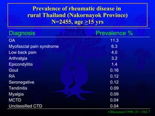
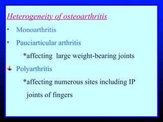
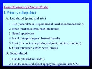
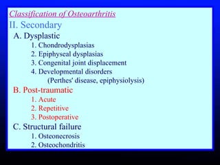
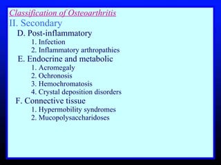
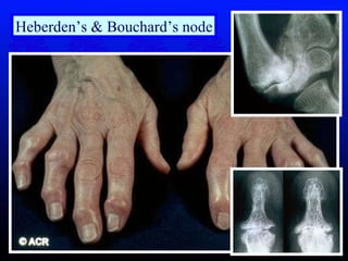
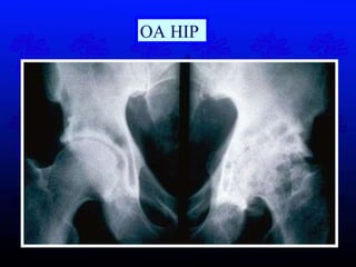
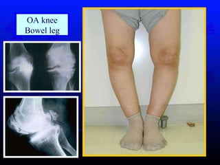
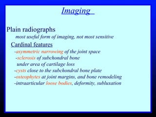
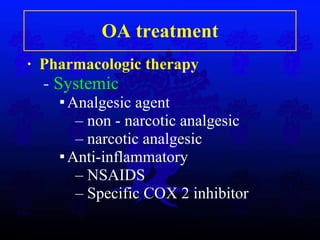
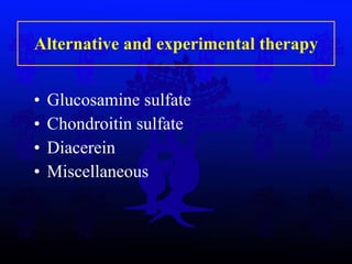
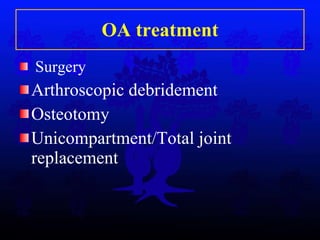
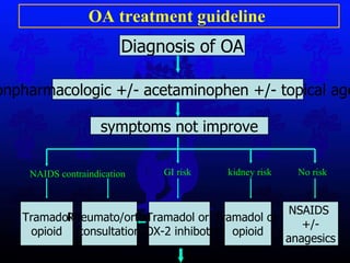
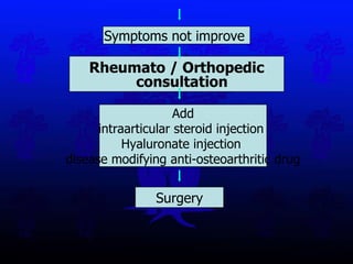
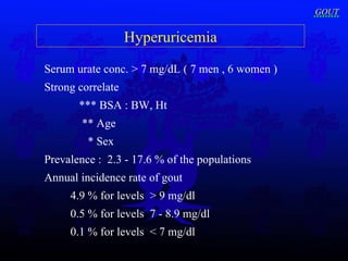
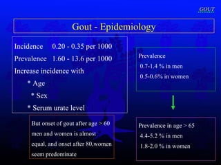
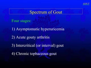
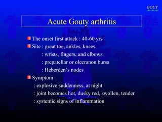
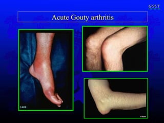
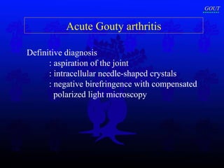
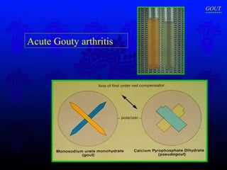
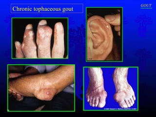
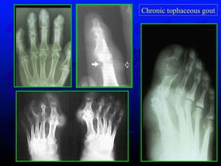
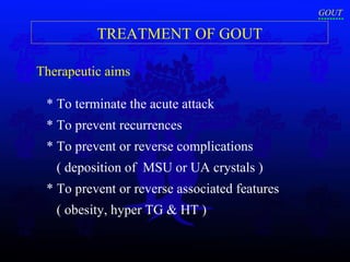
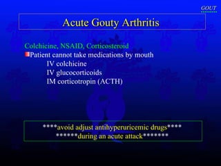
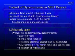
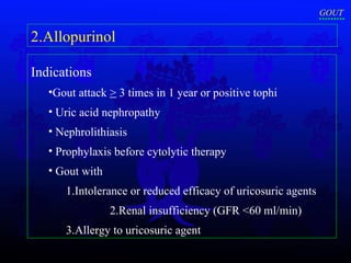
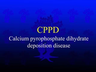
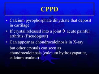
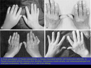
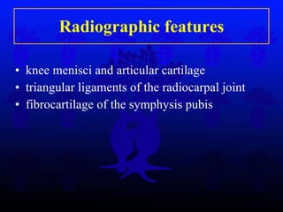
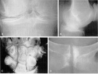
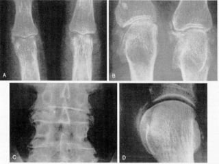
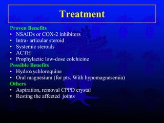
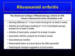
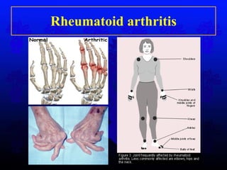
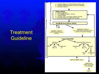
Common Rheumatological disorders in Orthopaedics Department of Orthopaedic Surgery  Faculty of Medicine Siriraj Hospital Mahidol University
Contents Osteoarthritis Gouty Arthritis CPPD Rheumatoid Arthritis
Prevalence of rheumatic disease in  rural Thailand (Nakornayok Province) N=2455, age  > 15 yrs J Rheumatol 1998; 25: 1382-7 Diagnosis Prevalence % OA Myofascial pain syndrome Low back pain Arthralgia Epicondylitis Gout RA Seronegative Tendinitis Myalgia MCTD Unclassified CTD 11.3 6.3 4.0 3.2 1.4 0.16 0.12 0.12 0.09 0.09 0.04 0.04
Osteoarthritis
Heterogeneity of osteoarthritis   Monoarthritis  Pauciarticular arthritis  *affecting  large weight-bearing joints Polyarthritis  *affecting numerous sites including IP      joints of fingers
Classification of Osteoarthritis I. Primary (idiopathic)     A. Localized (principal site)       1 .  Hip (superolateral, superomedial, medial, inferoposterior)       2. Knee (medial, lateral, patellofemoral)      3. Spinal apophyseal      4. Hand (interphalangeal, base of thumb)       5. Foot (first metatarsophalangeal joint, midfoot, hindfoot)      6. Other (shoulder, elbow, wrist, ankle)     B. Generalized       1. Hands (Heberden's nodes)       2. Hands, knees and spinal apophyseal (generalized OA)
Classification of Osteoarthritis II. Secondary     A. Dysplastic       1. Chondrodysplasias       2. Epiphyseal dysplasias       3. Congenital joint displacement       4. Developmental disorders    (Perthes' disease, epiphysiolysis)      B. Post-traumatic       1. Acute       2. Repetitive       3. Postoperative      C. Structural failure      1. Osteonecrosis      2. Osteochondritis
Classification of Osteoarthritis II. Secondary        D. Post-inflammatory       1. Infection      2. Inflammatory arthropathies  E. Endocrine and metabolic      1. Acromegaly       2. Ochronosis       3. Hemochromatosis       4. Crystal deposition disorders      F. Connective tissue       1. Hypermobility syndromes       2. Mucopolysaccharidoses   
CLINICAL FEATURES General Presentation  middle or later years, overweight  History  1) Pain  : localized or refer pain   - according to amount of exertion  - exacerbated by movement & weight bearing    - relieved by rest (except in advanced stages)
CLINICAL FEATURES 2) Stiffness   Initially, intermittent  < 30 minutes 3) Swelling and deformity   Superficial joints (knee or IP joints)  * Bowlegs or knock knees
CLINICAL FEATURES 4)  Loss of function   Specific to the site involved *  Poor grip : osteoarthritis of hands *Difficult grooming : osteoarthritis of the shoulders *Restricted walking distance, limp, fatigue : osteoarthritis of the hips and knees
CLINICAL FEATURES Examination  1) Disturbance of gait     *often noticed first osteoarthritis of the hip or knee    *real or apparent shortening of one of lower limbs 2) Swelling and deformity   3)  Muscle wasting   4) Tenderness  5) Synovial thickening 6) intra-articular fluid, marginal osteophytes
Heberden’s & Bouchard’s node
OA HIP
OA knee Bowel leg
Imaging  Plain radiographs  most useful form of imaging, not most sensitive  Cardinal features   - asymmetric narrowing  of the joint space - sclerosis  of subchondral bone    under area of cartilage loss - cysts  close to the subchondral bone plate - osteophytes  at joint margins, and bone remodeling -intraarticular  loose bodies , deformity, subluxation
Laboratory Investigations  **No reliable diagnostic test  Synovial fluid   Nonspecific : non- inflammation *increased volume, decreased viscosity *mild pleocytosis, and slight increase in protein
General principles of OA treatment Relieving symptoms Maintaining and/or improving functions L imit physical disability Avoiding drug toxicity
OA treatment   Non pharmacologic therapy Pharmacologic therapy Surgery
OA treatment Non pharmacologic therapy  P atient education  Physical therapy  Occupational therapy  Weight  reduction
OA treatment Pharmacologic therapy   -  Systemic Analgesi c  agent  non - narcotic analgesic narcotic analgesi c Anti-inflammatory  NSAIDS Specific COX  2  inhibitor
OA treatment -  Local Intraartricular  :-C orticosteroid injection  :- Viscosupplementation - hyaluronan injection  Topical therap y :- Capsaici n :- Tropical NSAIDs
Alternative and experimental therapy Glucosamine sulfate C hondroitin sulfate Diacerein Miscellaneous
OA treatment Surgery Arthroscopic debridement Osteotomy Unicompartment/Total joint replacement
OA treatment guideline  Tramadol opioid Rheumato/ortho consultation Tramadol or  COX-2 inhibotor Tramadol or  opioid NSAIDS  +/- anagesics Diagnosis of OA nonpharmacologic +/- acetaminophen +/- topical agent symptoms not improve NAIDS contraindication GI risk kidney risk No risk
Rheumato / Orthopedic consultation Symptoms not improve  Add intraarticular steroid injection  Hyaluronate injection  disease modifying anti-osteoarthritic drug Surgery
GOUT
Hyperuricemia Serum urate conc. > 7 mg/dL ( 7 men , 6 women ) Strong correlate  *** BSA : BW, Ht   ** Age   * Sex   Prevalence :  2.3 - 17.6 % of the populations   Annual incidence rate of gout  4.9 % for levels  > 9 mg/dl 0.5 % for levels  7 - 8.9 mg/dl 0.1 % for levels  < 7 mg/dl GOUT
Incidence  0.20 - 0.35 per 1000 Prevalence  1.60 - 13.6 per 1000 Increase incidence with  * Age * Sex * Serum urate level Gout - Epidemiology Prevalence  0.7-1.4 % in men  0.5-0.6% in women Prevalence  in age > 65 4.4-5.2 % in men  1.8-2.0 % in women But onset of gout after age > 60   men and women is almost equal, and onset after 80,women seem predominate   GOUT
Four stages: 1) Asymptomatic hyperuricemia 2) Acute gouty arthritis  3) Intercritical (or interval) gout 4) Chronic tophaceous gout Spectrum of Gout GOUT
M ost : asymptomatic throughout their lifetime Risk of  acute gout increases with  *  serum urate concentration R isk of nephrolithiasis increases with  *  serum urate level  *  daily urinary uric acid excretion > 20 years of sustained hyperuricemia  - Gout  Asymptomatic Hyperuricemia GOUT
Acute Gouty arthritis The onset first attack : 40-60 yrs Site  : great toe, ankles, knees  : wrists, fingers, and elbows : prepatellar or olecranon bursa : Heberden’s nodes Symptom : explosive suddenness, at night : joint becomes hot, dusky red, swollen, tender : systemic signs of inflammation GOUT
Acute Gouty arthritis GOUT
Definitive diagnosis  : aspiration of the joint  : intracellular needle-shaped crystals : negative birefringence with compensated    polarized light microscopy Acute Gouty arthritis GOUT
Acute Gouty arthritis GOUT
Acute Gouty arthritis Strongly negative birefringent crystal GOUT
Acute Gouty arthritis S ubside several hours to weeks  B ecomes asymptomatic (intercritical period) Provocative factors   Drugs : anti-hyperuricemic therapy : drug-induced hyperuricemia Infections Trauma, surgery, hemorrhage Alcohol ingestion  Dietary excess GOUT
Acute Gouty arthritis GOUT
Intercritical Gout Second  attack  within 6 mo - 2 yrs  Diagnosis : difficult or inconclusive GOUT
Characters * chronic polyarticular gout  * without pain-free intercritical periods * average of 11.6 years  * severe crippling disease Chronic tophaceous gout GOUT
Chronic tophaceous gout GOUT
Chronic tophaceous gout
Therapeutic aims  * To terminate the acute attack * To prevent recurrences         * To prevent or reverse complications  ( deposition of  MSU or UA crystals )      * To prevent or reverse associated features ( obesity, hyper TG & HT ) TREATMENT OF GOUT GOUT
Acute Gouty Arthritis  Colchicine, NSAID, Corticosteroid Patient cannot take medications by mouth  IV colchicine IV glucocorticoids IM corticotropin (ACTH)  **** avoid adjust antihyperuricemic drugs ****   ****** during an acute attack *******  GOUT
Prophylaxis against acute attacks of gout  Colchicine 0.6 mg once or twice a day  ***Continued until the serum urate value  maintained well within the normal range, no tophi  and no acute attacks for 3 to 6 months***  GOUT
Control of Hyperuricemia in MSU Deposit Indication: Gout attack  >  3 times in 1 year   In general, the aim of anti- hyperuricemic therapy  Reduce the serum urate  < 5.0- 6.0 mg/dl by allopurinol or a uricosuric agent  1.Uricosuric agent  Probenecid, Sulfinpyrazone, Benzbromarone  * age < 60 years  * normal renal function  (creatinine clearance greater than 80 ml/minute) * UA excretion < 700 mg/24 hours on a general diet * no history of renal calculi  GOUT
Indications   Gout attack  >  3 times in 1 year or positive tophi Uric acid nephropathy Nephrolithiasis Prophylaxis before cytolytic therapy Gout with 1.Intolerance or reduced efficacy of uricosuric agents  2.Renal insufficiency (GFR <60 ml/min) 3.Allergy to uricosuric agent 2.Allopurinol GOUT
CPPD   Calcium pyrophosphate dihydrate  deposition disease
CPPD Calcium pyrophosphate dihydrate that deposit in cartilage If crystal released into a joint   acute painful arthritis (Pseudogout) Can appear as chondrocalcinosis in X-ray but other crystals can seen as chondrocalcinosis (calcium hydroxyapatite, calcium oxalate)
Prevalence  Common in elderly Most studies used radiologic or anatomically defined articular calcification as a marker Prevalence 8% (age 63-93) 15% in age 65-74, 36% in age 75-84 22% in excised cadaver menisci
Clinical feature M:F = 1.5:1 Mean age at time of diagnosis = 72 years Feature Pseudogout Pseudo-RA Pseudo-OA
Pseudogout Found 25% Acute or subacute arthritis(last 1-28 days) Involve one or few joints Most common in knee joint May severe as gout, longer time to peak intensity Less painful and disabling 20% hyperuricemia, 5% MSU deposit
Pseudo-RA Found 5-10 % Subacute attacks (4 weeks-several months) Non-specific symptoms (morning stiffness, fatigue) Large joint common: knee, elbow, ankle But also small joints of hands and feet Synovial thickening, localized  pitting edema, limit joint motion, flexion contracture,   ESR Positive RF 10%, 3% RA have calcified cartilage
Pseudo-RA Differentiate from RA   1. difference severity of multiple joints 2. asymmetrical involvement 3. osteophyte positive  4. positive chondrocalcinosis 5. no periarticular osteoporosis 6. no bony erosion But CPPD with RA co-exist 1%
Pseudo-OA Found 34-50 %  Women predominate Most common :knee > wrist >hip, shoulder, elbow, ankle, and spine Chronic joint pain Generally symmetric, with further advanced on one side, flexion contracture 50% episodic attacks, some time severe as gout Concomitant Heberden’s and Bouchard’s nodes
Pseudo-OA Differentiate from primary OA  1. severe degenerative change 2. atypical joints: MCP, wrist,       elbow, shoulder  3. unilateral or bilateral genu valgus     changes  4. isolated patellofemoral OA. 5. Flexion contractures 6. more likely to symmetric than primary OA 7. Radiographs: Chondrocalcinosis and     exuberant osteophyte formation
Asymptomatic May be the most common clinical presentation Incidental finding from radiograph
A , Acute pseudogout.  B , Pseudo-osteoarthritis.  C , Pseudo-rheumatoid arthritis with boutonniere deformity.  D , Pseudo-rheumatoid arthritis showing ulnar deviation, interosseous muscle atrophy, and metacarpophalangeal and wrist joint involvement. The patients in  A ,  C , and  D  are siblings.
Radiographic features knee menisci and articular cartilage triangular ligaments of the radiocarpal joint fibrocartilage of the symphysis pubis
 
 
Synovial fluid findings usually cloudy, WBC 2,000-80,000/mm 3 , 80% PMN with intracellular and extracellular crystals resolving arthritis, the WBC usually <2,000 /mm 3 , MNC>PMN rhomboid or rod-shaped, weakly birefringent crystals with positive elongation cannot exclude other causes of acute or chronic inflammation such as infection, rheumatoid arthritis
Treatment Proven Benefits NSAIDs or COX-2 inhibitors Intra- articular steroid Systemic steroids ACTH Prophylactic low-dose colchicine Possible Benefits Hydroxychloroquine Oral magnesium (for pts. With hypomagnesemia) Others Aspiration, removal CPPD crystal Resting the affected  joints
Rheumatoid Arthritis
Rheumatoid arthritis
Treatment Guideline

ortho 05 common rheumatic dx rx

  • 1.
    Common Rheumatological disordersin Orthopaedics Department of Orthopaedic Surgery Faculty of Medicine Siriraj Hospital Mahidol University
  • 2.
    Contents Osteoarthritis GoutyArthritis CPPD Rheumatoid Arthritis
  • 3.
    Prevalence of rheumaticdisease in rural Thailand (Nakornayok Province) N=2455, age > 15 yrs J Rheumatol 1998; 25: 1382-7 Diagnosis Prevalence % OA Myofascial pain syndrome Low back pain Arthralgia Epicondylitis Gout RA Seronegative Tendinitis Myalgia MCTD Unclassified CTD 11.3 6.3 4.0 3.2 1.4 0.16 0.12 0.12 0.09 0.09 0.04 0.04
  • 4.
  • 5.
    Heterogeneity of osteoarthritis Monoarthritis Pauciarticular arthritis *affecting large weight-bearing joints Polyarthritis *affecting numerous sites including IP joints of fingers
  • 6.
    Classification of OsteoarthritisI. Primary (idiopathic)    A. Localized (principal site)      1 . Hip (superolateral, superomedial, medial, inferoposterior)      2. Knee (medial, lateral, patellofemoral)     3. Spinal apophyseal      4. Hand (interphalangeal, base of thumb)      5. Foot (first metatarsophalangeal joint, midfoot, hindfoot)     6. Other (shoulder, elbow, wrist, ankle)    B. Generalized      1. Hands (Heberden's nodes)      2. Hands, knees and spinal apophyseal (generalized OA)
  • 7.
    Classification of OsteoarthritisII. Secondary    A. Dysplastic      1. Chondrodysplasias      2. Epiphyseal dysplasias      3. Congenital joint displacement      4. Developmental disorders (Perthes' disease, epiphysiolysis)     B. Post-traumatic      1. Acute      2. Repetitive      3. Postoperative     C. Structural failure      1. Osteonecrosis     2. Osteochondritis
  • 8.
    Classification of OsteoarthritisII. Secondary       D. Post-inflammatory      1. Infection      2. Inflammatory arthropathies E. Endocrine and metabolic      1. Acromegaly      2. Ochronosis      3. Hemochromatosis      4. Crystal deposition disorders    F. Connective tissue      1. Hypermobility syndromes      2. Mucopolysaccharidoses  
  • 9.
    CLINICAL FEATURES GeneralPresentation middle or later years, overweight History 1) Pain : localized or refer pain - according to amount of exertion - exacerbated by movement & weight bearing - relieved by rest (except in advanced stages)
  • 10.
    CLINICAL FEATURES 2)Stiffness Initially, intermittent < 30 minutes 3) Swelling and deformity Superficial joints (knee or IP joints) * Bowlegs or knock knees
  • 11.
    CLINICAL FEATURES 4) Loss of function Specific to the site involved * Poor grip : osteoarthritis of hands *Difficult grooming : osteoarthritis of the shoulders *Restricted walking distance, limp, fatigue : osteoarthritis of the hips and knees
  • 12.
    CLINICAL FEATURES Examination 1) Disturbance of gait *often noticed first osteoarthritis of the hip or knee *real or apparent shortening of one of lower limbs 2) Swelling and deformity 3) Muscle wasting 4) Tenderness 5) Synovial thickening 6) intra-articular fluid, marginal osteophytes
  • 13.
  • 14.
  • 15.
  • 16.
    Imaging Plainradiographs most useful form of imaging, not most sensitive Cardinal features - asymmetric narrowing of the joint space - sclerosis of subchondral bone under area of cartilage loss - cysts close to the subchondral bone plate - osteophytes at joint margins, and bone remodeling -intraarticular loose bodies , deformity, subluxation
  • 17.
    Laboratory Investigations **No reliable diagnostic test Synovial fluid Nonspecific : non- inflammation *increased volume, decreased viscosity *mild pleocytosis, and slight increase in protein
  • 18.
    General principles ofOA treatment Relieving symptoms Maintaining and/or improving functions L imit physical disability Avoiding drug toxicity
  • 19.
    OA treatment Non pharmacologic therapy Pharmacologic therapy Surgery
  • 20.
    OA treatment Nonpharmacologic therapy P atient education Physical therapy Occupational therapy Weight reduction
  • 21.
    OA treatment Pharmacologictherapy - Systemic Analgesi c agent non - narcotic analgesic narcotic analgesi c Anti-inflammatory NSAIDS Specific COX 2 inhibitor
  • 22.
    OA treatment - Local Intraartricular :-C orticosteroid injection :- Viscosupplementation - hyaluronan injection Topical therap y :- Capsaici n :- Tropical NSAIDs
  • 23.
    Alternative and experimentaltherapy Glucosamine sulfate C hondroitin sulfate Diacerein Miscellaneous
  • 24.
    OA treatment SurgeryArthroscopic debridement Osteotomy Unicompartment/Total joint replacement
  • 25.
    OA treatment guideline Tramadol opioid Rheumato/ortho consultation Tramadol or COX-2 inhibotor Tramadol or opioid NSAIDS +/- anagesics Diagnosis of OA nonpharmacologic +/- acetaminophen +/- topical agent symptoms not improve NAIDS contraindication GI risk kidney risk No risk
  • 26.
    Rheumato / Orthopedicconsultation Symptoms not improve Add intraarticular steroid injection Hyaluronate injection disease modifying anti-osteoarthritic drug Surgery
  • 27.
  • 28.
    Hyperuricemia Serum urateconc. > 7 mg/dL ( 7 men , 6 women ) Strong correlate *** BSA : BW, Ht ** Age * Sex Prevalence : 2.3 - 17.6 % of the populations Annual incidence rate of gout 4.9 % for levels > 9 mg/dl 0.5 % for levels 7 - 8.9 mg/dl 0.1 % for levels < 7 mg/dl GOUT
  • 29.
    Incidence 0.20- 0.35 per 1000 Prevalence 1.60 - 13.6 per 1000 Increase incidence with * Age * Sex * Serum urate level Gout - Epidemiology Prevalence 0.7-1.4 % in men 0.5-0.6% in women Prevalence in age > 65 4.4-5.2 % in men 1.8-2.0 % in women But onset of gout after age > 60 men and women is almost equal, and onset after 80,women seem predominate GOUT
  • 30.
    Four stages: 1)Asymptomatic hyperuricemia 2) Acute gouty arthritis 3) Intercritical (or interval) gout 4) Chronic tophaceous gout Spectrum of Gout GOUT
  • 31.
    M ost :asymptomatic throughout their lifetime Risk of acute gout increases with * serum urate concentration R isk of nephrolithiasis increases with * serum urate level * daily urinary uric acid excretion > 20 years of sustained hyperuricemia - Gout Asymptomatic Hyperuricemia GOUT
  • 32.
    Acute Gouty arthritisThe onset first attack : 40-60 yrs Site : great toe, ankles, knees : wrists, fingers, and elbows : prepatellar or olecranon bursa : Heberden’s nodes Symptom : explosive suddenness, at night : joint becomes hot, dusky red, swollen, tender : systemic signs of inflammation GOUT
  • 33.
  • 34.
    Definitive diagnosis : aspiration of the joint : intracellular needle-shaped crystals : negative birefringence with compensated polarized light microscopy Acute Gouty arthritis GOUT
  • 35.
  • 36.
    Acute Gouty arthritisStrongly negative birefringent crystal GOUT
  • 37.
    Acute Gouty arthritisS ubside several hours to weeks B ecomes asymptomatic (intercritical period) Provocative factors Drugs : anti-hyperuricemic therapy : drug-induced hyperuricemia Infections Trauma, surgery, hemorrhage Alcohol ingestion Dietary excess GOUT
  • 38.
  • 39.
    Intercritical Gout Second attack within 6 mo - 2 yrs Diagnosis : difficult or inconclusive GOUT
  • 40.
    Characters * chronicpolyarticular gout * without pain-free intercritical periods * average of 11.6 years * severe crippling disease Chronic tophaceous gout GOUT
  • 41.
  • 42.
  • 43.
    Therapeutic aims * To terminate the acute attack * To prevent recurrences        * To prevent or reverse complications ( deposition of MSU or UA crystals )    * To prevent or reverse associated features ( obesity, hyper TG & HT ) TREATMENT OF GOUT GOUT
  • 44.
    Acute Gouty Arthritis Colchicine, NSAID, Corticosteroid Patient cannot take medications by mouth IV colchicine IV glucocorticoids IM corticotropin (ACTH) **** avoid adjust antihyperuricemic drugs **** ****** during an acute attack ******* GOUT
  • 45.
    Prophylaxis against acuteattacks of gout Colchicine 0.6 mg once or twice a day ***Continued until the serum urate value maintained well within the normal range, no tophi and no acute attacks for 3 to 6 months*** GOUT
  • 46.
    Control of Hyperuricemiain MSU Deposit Indication: Gout attack > 3 times in 1 year In general, the aim of anti- hyperuricemic therapy Reduce the serum urate < 5.0- 6.0 mg/dl by allopurinol or a uricosuric agent 1.Uricosuric agent Probenecid, Sulfinpyrazone, Benzbromarone * age < 60 years * normal renal function (creatinine clearance greater than 80 ml/minute) * UA excretion < 700 mg/24 hours on a general diet * no history of renal calculi GOUT
  • 47.
    Indications Gout attack > 3 times in 1 year or positive tophi Uric acid nephropathy Nephrolithiasis Prophylaxis before cytolytic therapy Gout with 1.Intolerance or reduced efficacy of uricosuric agents 2.Renal insufficiency (GFR <60 ml/min) 3.Allergy to uricosuric agent 2.Allopurinol GOUT
  • 48.
    CPPD Calcium pyrophosphate dihydrate deposition disease
  • 49.
    CPPD Calcium pyrophosphatedihydrate that deposit in cartilage If crystal released into a joint  acute painful arthritis (Pseudogout) Can appear as chondrocalcinosis in X-ray but other crystals can seen as chondrocalcinosis (calcium hydroxyapatite, calcium oxalate)
  • 50.
    Prevalence Commonin elderly Most studies used radiologic or anatomically defined articular calcification as a marker Prevalence 8% (age 63-93) 15% in age 65-74, 36% in age 75-84 22% in excised cadaver menisci
  • 51.
    Clinical feature M:F= 1.5:1 Mean age at time of diagnosis = 72 years Feature Pseudogout Pseudo-RA Pseudo-OA
  • 52.
    Pseudogout Found 25%Acute or subacute arthritis(last 1-28 days) Involve one or few joints Most common in knee joint May severe as gout, longer time to peak intensity Less painful and disabling 20% hyperuricemia, 5% MSU deposit
  • 53.
    Pseudo-RA Found 5-10% Subacute attacks (4 weeks-several months) Non-specific symptoms (morning stiffness, fatigue) Large joint common: knee, elbow, ankle But also small joints of hands and feet Synovial thickening, localized pitting edema, limit joint motion, flexion contracture,  ESR Positive RF 10%, 3% RA have calcified cartilage
  • 54.
    Pseudo-RA Differentiate fromRA  1. difference severity of multiple joints 2. asymmetrical involvement 3. osteophyte positive 4. positive chondrocalcinosis 5. no periarticular osteoporosis 6. no bony erosion But CPPD with RA co-exist 1%
  • 55.
    Pseudo-OA Found 34-50% Women predominate Most common :knee > wrist >hip, shoulder, elbow, ankle, and spine Chronic joint pain Generally symmetric, with further advanced on one side, flexion contracture 50% episodic attacks, some time severe as gout Concomitant Heberden’s and Bouchard’s nodes
  • 56.
    Pseudo-OA Differentiate fromprimary OA  1. severe degenerative change 2. atypical joints: MCP, wrist, elbow, shoulder 3. unilateral or bilateral genu valgus changes 4. isolated patellofemoral OA. 5. Flexion contractures 6. more likely to symmetric than primary OA 7. Radiographs: Chondrocalcinosis and exuberant osteophyte formation
  • 57.
    Asymptomatic May bethe most common clinical presentation Incidental finding from radiograph
  • 58.
    A , Acutepseudogout. B , Pseudo-osteoarthritis. C , Pseudo-rheumatoid arthritis with boutonniere deformity. D , Pseudo-rheumatoid arthritis showing ulnar deviation, interosseous muscle atrophy, and metacarpophalangeal and wrist joint involvement. The patients in A , C , and D are siblings.
  • 59.
    Radiographic features kneemenisci and articular cartilage triangular ligaments of the radiocarpal joint fibrocartilage of the symphysis pubis
  • 60.
  • 61.
  • 62.
    Synovial fluid findingsusually cloudy, WBC 2,000-80,000/mm 3 , 80% PMN with intracellular and extracellular crystals resolving arthritis, the WBC usually <2,000 /mm 3 , MNC>PMN rhomboid or rod-shaped, weakly birefringent crystals with positive elongation cannot exclude other causes of acute or chronic inflammation such as infection, rheumatoid arthritis
  • 63.
    Treatment Proven BenefitsNSAIDs or COX-2 inhibitors Intra- articular steroid Systemic steroids ACTH Prophylactic low-dose colchicine Possible Benefits Hydroxychloroquine Oral magnesium (for pts. With hypomagnesemia) Others Aspiration, removal CPPD crystal Resting the affected joints
  • 64.
  • 65.
  • 66.
  • 67.