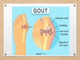
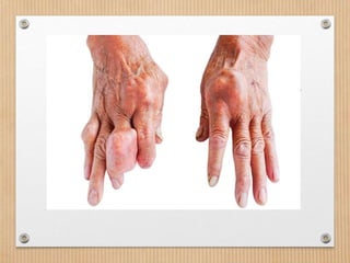
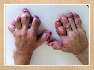
Pathology
JOINT DISEASES
ARTHRITIS
What is arthritis?
• Arthritis literally means “joint inflammation” redness
and warmth within the joint.
• Because inflammation is painful, arthritis limits
movement
TYPES OF ARTHRITIS
The three most common types of arthritis are:
• Osteoarthritis
• Rheumatoid arthritis
• Gout
Osteoarthritis
intro:
• OA is the “wear-and-tear arthritis,” is also called
degenerative joint disease.
• OA is the most common form of arthritis and the
most common joint disease
• It usually develops after age 45 and progresses slowly,
often over a decade.
• Typically, the hips, hands, knees, lower back and/or
neck are affected
Cont..
• Osteoarthritis is an idiopathic disease
characterized by degeneration of
articular cartilage leads to wear and tear,
gross ulceration and finally
disappearance of the full thickness of
articular cartilage
Cont..
• As defined by the American College of
Rheumatology (ACR), OA is a
heterogeneous group of condition that
leads to joint signs and symptoms
which are associated with defective
integrity of articular cartilage, in addition
to related changes in the underlying
bone at the joint margins
OA
• OA is a disease of
joints that affects
all of the weight-
bearing
components of the
joint:
•Articular cartilage
•Menisci
•Bone
Commonly Affects
• – Hips
• –Knees
• – Feet
• – Spine
• – Hands (Interphalangeal joints)
Uncommonly Affected
Joints
• • Shoulder
• • Wrist
• • Elbow
• • Metacarpophalangeal joint
• • TMJ
• • SI
• • Ankle
Incidence
• Osteoarthritis or OA, is the most common type of
arthritis
• It affects up to 27 million Americans.
• More common in women than men
• Majority of patients are over 65.
Pathophysiology
• The disease affects the joint cartilages.
• The cartilage helps bones glide over one another.
• In OA, cartilage within the joint gradually thins and wears away.
• As a result, bones rub against each other causing pain, swelling,
and loss of motion.
• Osteophytes (bone overgrowth) may develop when the ends of
the bone touch each other, and the joint can eventually become
deformed.
• When cartilage wears away, allowing bone to touch bone
completely, advanced or end-stage OA is diagnosed
CAUSES
• The root cause of OA is unknown
• the means of developing symptomatic OA is
influenced by multiple risk factors
Risk factors
• Age
• Age is the strongest risk factor for OA. Although OA
can start in young adulthood, if you are over 45 years
old, you are at higher risk.
• Female gender
• In general, arthritis occurs more frequently in women
than in men. Before age 45, OA occurs more frequently
in men; after age 45, OA is more common in women.
OA of the hand is particularly common among women.
Cont..
• Joint alignment
• •People with joints that move or fit together incorrectly, such as
bow legs, a dislocated hip, or double-jointedness, are at higher
risk
• Obesity/ overweight
• Being overweight during midlife or the later years is among the
strongest risk factors for OA of the knee.
• Trauma-Exercise or sports-generated stress placed on joint
• Sometimes family history
Signs and symptoms
• Depending on the joint affected, you may
experience:
• Pain
• Stiffness (especially after periods of inactivity)
• Swelling or tenderness in the joints
• Osteophytes (bony enlargement over the joint)
• Crepitus (crackling sound with joint movement)
Diagnosis
• OA is diagnosed with a thorough family history and
physical exam.
• X-rays and magnetic resonance imaging (MRI): help
determine the cause of joint pain and the extent of
the damage.
• If fluid is present in the joint, the doctor may
aspirate (drain the fluid from) a swollen joint to
evaluate it in the laboratory.
OA – Disease Management
• OA is a condition which progresses slowly over a period of
many years and cannot be cured
•Treatment is directed at decreasing the symptoms of the
condition, and slowing the progress of the condition
•Functional treatment goals:
•Limit pain
•Increase range of motion
•Increase muscle strength
Management cont…
•Pain medications
•Physical therapy
•Walking aids
•Shock absorption
•Re-alignment through orthotics
•Limit strain to affected areas
Treatment
Nondrug
therapies for
OA
Drug therapies for OA Nutritional
Supplements
Weight Loss Nonsteroidal anti-inflammatory
drugs (NSAIDs) such as aspirin,
acetaminophen and ibuprofen
Glucosamine
supplements
Physical
therapy and
exercise
Steroid injections when necessary
Education
about lifestyle
modification
Hyaluronan injections(a substance
found in normal joint fluid, can be injected into
arthritic knee joints. Viscosupplementation, may
effectively alleviate pain and increase range of
motion in some patients with OA of the knee
Bracing as
needed
End-stage OA calls for surgery
• Total joint replacement.
• Partial knee replacement may be an option when only
one compartment of the knee joint is affected.
Partial knee replacement
• Partial knee
replacement may be an
option when only one
compartment of the
knee joint is affected.
Total joint replacement
Group assignment/ presentation
4 groups of 3
• Topics
 Osteomyelitis
 Alzhemer’s disease
 Ankylosing spondylitis
 Myositis
• Subtopics
 Introduction/definition of term
 Etiology
 Pathogenesis/pathophysiology
 Diagnosis
 Risk factors
 Signs and symptoms
 Treatment/management
 Prevention
 References
Rheumatoid arthritis
Definition:
• Rheumatoid arthritis is a type of chronic (ongoing) arthritis that
occurs in joints on both sides of the body (for instance, both
hands, wrists, and/or knees)
• Rheumatoid arthritis is an autoimmune disease, meaning that
patient’s immune system (the body’s infection-fighting system) is
overreacting against itself.
• While OA is confined to the joint(s), RA is systemic and can
affect the entire body.
Rheumatoid arthritis
• RA commonly affects small joints such as the fingers
and toes, and can progress to large ones such as the
knees and shoulders.
• Typically, joints on both sides of the body are
similarly affected, but RA can involve a single joint
anywhere in the body
other parts of the body affected
• Skin
• Eyes
• Lungs
• Heart
• Blood
• Nerves
• Kidneys.
Incidence
• RA affects about 1% of the US population (about
2.1 million people).
• Often begins in middle age
• More frequent in the older generation
• RA can develop at any age but is most common between
ages 30 and 60, or during childhood.
Pathophysiology
• In RA, the immune system loses its ability to distinguish between
healthy joint cells and “foreign invaders.”
• The immune system attacks healthy cells causing inflammation in
the joint.
• The inflamed joint then starts attracting more immune cells
which results in swelling and fluid buildup at the joint.
• As the disease progresses, joint cartilage then wears away.
• Without treatment, joint destruction may occur quickly.
• Fortunately, the chances of maintaining joint function and
mobility in RA are very good with early treatment.
• Exposure to bacterial or viral infections and increased stress can
worsen RA.
Risk factors
• Family history of RA
• Age between 30 and 60
• Being female (70 percent of sufferers are women)
• Smoking
Obesity and prior joint injury will not increase your
risk of developing RA
Diagnosis
• Evaluation for RA involves a thorough history and
physical exam
• blood tests to detect antibodies.
• The two blood tests that are helpful in diagnosing
RA are:
• rheumatoid factor
• anti-CCP(Anti-cyclic citrullinated peptide is an antibody
present in most rheumatoid arthritis patients).
Common symptoms
• Fatigue
• Low-grade fevers
• Warm, tender, stiff joints
• Joints on both sides of the body affected (usually, but not always)
• Bumps under the skin (rheumatoid nodules)
Medical treatment
• hydroxychloroquine
• methotrexate
• sulfasalazine
• leflunomide (Arava®)
Surgical management
• Total joint replacement.
Gout
• Gout is an increasingly common form of arthritis in
people over 40 years of age.
• It involves sudden severe inflammation of a joint in which
uric acid crystals have collected
• Gout pain is described as being so intense that even
placing a sheet over the affected joint seems intolerable.
• Gout typically affects a single joint such as the big toe,
ankle or knee.
• The attacks may last from several days to two weeks and
can recur if not treated.
Pathophysiology
• Gout is caused by an inherited enzyme defect that
prevents the body from eliminating uric acid.
• Uric acid deposits that accumulate can cause white
bumps around the joint called tophi, usually in more
advanced disease.
Incidence
• Gout is 10 times more common in men than in
women
Risk factors
• Frequent alcohol consumption
• Use of certain medications (thiazide diuretics to control
high blood pressure)
• Being male (gout is 10 times more common in men than
in women)
• Certain medical conditions (metabolic syndrome — the
combination of high blood pressure, high cholesterol,
diabetes and obesity — or heart failure)
• A family history of gout
Diagnosis
• The diagnosis of gout is based on a thorough history
and physical examination (the history is especially
helpful).
• To confirm the diagnosis, fluid may be aspirated
from the joint and examined under a microscope for
uric acid crystals.
Signs and symptoms
• Sudden onset of joint pain
• Warmth and swelling in the joint
• Affected area turns red or purple
• Usually just one joint is affected
Lifestyle modification.
• Drinking less alcohol and avoiding red meats, organ
meats and other foods that are rich in uric acid can
help reduce gout to a lesser degree.
Medications
• NSAIDs
• Steroids
• Allopurinol and febuxostat are medications used to
control uric acid levels and prevent or reduce the number
of gout attacks.
• Probenecid, a medication used to prevent the formation
of kidney stones from uric acid are also helpful.
• Colchicine may be used as well.
•THANK
YOU

ARTHRITIS.pptx

  • 1.
  • 2.
    What is arthritis? •Arthritis literally means “joint inflammation” redness and warmth within the joint. • Because inflammation is painful, arthritis limits movement
  • 4.
    TYPES OF ARTHRITIS Thethree most common types of arthritis are: • Osteoarthritis • Rheumatoid arthritis • Gout
  • 5.
    Osteoarthritis intro: • OA isthe “wear-and-tear arthritis,” is also called degenerative joint disease. • OA is the most common form of arthritis and the most common joint disease • It usually develops after age 45 and progresses slowly, often over a decade. • Typically, the hips, hands, knees, lower back and/or neck are affected
  • 6.
    Cont.. • Osteoarthritis isan idiopathic disease characterized by degeneration of articular cartilage leads to wear and tear, gross ulceration and finally disappearance of the full thickness of articular cartilage
  • 7.
    Cont.. • As definedby the American College of Rheumatology (ACR), OA is a heterogeneous group of condition that leads to joint signs and symptoms which are associated with defective integrity of articular cartilage, in addition to related changes in the underlying bone at the joint margins
  • 8.
    OA • OA isa disease of joints that affects all of the weight- bearing components of the joint: •Articular cartilage •Menisci •Bone
  • 9.
    Commonly Affects • –Hips • –Knees • – Feet • – Spine • – Hands (Interphalangeal joints)
  • 10.
    Uncommonly Affected Joints • •Shoulder • • Wrist • • Elbow • • Metacarpophalangeal joint • • TMJ • • SI • • Ankle
  • 12.
    Incidence • Osteoarthritis orOA, is the most common type of arthritis • It affects up to 27 million Americans. • More common in women than men • Majority of patients are over 65.
  • 13.
    Pathophysiology • The diseaseaffects the joint cartilages. • The cartilage helps bones glide over one another. • In OA, cartilage within the joint gradually thins and wears away. • As a result, bones rub against each other causing pain, swelling, and loss of motion. • Osteophytes (bone overgrowth) may develop when the ends of the bone touch each other, and the joint can eventually become deformed. • When cartilage wears away, allowing bone to touch bone completely, advanced or end-stage OA is diagnosed
  • 14.
    CAUSES • The rootcause of OA is unknown • the means of developing symptomatic OA is influenced by multiple risk factors
  • 15.
    Risk factors • Age •Age is the strongest risk factor for OA. Although OA can start in young adulthood, if you are over 45 years old, you are at higher risk. • Female gender • In general, arthritis occurs more frequently in women than in men. Before age 45, OA occurs more frequently in men; after age 45, OA is more common in women. OA of the hand is particularly common among women.
  • 16.
    Cont.. • Joint alignment ••People with joints that move or fit together incorrectly, such as bow legs, a dislocated hip, or double-jointedness, are at higher risk • Obesity/ overweight • Being overweight during midlife or the later years is among the strongest risk factors for OA of the knee. • Trauma-Exercise or sports-generated stress placed on joint • Sometimes family history
  • 17.
    Signs and symptoms •Depending on the joint affected, you may experience: • Pain • Stiffness (especially after periods of inactivity) • Swelling or tenderness in the joints • Osteophytes (bony enlargement over the joint) • Crepitus (crackling sound with joint movement)
  • 18.
    Diagnosis • OA isdiagnosed with a thorough family history and physical exam. • X-rays and magnetic resonance imaging (MRI): help determine the cause of joint pain and the extent of the damage. • If fluid is present in the joint, the doctor may aspirate (drain the fluid from) a swollen joint to evaluate it in the laboratory.
  • 19.
    OA – DiseaseManagement • OA is a condition which progresses slowly over a period of many years and cannot be cured •Treatment is directed at decreasing the symptoms of the condition, and slowing the progress of the condition •Functional treatment goals: •Limit pain •Increase range of motion •Increase muscle strength
  • 20.
    Management cont… •Pain medications •Physicaltherapy •Walking aids •Shock absorption •Re-alignment through orthotics •Limit strain to affected areas
  • 21.
    Treatment Nondrug therapies for OA Drug therapiesfor OA Nutritional Supplements Weight Loss Nonsteroidal anti-inflammatory drugs (NSAIDs) such as aspirin, acetaminophen and ibuprofen Glucosamine supplements Physical therapy and exercise Steroid injections when necessary Education about lifestyle modification Hyaluronan injections(a substance found in normal joint fluid, can be injected into arthritic knee joints. Viscosupplementation, may effectively alleviate pain and increase range of motion in some patients with OA of the knee Bracing as needed
  • 22.
    End-stage OA callsfor surgery • Total joint replacement. • Partial knee replacement may be an option when only one compartment of the knee joint is affected.
  • 23.
    Partial knee replacement •Partial knee replacement may be an option when only one compartment of the knee joint is affected.
  • 25.
  • 26.
    Group assignment/ presentation 4groups of 3 • Topics  Osteomyelitis  Alzhemer’s disease  Ankylosing spondylitis  Myositis • Subtopics  Introduction/definition of term  Etiology  Pathogenesis/pathophysiology  Diagnosis  Risk factors  Signs and symptoms  Treatment/management  Prevention  References
  • 28.
    Rheumatoid arthritis Definition: • Rheumatoidarthritis is a type of chronic (ongoing) arthritis that occurs in joints on both sides of the body (for instance, both hands, wrists, and/or knees) • Rheumatoid arthritis is an autoimmune disease, meaning that patient’s immune system (the body’s infection-fighting system) is overreacting against itself. • While OA is confined to the joint(s), RA is systemic and can affect the entire body.
  • 30.
    Rheumatoid arthritis • RAcommonly affects small joints such as the fingers and toes, and can progress to large ones such as the knees and shoulders. • Typically, joints on both sides of the body are similarly affected, but RA can involve a single joint anywhere in the body
  • 31.
    other parts ofthe body affected • Skin • Eyes • Lungs • Heart • Blood • Nerves • Kidneys.
  • 32.
    Incidence • RA affectsabout 1% of the US population (about 2.1 million people). • Often begins in middle age • More frequent in the older generation • RA can develop at any age but is most common between ages 30 and 60, or during childhood.
  • 33.
    Pathophysiology • In RA,the immune system loses its ability to distinguish between healthy joint cells and “foreign invaders.” • The immune system attacks healthy cells causing inflammation in the joint. • The inflamed joint then starts attracting more immune cells which results in swelling and fluid buildup at the joint. • As the disease progresses, joint cartilage then wears away. • Without treatment, joint destruction may occur quickly. • Fortunately, the chances of maintaining joint function and mobility in RA are very good with early treatment. • Exposure to bacterial or viral infections and increased stress can worsen RA.
  • 36.
    Risk factors • Familyhistory of RA • Age between 30 and 60 • Being female (70 percent of sufferers are women) • Smoking Obesity and prior joint injury will not increase your risk of developing RA
  • 37.
    Diagnosis • Evaluation forRA involves a thorough history and physical exam • blood tests to detect antibodies. • The two blood tests that are helpful in diagnosing RA are: • rheumatoid factor • anti-CCP(Anti-cyclic citrullinated peptide is an antibody present in most rheumatoid arthritis patients).
  • 38.
    Common symptoms • Fatigue •Low-grade fevers • Warm, tender, stiff joints • Joints on both sides of the body affected (usually, but not always) • Bumps under the skin (rheumatoid nodules)
  • 39.
    Medical treatment • hydroxychloroquine •methotrexate • sulfasalazine • leflunomide (Arava®)
  • 40.
  • 41.
    Gout • Gout isan increasingly common form of arthritis in people over 40 years of age. • It involves sudden severe inflammation of a joint in which uric acid crystals have collected • Gout pain is described as being so intense that even placing a sheet over the affected joint seems intolerable. • Gout typically affects a single joint such as the big toe, ankle or knee. • The attacks may last from several days to two weeks and can recur if not treated.
  • 44.
    Pathophysiology • Gout iscaused by an inherited enzyme defect that prevents the body from eliminating uric acid. • Uric acid deposits that accumulate can cause white bumps around the joint called tophi, usually in more advanced disease.
  • 46.
    Incidence • Gout is10 times more common in men than in women
  • 47.
    Risk factors • Frequentalcohol consumption • Use of certain medications (thiazide diuretics to control high blood pressure) • Being male (gout is 10 times more common in men than in women) • Certain medical conditions (metabolic syndrome — the combination of high blood pressure, high cholesterol, diabetes and obesity — or heart failure) • A family history of gout
  • 48.
    Diagnosis • The diagnosisof gout is based on a thorough history and physical examination (the history is especially helpful). • To confirm the diagnosis, fluid may be aspirated from the joint and examined under a microscope for uric acid crystals.
  • 49.
    Signs and symptoms •Sudden onset of joint pain • Warmth and swelling in the joint • Affected area turns red or purple • Usually just one joint is affected
  • 50.
    Lifestyle modification. • Drinkingless alcohol and avoiding red meats, organ meats and other foods that are rich in uric acid can help reduce gout to a lesser degree.
  • 51.
    Medications • NSAIDs • Steroids •Allopurinol and febuxostat are medications used to control uric acid levels and prevent or reduce the number of gout attacks. • Probenecid, a medication used to prevent the formation of kidney stones from uric acid are also helpful. • Colchicine may be used as well.
  • 52.