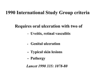
Beh ç et’s syndrome
Beh ç et’s syndrome 5th C BC  Description by Hippocrates 19th C   Early European literature 1930    Benedictos Adamantiades  1937   Hulusi Beh ç et
1990 International Study Group criteria  Requires oral ulceration with two of  -  Uveitis, retinal vasculitis   -  Genital ulceration -  Typical skin lesions -  Pathergy Lancet 1990 335: 1078-80
Epidemiology of Behçet’s syndrome  “ The Silk Route disease” Japan 1:10,000 Turkey 3-8:10,000 UK ? about 2000 cases
oral   ulcers Commonly present in childhood or adolescence then gap of 10-15 years Minor (80%), major (10%) and herpetiform (10%) Predominantly on non-keratinized mucosa May get submandibular LNs
Differential diagnosis of oral ulceration Benign recurrent oral ulceration Deficiencies (haematinics, B group vitamins) GIT disease (coeliac disease, IBD) Rheumatological (Beh ç et’s, Reiter’s, SLE) Infection (EBV, HSV, HIV) Neutropenia (drugs, cyclic) Imuune-deficiency (eg IgA)
Skin Papules and pustules Ulcers and Pyoderma  Erythema nodosum / nodular vasculitis Pathergy
PATHERGY Inappropriate excessive response to injury  Very suggestive of Beh çet’s syndrome Low sensitivity in UK Probably associated with disease severity/activity
Eye involvement Anterior uveitis with hypopyon Posterior uveitis Retinal vasculitis Retinal artery or vein occlusion  Optic neuritis leading to optic atrophy Secondary cataracts, glaucoma, blindness
neurological manifestations Headaches -  +  migraine -  Intracranial hypertension Meningoencephalitis Space occupying lesions Stroke syndrome -  Cortical - Brain-stem  - Spinal cord Audiovestibular
headaches : 7 year follow-up Presented with headache only 27 No neurologic involvement 18 Silent involvement     7  Neurologic attack   2 Objective neurologic involvement  15 Stationary      7 Progressive     8 Akman-Demir et al 1996 Arch Neurol 53:691
intracranial hypertension Aged 31 recurrent oral and vulval ulcers Aged 36 Frontal headache and blurred vision  Acuities normal but papilloedema and R VI palsy MRI failed to demonstrate venous sinus thrombosis  CSF opening pressure 30 cms H 2 0 Aged 42  Represented with intermittent  headaches  Bilateral optic atrophy CSF opening pressure 57 cms H 2 0 Deteriorating vision resulted in R optic nerve fenestration Temporary lumbo-peritoneal shunt
Auditory symptoms : 58% Auditory test abnormalities: 52% Bilateral: 77% Cochlear: 85% Vestibular symptoms: 60% Vestibular test abnormalities: 37% Unilateral: 80% Peripheral: 91% audio-vestibular manifestations
vascular manifestations Venous Superficial thrombophlebitis Thrombosis Systemic artery  Thrombosis and stenosis Aneurysm Pulmonary artery  Thrombosis  Aneurysm
Assessing risk of DVT Greater risk of thrombosis if: Age less than 31 years Within two years of diagnosis Eye disease Demiroglu et al 1996 Thrombosis Research 84:297
Gastrointestinal involvement Ulceration (oral to anal) Abdominal pain Diarrhoea Bleeding Pancreatitis
Beh ç et’s arthritis Asymmetrical Flitting and usually non-erosive Oligoarticular  Knees, ankles, wrists, elbows
REITER’S BEHCET’S Oral ulcers “ painless” painful Urogental prostatitis urethritis ulcers epididymitis Skin keratoderma psoriasiform lesions pustules nodules ulcers Arthritis persistent, erosive axial and peripheral transient peripheral
REITER’S BEH Ç ET’S Nervous system rare 20% Large vessels aortitis thrombophlebitis aneurysms Eye conjunctivitis anterior uveitis panuveitis retinal vasculitis HLA Class I B27 B51
Beh ç et’s syndrome Oral and genital ulcers Ocular inflammation  Skin lesions Arthralgias, arthritis Vascular lesions Neurological lesions Gastrointestinal Urogenital  Systemic features
non-specific features Fatigue Joint pains Muscle pains Headaches Fevers
routine investigations modest    WCC modest    CRP and ESR modest    immunoglobulins normal complement usually no autoantibodies
Behçet’s syndrome Autoimmunity Genetic Vascular dysfunction Prolonged inflammation Thrombosis Ulceration
Evidence for cell mediated autoimmunity in Beh çet’s disease Response to immunosuppressive medication  Presence of T lymphocytes in lesions Immunogenetic associations Th1 profile of cytokines released from blood T cells (IFN  , IL-2, IL-12) Autoreactive T cells (eg anti-hsp-60)
Prognosis Kural- Seyahi et al 2003 Medicine 82:60  20 year follow-up of 387 (90%) of cohort of 428 patients (262 M, 125 F) enrolled from 1977-1983 42 (9.8%; 39 M, 3F) died – from major vessel and neurological disease Mortality and morbidity decreased with time Disease burden tends to be greatest in early years and may burn out … .but major vessel and neurological involvement may occur late
problems with treatment Uncertain aetiology and pathogenesis Heterogeneity of manifestations Rarity Few laboratory indices of activity
Management of oral ulceration Oral hygiene Chlorhexidine mouth washes Topical steroids Beclomethasone inhaler  Triamcinolone (Adcortyl) in Orabase Hydrocortisine lozenges (Corlan) Betamethasone (500  g) and doxycycline (100mg) in 10 ml water as mouth wash  Systemic corticosteroids other
Colchicine Commonly prescribed for mucocutaneous manifestations Aktulga et al 1980 – limited usefulness  Randomised placebo-controlled trial:  reduced arthritis in men and women reduced GU and EN in women only. Yurdakul et al (2001) Arthritis Rheum 2001; 44:2686
Immunosuppressants Azathioprine Mycophenolate mofetil Methotrexate  Cyclosporin A Tacrolimus Rapamycin
Thalidomide May act by inhibiting TNF   release Randomised placebo-controlled trial showed efficacy for oral ulcers, genital ulcers and pustules  (Hamuryudan et al 1998 Ann Intern Med 128:443)  May exacerbate nodular vasculitis Beware teratogenesis and peripheral neuropathy
Hamuryudan, V. et. al. Ann Intern Med 1998;128:443-450 Thalidomide 100mg/300mg daily vs placebo placebo 300mg/day 100mg/day treatment
Interferon alpha Many case-reports and small open studies Randomised placebo-controlled study showed significant benefit for OU, GU, papulopustular lesions  - underpowered for ocular inflammation  (Alpsoy et al 2002  Arch Dermatol 138:467)
TNF-antagonists Goossens et al 2001  ulceration, retinal vasculitis Hassard et al 2001  gastrointestinal disease Munoz-Fernandez  et al 2001 uveitis Sfikakis et al 2001 uveitis Travis et al 2001 gastrointestinal disease
Caution  Dentistry  Invasive investigations Surgery
References   Sakane T, Takeno M, Suzuki N, Inaba G (1999) Behcet's disease. N Engl J Med 341: 1284-91   Pickering MC, Haskard DO. Behcet's Syndrome - a review of 230 referrals. Journal of the Royal College of Physicians 2000; 34(2):169-177. Hirohata S, Kikuchi H. Behcet's disease. Arthritis Research 2003; 5:139-146. Yazici H. Behcet's Syndrome: An Update. Current Rheumatolology Reports 2003; 5(3):195-199.

Behcet

  • 1.
    Beh ç et’ssyndrome
  • 2.
    Beh ç et’ssyndrome 5th C BC Description by Hippocrates 19th C Early European literature 1930 Benedictos Adamantiades 1937 Hulusi Beh ç et
  • 3.
    1990 International StudyGroup criteria Requires oral ulceration with two of - Uveitis, retinal vasculitis - Genital ulceration - Typical skin lesions - Pathergy Lancet 1990 335: 1078-80
  • 4.
    Epidemiology of Behçet’ssyndrome “ The Silk Route disease” Japan 1:10,000 Turkey 3-8:10,000 UK ? about 2000 cases
  • 5.
    oral ulcers Commonly present in childhood or adolescence then gap of 10-15 years Minor (80%), major (10%) and herpetiform (10%) Predominantly on non-keratinized mucosa May get submandibular LNs
  • 6.
    Differential diagnosis oforal ulceration Benign recurrent oral ulceration Deficiencies (haematinics, B group vitamins) GIT disease (coeliac disease, IBD) Rheumatological (Beh ç et’s, Reiter’s, SLE) Infection (EBV, HSV, HIV) Neutropenia (drugs, cyclic) Imuune-deficiency (eg IgA)
  • 7.
    Skin Papules andpustules Ulcers and Pyoderma Erythema nodosum / nodular vasculitis Pathergy
  • 8.
    PATHERGY Inappropriate excessiveresponse to injury Very suggestive of Beh çet’s syndrome Low sensitivity in UK Probably associated with disease severity/activity
  • 9.
    Eye involvement Anterioruveitis with hypopyon Posterior uveitis Retinal vasculitis Retinal artery or vein occlusion Optic neuritis leading to optic atrophy Secondary cataracts, glaucoma, blindness
  • 10.
    neurological manifestations Headaches- + migraine - Intracranial hypertension Meningoencephalitis Space occupying lesions Stroke syndrome - Cortical - Brain-stem - Spinal cord Audiovestibular
  • 11.
    headaches : 7year follow-up Presented with headache only 27 No neurologic involvement 18 Silent involvement 7 Neurologic attack 2 Objective neurologic involvement 15 Stationary 7 Progressive 8 Akman-Demir et al 1996 Arch Neurol 53:691
  • 12.
    intracranial hypertension Aged31 recurrent oral and vulval ulcers Aged 36 Frontal headache and blurred vision Acuities normal but papilloedema and R VI palsy MRI failed to demonstrate venous sinus thrombosis CSF opening pressure 30 cms H 2 0 Aged 42 Represented with intermittent headaches Bilateral optic atrophy CSF opening pressure 57 cms H 2 0 Deteriorating vision resulted in R optic nerve fenestration Temporary lumbo-peritoneal shunt
  • 13.
    Auditory symptoms :58% Auditory test abnormalities: 52% Bilateral: 77% Cochlear: 85% Vestibular symptoms: 60% Vestibular test abnormalities: 37% Unilateral: 80% Peripheral: 91% audio-vestibular manifestations
  • 14.
    vascular manifestations VenousSuperficial thrombophlebitis Thrombosis Systemic artery Thrombosis and stenosis Aneurysm Pulmonary artery Thrombosis Aneurysm
  • 15.
    Assessing risk ofDVT Greater risk of thrombosis if: Age less than 31 years Within two years of diagnosis Eye disease Demiroglu et al 1996 Thrombosis Research 84:297
  • 16.
    Gastrointestinal involvement Ulceration(oral to anal) Abdominal pain Diarrhoea Bleeding Pancreatitis
  • 17.
    Beh ç et’sarthritis Asymmetrical Flitting and usually non-erosive Oligoarticular Knees, ankles, wrists, elbows
  • 18.
    REITER’S BEHCET’S Oralulcers “ painless” painful Urogental prostatitis urethritis ulcers epididymitis Skin keratoderma psoriasiform lesions pustules nodules ulcers Arthritis persistent, erosive axial and peripheral transient peripheral
  • 19.
    REITER’S BEH ÇET’S Nervous system rare 20% Large vessels aortitis thrombophlebitis aneurysms Eye conjunctivitis anterior uveitis panuveitis retinal vasculitis HLA Class I B27 B51
  • 20.
    Beh ç et’ssyndrome Oral and genital ulcers Ocular inflammation Skin lesions Arthralgias, arthritis Vascular lesions Neurological lesions Gastrointestinal Urogenital Systemic features
  • 21.
    non-specific features FatigueJoint pains Muscle pains Headaches Fevers
  • 22.
    routine investigations modest  WCC modest  CRP and ESR modest  immunoglobulins normal complement usually no autoantibodies
  • 23.
    Behçet’s syndrome AutoimmunityGenetic Vascular dysfunction Prolonged inflammation Thrombosis Ulceration
  • 24.
    Evidence for cellmediated autoimmunity in Beh çet’s disease Response to immunosuppressive medication Presence of T lymphocytes in lesions Immunogenetic associations Th1 profile of cytokines released from blood T cells (IFN  , IL-2, IL-12) Autoreactive T cells (eg anti-hsp-60)
  • 25.
    Prognosis Kural- Seyahiet al 2003 Medicine 82:60 20 year follow-up of 387 (90%) of cohort of 428 patients (262 M, 125 F) enrolled from 1977-1983 42 (9.8%; 39 M, 3F) died – from major vessel and neurological disease Mortality and morbidity decreased with time Disease burden tends to be greatest in early years and may burn out … .but major vessel and neurological involvement may occur late
  • 26.
    problems with treatmentUncertain aetiology and pathogenesis Heterogeneity of manifestations Rarity Few laboratory indices of activity
  • 27.
    Management of oralulceration Oral hygiene Chlorhexidine mouth washes Topical steroids Beclomethasone inhaler Triamcinolone (Adcortyl) in Orabase Hydrocortisine lozenges (Corlan) Betamethasone (500  g) and doxycycline (100mg) in 10 ml water as mouth wash Systemic corticosteroids other
  • 28.
    Colchicine Commonly prescribedfor mucocutaneous manifestations Aktulga et al 1980 – limited usefulness Randomised placebo-controlled trial: reduced arthritis in men and women reduced GU and EN in women only. Yurdakul et al (2001) Arthritis Rheum 2001; 44:2686
  • 29.
    Immunosuppressants Azathioprine Mycophenolatemofetil Methotrexate Cyclosporin A Tacrolimus Rapamycin
  • 30.
    Thalidomide May actby inhibiting TNF  release Randomised placebo-controlled trial showed efficacy for oral ulcers, genital ulcers and pustules (Hamuryudan et al 1998 Ann Intern Med 128:443) May exacerbate nodular vasculitis Beware teratogenesis and peripheral neuropathy
  • 31.
    Hamuryudan, V. et.al. Ann Intern Med 1998;128:443-450 Thalidomide 100mg/300mg daily vs placebo placebo 300mg/day 100mg/day treatment
  • 32.
    Interferon alpha Manycase-reports and small open studies Randomised placebo-controlled study showed significant benefit for OU, GU, papulopustular lesions - underpowered for ocular inflammation (Alpsoy et al 2002 Arch Dermatol 138:467)
  • 33.
    TNF-antagonists Goossens etal 2001 ulceration, retinal vasculitis Hassard et al 2001 gastrointestinal disease Munoz-Fernandez et al 2001 uveitis Sfikakis et al 2001 uveitis Travis et al 2001 gastrointestinal disease
  • 34.
    Caution Dentistry Invasive investigations Surgery
  • 35.
    References Sakane T, Takeno M, Suzuki N, Inaba G (1999) Behcet's disease. N Engl J Med 341: 1284-91 Pickering MC, Haskard DO. Behcet's Syndrome - a review of 230 referrals. Journal of the Royal College of Physicians 2000; 34(2):169-177. Hirohata S, Kikuchi H. Behcet's disease. Arthritis Research 2003; 5:139-146. Yazici H. Behcet's Syndrome: An Update. Current Rheumatolology Reports 2003; 5(3):195-199.

Editor's Notes

  • #3 463 History of Behcet's Syndrome Benedictos Adamantiades and Hulusi Behcet were first to put the syndrome as a distinct pathological entity. Benedictos Adamantiades was a Greek dermatologist who clearly recognised association of ROU, genital ulcers and ocular lesions but published in the Greek rather than international literature. ? Get copy of publication Hulusi Behcet described ROU, genital ulcers and ocular lesions with hypopyon. This was published in the German literature and the syndrome became widely adopted. In those days publishing in a German journal would have been similar to publishing in an American journal today, so probably nothing much has changed. ?Skin lesions in initial description Since Behcet’s initial description many other manifestations have been described and these include…
  • #4 To classify a patient as definitely having Behcet’s syndrome, the patient should have recurrent oral ulceration, which means more than three attacks in a twelve month period, and at least two of the other major manifestations which include anterior or posterior uveitis, cells in the vitreous on slit lamp examination or retinal vasculitis; recurrent genital ulceration; typical skin lesions (which I shall come on to) or the cutaneous hypersensitivity reaction known as “pathergy”, which is elicited by insertion of a 20G or smaller needle obliquely into the skin under sterile conditions and read at 24-28 hours. Those of you who do not know about pathergy should not worry as I plan to discuss it in more detail later in the talk My own interest in Behcet’s syndrome dates back to 1994, and is entirely due to being asked to give a lecture to this gathering. I am a rheumatologist with particular research experience in inflammation of blood vessels, and it was because of this rather than because of any particular experience with Behcet’s syndrome that I was asked. Since then I have been asked to see seen quite a large number of patients suspected of having Behcet’s syndrome. I say “suspected”, as no one ever seems very certain, and referrals usually contain statements such “ I think this patient may have Behcet’s syndrome”, and the diagnosis on discharge summaries usually has a ? before the Behcet’s syndrome. Yazici group – reassessment of ISG criteria in Seoul
  • #6 For review of ROU see Woo et al 1996 JADA 127:1202 {add} Major: >1 cm and may last weeks-months and heal with scarring Minor: Usualy < 1 cm and heal within 14 days without treatment and withoutr scarring Herpetiform: Like Herpes Non-keratinized mucosa: excludes attached gingivae, dorsum of tongue and hard palate Aetiological factors of benign ROU include Genetics: Present in 20% of population. 50% of first degree relatives have condition. Food hypersensitivities as always difficult to determine importance but worth thinking about gluten-sensitivity. Menses, hormones etc again no firm evidence Trauma: as in BS, trauma may precipitate an ulcer. Smoking: tends to be negatively associated, possibly through increasing mucosal keratinization and/or effects of nicotine (nicotine replacement therapy seems to protect) Infection: Strep, Herpes. Many attending oral medicine clinics have an unclassifyable mono-or oligo-arthropathy (Livneh et al 1996 Clin Exp Rheumatol 14:407 {add})
  • #7 407 Differential diagnosis of oral ulcers Haematinics include FE, B12 and folate Neutropenia functionl causes include CGD, Chediak-Higashi disease, Job’s syndrome, Tyupe 1b glycogenosis, LAD.
  • #8 408 Skin manifestations of BS Give break-down of proportions Differential diagnosis of skin lesions in presence of oral + genital ulcers is Stevens-Johnson Syndrome ? does sarcoid cause OU
  • #9 Make slide from Yazici MSU papers
  • #10 299: Eye involvement
  • #11 Slide 298: Neurological features of BS
  • #12 404 Outcome of headaches Akman-Demir et al 1996 Arch Neurol 53:691{add} abnormal CSF marked those with a progressive course. The 2 pts with headaches who went on to get attacks had dural inus thromvboss (1) and a brain stem attack (1) .Silent involvement mainly detected by neuropsychiatric or evoked response testing.
  • #13 405 Intracranial hypertension Mrs Vanessa Edwards (also vaginal-rectal fistula) Severe headache with vomiting Papilloedema, blurred vision, photophobia, transient visual obscurations Sixth nerve palsies and/or diplopia Optic atrophy with reduced visual fields LP: raised pressure but usually normal fluid dd superior vena cava occlusion, space occupying lesion, drugs OR may be more indolent subacute onset with headaches This case illustrtaes both presentations Diagnosis: CT insensitive. MRI very sensitive and may be supported by MR venography Akman-Demir et al 1996 Eur J Neurol 3:66 {add}main cause is cerebral venous thrombosis. Not usually associated with other forms of neurological involvement or uveitis, but associated with thrombophlebitis. Therefore may be better classified as vasularBS than neuroBS. Usually has a better outcome than other forms of neuroBS. 50% settle after one attack, whilst the rest have further attacks. CHECK ANATOMY OF ARACHNOID GRANULATIONS AND PATHOGENESIS Treatment: Steroids. Anticoagulants controversial Acetazolamide, repeated LPs.
  • #14 470 Audiovestibular manifestations of Behcet's
  • #15 Slide 300 : Vascular manifestations of Beh çet’s ring out venous nature of disease Bring out lack of serious renal involvement Opposite of atherosclerosis and of necrotising arteritides Discuss differential diagnosis Thrombophilia Protein C, protein S, anti-thrombin III def Factor V Leiden Raised Factor VIII Anti-phospholipid syndrome Other forms of vasculitis Malignancy Discuss presentation of pulm A disease: chest pain, haemoptysis, X-Ray opacities. Bring out importance of diagnosis from point of view of anti-coagulation
  • #16 Slide 327: Assessing the risk of DVT
  • #17 461 Gastro manifestaions of Behcet's Case histories of Carmel Foley Vevien Brister Daphne Spink
  • #18 Slide 313: Arthritis in Behcets Sacroiliitis: Was described as a feature by Dilsen but most people would say that sacroiliitis is probably not a feature classical BS, if there is any such thing (Yazici et al 1981 ARD 40:558). The connection probably reflects the clinical overlap that exists with B27-related diseases and inflammatory bowel disease.
  • #19 459 Differences between Behçet's and Reiter's syndromes
  • #20 459 Differences between Behçet's and Reiter's syndromes
  • #21 464 Behcet's Syndrome - list of overall manifestations Put skin manifestations after systemic features if Behcet did not describe them It is important to stress the systemic manifestations which may be a major feature in some patients and which are present in the large majority. This is probably no different from patients with other inflammatory disorders. fatigue sweats and fevers, usually at night. AS most patients do not take their temperature it is difficult to know how closely the two are related!#
  • #22 257 Beh ç et’s Syndrome – non-specific features In our experience non-specific symptoms such as fatigue, arthalgias and myalgias are almost universal, and patients often say they get fevers although they never seem to take their own temperature. A recent survey of patients experiences performed in collaboration with the American Behcets’s Association found that apart from the oral ulcers, pain and fatigue were the symptoms most hard to cope with and that these tended to persist despite apparent remissions of other symptoms. We view these non-specific symptoms as a very important part of the syndrome, since they are often so severe that patients are unable to work
  • #23 Moving on then to common laboratory investigations (Slide 33), these need to be done to exclude other pathologies but are usually rather unhelpful. The patients do not usually become anaemic, but often have slightly raised neutrophil and platelet counts. The immunoglobulins are usually normal, although occasionally the IgA is raised. Complement factors are normal and the autoantibodies we are used to looking for in vasculitic syndromes are absent. Proen ça et al (Portugal) – presented in Seoul the value of highly sensitive CRP
  • #24 5 5
  • #25 Make slide from Yazici MSU papers
  • #31 COMMENT ON DILLON PAPER
  • #35 466 Caution in Behcet's