SlideShare a Scribd company logo
1 of 85
ARTHRITIS
MOHD NUR HAZIQ BIN NOOR HAMIZAM SHAH
Arthritis
is a disorder of the joint characterized by pain, swelling
and limitation of movement.
Arthralgia
is pain of a joint, without any associated signs of
inflammation.
Essential Orthopaedics 4th edition
ANATOMY OF A
SYNOVIAL JOINT
ARTICULARCARTILAGE:
-‐ Covered by Hyaline
cartilage.
-‐ Covers weight-‐bearing
surfaces of bones.
-‐ Transmits load
uniformly to bone.
-‐ Decreasesfriction
https://www.orthobullets.com/basic-science/9017/articular-cartilage?expandLeftMenu=true
• ARTICULAR CARTILAGE:
composedof -‐
1. Extracellular matrix
2. Cells
Extracellularmatrix
- matrix of proteogylcans, collagen &
glycoproteins
- makes up the dry weight
- mainly type II collagen & proteoglycan
- provides compressive and tensile strength
- atracts and retains water incartilage
Water
- about 65‐80%of the cartilage mass
- Distribute nutrients to chondrocytes
-providing lubrication
Condrocytes
• produce collagen, proteoglycans and
enzyme
• derive from chondroblasts that are
trapped in lacunae and become
chondrocytes.
Sophia Fox, A. J., Bedi, A., & Rodeo, S. A. (2009). The basic science of articular
cartilage: structure, composition, and function. Sports health, 1(6), 461-468.
ARTICULAR CARTILAGE:nutrition-‐
-‐ Articular cartilage is avascular and
aneural tissue.
-‐ Nourished by diffusion from
synovial fluid at surface
-‐ Nourished by subchrondal bone at
the base
SYNOVIUM:
-‐ Lining the cavity of the joint
-‐ Highly vascularized structure
-‐ Produces synovial fluid responsible
for nutrition and lubrication
-‐ Synovial fluid rich in hyaluronin
and lubricin
CAPSULE &LIGAMENTS:
-‐ The tough fibrous material that covers the entire joint
-‐ Stabilizers of joints
-‐Prevent excessive movement in a joint
-‐Have mechanoreceptors and free nerve endings that help with joint proprioception
https://www.orthobullets.com/basic-science/9016/ligaments?expandLeftMenu=true
OSTEOARTHRTIS
• A chronic jointdisorder in which there is
progressive softening and disintegration of
articular cartilage accompanied by new growth
of cartilage and bone at the joint margins and
capsular fibrosis.
Apley's System of Orthopaedics. and Fractures, Ninth Edition
• Cardinal features are:
1. Progressive loss of articular cartilage thickness
2. Subarticular cyst formation and sclerosis
3. Remodelling of the bone ends and osteophytes formation
4. Synovial irritation
5. Capsular fibrosis
OA is a dynamic phenomenon, which involves both destruction and
repair
Apley's System of Orthopaedics. and Fractures, Ninth Edition
PATHOPHYSIOLOGY
Softening of the articular cartilage (normally smooth and glistening surface
become frayed and fibrillated)
Articular cartilage worn away and expose the underlying bone
Subarticular bone changes (attempts to remodel)
High stress area – cyst appear and surrounding trabecular thickened or sclerotic,
vascular congestion and increased intraosseous pressure rise
Low stress area – cartilage proliferate and ossifies producing bony outgrowth
(osteophytes)
Shedding of the fibrilated articular cartilage as well as release of enzyme from
damaged cells lead to synovitis
Later stage, capsular fibrosis lead to joint stiffness
Apley's System of Orthopaedics. and Fractures, Ninth Edition
PREVALENCE & RISK FACTORS
• Patients usually present after middle
age, though cartilage changes start
10 - 20 years before.
• Most affectedjoints:
– Knee
– Hip
– Hand
– Spine
Apley's System of Orthopaedics. and Fractures, Ninth Edition
RISK FACTORS
• Non-modifiable
• • Advancing age
• • Female
• • Genetic influence on hand and knee OA in women
• Heberden’s nodes in hand
• Modifiable
• Previous knee injury/articular trauma
• Occupation, repetitive knee bending
• Muscle weakness
• BMI >25kg/m2
CPG Management of Osteoarthritis ( Second Edition) 2013
CLASSIFICATION
PRIMARY
No identifiable cause.
A diagnosis by exclusion.
SECONDARY
Has an identifiable cause.
-‐Dysplastic joint.
-‐Trauma/ Infection/
Inflammatory arthritis.
MONO/PAUCI-‐ARTICULAR-‐
Involves only1-‐2joints.
-‐Large weight-‐bearingjoints. (hip,
knees)
-‐Has an underlying bone
dysplasia.
POLYARTICULAR-‐Multiplesmall
jointsinvolved.
eg: hands, MTP, knees, lumbar
facet
Apley's System of Orthopaedics. and Fractures, Ninth Edition
SYMPTOMS
• JOINT PAIN
– Insidious onset
– intermittent & relapsing
– increased by joint use & impact
– relieved by rest
– night pain in severe OA
• Stiffness
– due to due to capsular fibrosis, muscle spasms
– only last a few minutes; always < 30minutes
– usually occurs after inactivity
CPG Management of Osteoarthritis ( Second Edition) 2013
• Swelling
– with or without associated warmth & loss of function
– Bony swelling: in hand OA, hypertrophic bone formation in the IP joint may
result in reduced dexterity & difficulty in performing fine movements such
as sewing
– OA of the first CMC joint may result in writing difficulties
• Gait disturbance
– on weight-bearing joints (hip/knee)
– can produce a prominent limp
– impaired function of a weight-bearing joint will cause added stress on the
contralateral weight-bearing joint e.g. a patient with impaired right knee
function & pain will have difficulty with the left hip & vice versa
CPG Management of Osteoarthritis ( Second Edition) 2013
• Loss of muscle bulk
– inactivity secondary to pain, may lead to significant
weakness & loss of quadriceps muscle bulk
• Limb deformity
– knock knees’ (valgus) or ‘bowing’ (varus)
• Clicking/ grinding sensation
– clicking or grinding sensation with joint motion
resulting in discomfort or pain
• Instability
– instability in the knee or hip may cause the patient to
seek assistance in ambulation e.g. using a cane or
crutch
CPG Management of Osteoarthritis ( Second Edition) 2013
SIGNS OF OA
– Look
• Alignment,symmetry
• Muscle wasting
• Swelling
• Scar
• Deformities (Heberden’s
nodes, Bouchard’s
nodes, genu varus,genu
valgus)
• Gait
– Feel
• Tenderness
• Warmness
• Jointeffusions
• Crepitus
– Move
• Limited joint
movement
• Locking
CPG Management of Osteoarthritis ( Second Edition) 2013
• SWELLING-
• Fluctuant (effusion)-‐ fluid shift/ patella tap/
fluid thrill.
• Doughy (synovial hypertrophy).
• Hard immobile (osteophytes).
DEFORMITY-‐HAND-‐
Heberden’s nodes/
Bouchard’s nodes.
DIAGNOTIC CRITERIA
• The diagnostic criteria for classification of OA
are based on the American College of
Rheumatology (ACR) criteria
CPG Management of Osteoarthritis ( Second Edition) 2013
KNEE OA
• can also be diagnosed using evidence based
recommendations by European League
Against Rheumatism (EULAR) 2010
CPG Management of Osteoarthritis ( Second Edition) 2013
• In the presence of 3/10 at least 1 point from Domain
II along with all entry criteria, the diagnosis of Knee
OA can be established
2016 ACR REVISED CRITERIA FOR EARLY DIAGNOSIS OF KNEE OA
http://aperito.org/uploads/pdf/ADTAOA-3-118.pdf
INVESTIGATIONS
• Diagnosis of OA is mainly clinical
• Blood investigations - ESR/CRP normal
• Synovial fluid analysis - normal & seldom
required except to exclude septic,
inflammatory & crystal arthropathy
• Plain radiography is the investigation of choice
– should be done in weight bearing position
CPG Management of Osteoarthritis ( Second Edition) 2013
PLAIN RADIOGRAPH
• Classical features of OA
– narrowed joint space
– subchondral bone
sclerosis
– osteophytes
– subchondral cysts
CPG Management of Osteoarthritis ( Second Edition) 2013
MANAGEMENT
• NON-PHARMACOLOGICAL TREATMENT
– Patient Education
• nature of the disease
• therapeutic options
• importance of patient participation
– Lifestyle Modification
• weight loss programs
• exercise
– Physiotherapy
– Occupational Therapy
– Orthoses
CPG Management of Osteoarthritis ( Second Edition) 2013
Physiotherapy:
– improve muscle strength, balance, coordination and joint
mobility
TENS: Trancutaneous Electrostimulation
• patients with chronic moderate to severe pain who are not suitable
for total knee arthroplasty. (ACR 2012)
OCT:
– To improve health, prevent disability & help individuals to
achieve their optimum functional level & independence in
performing ADL
– Joint protection exercises, splinting, assistive devices (walking
stick, tap turners)
PHARMACOLOGICAL
• Oral medication
• Simple analgesics – paracetamol (acetaminophen)
• Weak opioid analgesics – tramadol (combined with pcm or nsaids)
• NSAIDs and COX-2 Inhibitors (diclofenac, celecoxib)
• Nutraceutical - glucosamine, chondroitin, diacerein
• Intraarticular injections
• Steroids – for short term pain relief
• Viscosupplementation – to replace hyaluronic acid in joints,
controversial
• Topical cream
• NSAIDS and LMS cream
CPG Management of Osteoarthritis ( Second Edition) 2013
SURGICAL MANAGEMENT
• Considered if symptoms are significant:
– Pain
– Limitation of ADL
– Psychosocial health (psychological well being)
– Economic impact
– Recent deterioration
OPERATIVE
• Arthroscopic surgery
– indicated in patients with OA associated with
mechanical symptoms such as locking, catching or
giving way of the joint caused by presence of loose
bodies or flaps of meniscus or cartilage
• High Tibial Osteotomy
• Total Joint Replacement
• Partial Joint Replacement
• Arthrodesis
- Osteotomised at its upper end and repositioned
- To realign the mechanical axis of the limb away from the diseased area
- Allows the joint to glide freely and distribute weight evenly
In isolated medial compartment knee OA or varus malalingment
• <50 years old
• Good knee ROM (>120⁰)
• Realign to correct mechanical axis
• Load distributed more on normal knee compartment
High Tibial Osteotomy
ARTHROPLASTY
-‐ Replacing the joint with implant
-‐ Results in dramatic reduction of pain and
-‐ Causes significant improvement in ADL
-‐ Active infection is absolute contraindication
INDICATIONS FOR JOINT REPLACEMENT
• Pain
• Severe disability
• Deformity
• Limited function (ADL)
• Fail conservative treatment
CPG Management of Osteoarthritis ( Second Edition) 2013
ARTHRODESIS
• Surgical fusion of the involvedjoint in optimal
position
• Provides pain relief &stability despite stiff joint
Indications:
• Young active labourer with
unilateral painful OA
• Severe OA with combined
ligamentous injury
• Uncontrolled septic arthritis &
complete joint destruction
• Failed and/or infected
arthroplasty
Advantages:
• Stable pain free knee allowing
long-term ambulation
Disadvantages:
• Stiff joint
• Abnormal gait after surgery
• Late back pain
• Conversion to TKR or THR give
unpredictable result
CPG Management of Osteoarthritis ( Second Edition) 2013
(a)High Tibial Osteotomy.
(b)Arthroplasty.
(c)Arthrodesis.
RHEUMATOID ARTHRITIS
• Rheumatoid arthritis (RA) is a chronic
inflammatory disease characterized by joint
swelling, joint tenderness, and destruction of
synovial joints, leading to severe disability and
premature mortality
Aletaha, D., Neogi, T., Silman, A. J., Funovits, J., Felson, D. T., Bingham, C. O., ... & Combe, B. (2010). 2010
rheumatoid arthritis classification criteria: an American College of Rheumatology/European League Against
Rheumatism collaborative initiative. Arthritis & Rheumatology, 62(9), 2569-2581.
RISK FACTORS
1. Gender
Women are affected 3 times more often than men
2. Genetic factors
Human Leucocyte Antigen (HLA)- DR4 and HLA-DR1
confer susceptibility to RA and are associated with
development of more severe erosive disease
3. Agents such as mycoplasma, clostridium and EB
virus
Exposure of genetically predisposed individual to infectious agent
Formation of immune complexes with IgM antibodies in the serum
Immune complexes deposited in synovial membrane and initiate
chronic granulamatous inflamation
Synovium is edematous and filled with fibrin exudates and cellular
infiltrates
Synovium becomes hypertrophied and has effusion (reversible)
Synovium surrounds articular cartilage to form pannus which cause
reduction in joint space, early cartilage erosions and soft tissue
destruction
(controllable but irreversible)
Pannus ultimately destroys articular cartilage, burrows into the
subchondral bone to erode it (irreversible)
Fibrous ankylosis and joint deformity
Essential Orthopaedics 4th edition
CLINICAL FEATURES
Symptoms
• insidious onset
• morning stiffness >1 hour
• polyarthropathy
• usually affects hands and feet
– (DIP joint of hand is usually
spared)
• may also affect other joints
https://www.orthobullets.com/basic-science/9085/rheumatoid-arthritis
ARTICULAR SIGNS
• Red, warm, tender and swollen small joints of the
hands and feet, excluding DIP, first MTP, and first
CMC joint.
• Symmetrical
• Polyarthritic.
Atlas of rheumatoid arthritis 2015
Rheumatoid nodulesPIP Swelling
Forefoot
-Hyperpronation of
rheumatoid foot
-Metatarsalgia
-Claw toes
-Hallux valgus
Midfoot
-Talonavicular arthritis
-Midfoot hyperpronation
Hindfoot
-Calcaneovalgus
Atlas of rheumatoid arthritis 2015
EXTRA-ARTICULAR MANIFESTATION
• Systemic symptoms
• fever
• fatigue
• malaise
• weight loss
• Musculoskeletal
• osteoporotic #
• tenosynovitis and
bursitis
• rupture of tendons &
ligaments
Face and neck
-Mouth dryness
-Atlanto axial
subluxation
Eyes
-Conjuctival pallor
-Schirmer’s filter paper
test
-Episcleritis
-Scleritis
-Cataracts
-Keratoconjuctivitis
sicca
Chest
-Pericardial rub +-
effusion
-Myocarditis
-Pleural rub +-
effusion
-Fine crepitus
-Vasculitis
Abdomen
-Splenomegaly
Legs
-Mononeuritis
multiplex
Atlas of rheumatoid arthritis 2015
DIAGNOSTIC
CRITERIA
JOINT DISTRIBUTION (0-5)
1 large joint 0
2-10 large joints 1
1-3 small joints (large joints not counted) 2
4-10 small joints (large joints not counted) 3
>10 joints (at least one small joint) 5
SEROLOGY (0-3)
Negative RF AND negative ACPA 0
Low positive RF OR low positive ACPA 2
High positive RF OR high positive ACPA 3
SYMPTOM DURATION (0-1)
<6 weeks 0
≥6 weeks 1
ACUTE PHASE REACTANTS (0-1)
Normal CRP AND normal ESR 0
Abnormal CRP OR abnormal ESR 1
2010 ACR/ EULAR
CLASSIFICATION CRITERIA
≥6 = Definite RA
Aletaha, D., Neogi, T., Silman, A. J.,
Funovits, J., Felson, D. T., Bingham, C. O.,
... & Combe, B. (2010). 2010 rheumatoid
arthritis classification criteria: an
American College of
Rheumatology/European League Against
Rheumatism collaborative
initiative. Arthritis &
Rheumatology, 62(9), 2569-2581.
Definition of “JOINT INVOLVEMENT”
- Any swollen or tender joint (excluding DIP
of hand and feet, 1st MTP, 1st CMC)
- Additional evidence from MRI / US may be
used for confirmation of the clinical findings
Definition of “SMALL JOINT”
MCP, PIP, MTP 2-5, thumb IP, wrist
NOT: DIP, 1st CMC, 1st MTP
Definition of “LARGE JOINT”
Shoulder, elbow, hip, knee, ankles
Definition of “>10 JOINTS”
- At least one small joint
- Additional joints include:
temporomandibular, sternoclavicular,
acromioclavicular, and
others (reasonably expected in RA)
Definition of “SEROLOGY”
Negative: ≤ULN (for the respective lab)
Low positive: >ULN but ≤3xULN
High positive: >3xULN
Definition of “SYMPTOM DURATION”
Refers to the patient’s self-report on the
maximum duration of signs and
symptoms of any joint that is clinically
involved at the time of assessment.
INVESTIGATION
Subchondral
erosion
Periarticular
osteopenia
Reduced
joint space
Useful for diagnosis
• normochromic normocytic
anaemia
• elevated ESR
• elevated CRP
• anti-CCP (most sensitive &
specific)
• positive RF titer
• elevated in 75-80% of
patients with RA
• joint fluid testing
• decreased complement
• may have elevated RF levels
Useful for differential diagnosis
• serum uric acid
• antinuclear antibodies
Useful to detect complications
• chest xray
Investigations for follow up and
treatment
• ESR
• FBC
• RP
• LFT
• Chest xray
Categories of Synovial Fluid Based upon Clinical and Laboratory Findings
CPG Management of Osteoarthritis ( Second Edition) 2013
There is no single diagostic test for RA. Investigationsare used largely to support clinical
diagnosis and negative results does not exclude the diagnosis of RA.
MANAGEMENT
• Principle of treatment
• Induction of remission and it’s maintenance: disease
activity is brought under control by drugs
• Preservation of joint functions and prevention of
deformities during the activity of the disease and
thereafter, by physiotherapy and splinting
• Repair of joint damage which already exists, if it will
relieve pain or facilitate functions
TREATMENT MODALITIES FOR RA
• NSAIDS
• Steroids
• DMARDs
• Biological therapies
• Surgery
• Physiotherapy
• Occupational therapy
• Rehabilitation
• Treatment with DMARD should be initiated as soon as a
diagnosis of RA is made, with the aim of reaching a target
of remission or low disease activity
• EULAR recommends rheumatologists administer
methotrexate (MTX) or combination therapy of MTX with
other conventional synthetic DMARDs.
• Low-dose glucocorticoids should also be considered in
combination with DMARDs for up to six months, but should
be tapered as soon as clinically feasible
EULAR 2013 Rheumatoid Arthritis Management Recommendations, EULAR Data on File, 2013.
SURGERY
Preventive surgery:
– To prevent damage to the joint and nearby tendons by the inflamed,
hypertrophied synovium. It consists of synovectomy of the wrist, knee
and MP joints
Palliative surgery :
– Done in situations where condition of the patient doesn’t permit
corrective surgery, but some relief can be provided by bone block
operations or tendon lengthening
Reconstructive surgery :
– This has revolutionised the rehabilitation of patients with deformed and
painful joints. It includes tendon transfers and total joint replacement.
SEPTIC ARTHRITIS
Septic arthritis refers to infection in a joint; it is
usually caused by bacteria but can be caused by
fungi or mycobacteria.
https://www.uptodate.com/contents/septic-arthritis-in-adults
RISK FACTORS
Adults:
• Age > 80 years
• Medical conditions
• diabetes
• rheumatoid arthritis
• cirrhosis
• HIV
• History of crystal arthropathy
• Endocarditis or recent
bacteremia
• IV drug user
• Recent joint surgery
Neonates:
• Prematurity
• Cesarean section
• Patients treated in the NICU
• Invasive procedures leading to
transient bacteremia
https://www.orthobullets.com/trauma/1058/septic-arthritis--adult?expandLeftMenu=true
PATHOPHYSIOLOGY
Routes of inoculation
Direct inoculation from
trauma or surgery
Hematogenous seeding
(bacteremia)
Extension from adjacent bone
(osteomyelitis)
https://www.orthobullets.com/trauma/1058/septic-arthritis--adult?expandLeftMenu=true
Apley's System of Orthopaedics. and Fractures, Ninth Edition
a) In the early stage, there is an acute synovitis with a purulent joint effusion
b) Soon the articular cartilage is attacked by bacterial and cellular enzyme.
c) If infection is not arrested , the cartilage may be completely destroyed
d) Healing then leads to ankylosis
If left untreated, it will spread to the
underlying bone
and out of joint to form abscess and sinus.
Healing with:
1.Complete resolution
2.Partial loss of articular cartilage and fibrosis of
joint
3.Loss of articular cartilage and bony ankylosis
4.Bony destruction and permanent deformity
PHYSICAL EXAMINATIONS
• i)Temp. and vital signs
• Fever
• ii)Look
• Localized swelling and erythema
• Affected joint lies in a position of
ease
Joint Position of ease
Shoulder Adduction, internal rotation
Elbow Flexion, mid pronation
Wrist Flexion
Hip Flexion, abduction, ext rotation
(FABER)
Knee Flexion
Ankle Plantar-flexion
iii) Feel
Effusion
Warm to touch
Tenderness
iv) Move
Severe pain with passive
motion
Unwillingness to move joint
(pseudoparalysis)
INVESTIGATION
1. WBC raised (>10k with left shift)
2. ESR raised (>30)
• ESR is often elevated but may be normal early in process
• rises within 2 days of infection and can rise 3-5 days after initiation of
appropriate antibiotics, and returns to normal 3-4 weeks
3. CRP raised (>5)
• best way to judge efficacy of treatment, as CRP rises within few hours
of infection, and may normalize within 1 week of treatment
4. Joint aspiration (gold standard)
– should be analyzed for
• cell count with differential
• gram stain
• culture
• glucose level
• crystal analysis
More than 50k is considered diagnostic
5. USG (most reliable)
-widening of space between capsule and bone >2mm
-able to confirm effusion in large joint such as hip
-can be used in guiding aspiration
6. X-ray (AP & lateral of the joint, or frog leg lateral pelvic
xray in paeds)
-normal in early stages
-widening of radiographic joint space or effusion
-subluxation or dislocation
-bone lesions (assc. osteomyelitis)
7. MRI
-detects effusion and also adjacent bone
involvement
-helpful in determining operative treatment
MANAGEMENT
1. URGENT surgical I&D
2. IV Antibiotics
• Initiate empiric therapy prior to C&S based on age and risk
factors
• Transition to organism specific antibiotics after cultures obtained
• Monitor efficacy of treatment using WBC, ESR and CRP
• Paeds: Penicillinase-resistant penicillins+ 3rd generation
cephalopsporins
• Adults: Cloxacillin, or Flucloxacillin and Furisidic acid
3. Analgesia
4. Splintage: joint rested
Arthritis and Joint Anatomy

More Related Content

What's hot

approach a patient with low back pain
approach a patient with low back painapproach a patient with low back pain
approach a patient with low back painalyaqdhan
 
Ankle and foot injuries
Ankle and foot injuriesAnkle and foot injuries
Ankle and foot injuriesAmardeep kaur
 
Avascular necrosis ug class
Avascular necrosis ug classAvascular necrosis ug class
Avascular necrosis ug classArun K
 
Approach towards a case of musculoskeletal disorder.#
Approach towards a case of musculoskeletal disorder.#Approach towards a case of musculoskeletal disorder.#
Approach towards a case of musculoskeletal disorder.#sirmohit
 
Jones Fracture
Jones FractureJones Fracture
Jones Fracturejfreshour
 
Orthopaedics: historical perspective
Orthopaedics: historical perspectiveOrthopaedics: historical perspective
Orthopaedics: historical perspectiveDR. D. P. SWAMI
 
Ilizarov external fixator
Ilizarov external fixatorIlizarov external fixator
Ilizarov external fixatorMohamed Iraqi
 
Inflamatory arthritis
Inflamatory arthritisInflamatory arthritis
Inflamatory arthritisdrangelosmith
 
Approach to Low Back Pain
Approach to Low Back PainApproach to Low Back Pain
Approach to Low Back PainRK Dahal
 
2010 ACR/EULAR Criteria for RA
2010 ACR/EULAR Criteria for RA2010 ACR/EULAR Criteria for RA
2010 ACR/EULAR Criteria for RAYounis I Munshi
 
management Diabetic foot
management Diabetic foot management Diabetic foot
management Diabetic foot Drkabiru2012
 
Approach to low back pain
Approach to low back painApproach to low back pain
Approach to low back painSushil Sharma
 

What's hot (20)

approach a patient with low back pain
approach a patient with low back painapproach a patient with low back pain
approach a patient with low back pain
 
Ankle and foot injuries
Ankle and foot injuriesAnkle and foot injuries
Ankle and foot injuries
 
Osteoarthritis
OsteoarthritisOsteoarthritis
Osteoarthritis
 
Avascular necrosis ug class
Avascular necrosis ug classAvascular necrosis ug class
Avascular necrosis ug class
 
Orthopaedics history
Orthopaedics historyOrthopaedics history
Orthopaedics history
 
Approach towards a case of musculoskeletal disorder.#
Approach towards a case of musculoskeletal disorder.#Approach towards a case of musculoskeletal disorder.#
Approach towards a case of musculoskeletal disorder.#
 
Jones Fracture
Jones FractureJones Fracture
Jones Fracture
 
AVASCULAR NECROSIS
AVASCULAR NECROSISAVASCULAR NECROSIS
AVASCULAR NECROSIS
 
Orthopaedics: historical perspective
Orthopaedics: historical perspectiveOrthopaedics: historical perspective
Orthopaedics: historical perspective
 
Ilizarov external fixator
Ilizarov external fixatorIlizarov external fixator
Ilizarov external fixator
 
Diabetic foot
Diabetic footDiabetic foot
Diabetic foot
 
Groin Pain
Groin PainGroin Pain
Groin Pain
 
Inflamatory arthritis
Inflamatory arthritisInflamatory arthritis
Inflamatory arthritis
 
Approach to Low Back Pain
Approach to Low Back PainApproach to Low Back Pain
Approach to Low Back Pain
 
Teriparatide
TeriparatideTeriparatide
Teriparatide
 
Diabetic neuropathy
Diabetic neuropathyDiabetic neuropathy
Diabetic neuropathy
 
2010 ACR/EULAR Criteria for RA
2010 ACR/EULAR Criteria for RA2010 ACR/EULAR Criteria for RA
2010 ACR/EULAR Criteria for RA
 
Shoulder examination
Shoulder examination Shoulder examination
Shoulder examination
 
management Diabetic foot
management Diabetic foot management Diabetic foot
management Diabetic foot
 
Approach to low back pain
Approach to low back painApproach to low back pain
Approach to low back pain
 

Similar to Arthritis and Joint Anatomy

osteoarthritisknee-170221145316.pptx
osteoarthritisknee-170221145316.pptxosteoarthritisknee-170221145316.pptx
osteoarthritisknee-170221145316.pptxSourabh Jha
 
osteoarthritisknee-170221145316.pptx
osteoarthritisknee-170221145316.pptxosteoarthritisknee-170221145316.pptx
osteoarthritisknee-170221145316.pptxKareemElsharkawy6
 
Osteoarthritis joint pain of old age Dr. Parshant
Osteoarthritis joint pain of old age Dr. ParshantOsteoarthritis joint pain of old age Dr. Parshant
Osteoarthritis joint pain of old age Dr. ParshantPs Nadda
 
Osteoarthritis knee
Osteoarthritis  kneeOsteoarthritis  knee
Osteoarthritis kneeNarula Gandu
 
Degenerative dystrophic diseases of joints
Degenerative   dystrophic  diseases  of jointsDegenerative   dystrophic  diseases  of joints
Degenerative dystrophic diseases of jointsVakulychMyroslav
 
GENERAL ASPECTS OF FRACTURE.pptx
GENERAL ASPECTS OF FRACTURE.pptxGENERAL ASPECTS OF FRACTURE.pptx
GENERAL ASPECTS OF FRACTURE.pptxRakesh Singha
 
osteoarthritis-140614073518-phpapp02.pdf
osteoarthritis-140614073518-phpapp02.pdfosteoarthritis-140614073518-phpapp02.pdf
osteoarthritis-140614073518-phpapp02.pdfmunirmemon40
 
Osteoarthritis ppt
Osteoarthritis pptOsteoarthritis ppt
Osteoarthritis pptRupika Sodhi
 
Osteoarthritis – Knee
Osteoarthritis – KneeOsteoarthritis – Knee
Osteoarthritis – KneeJoe Antony
 
unilateral knee replacement vs high tibial osteotomy.
unilateral knee replacement vs high tibial osteotomy.unilateral knee replacement vs high tibial osteotomy.
unilateral knee replacement vs high tibial osteotomy.drabhichaudhary88
 
Musculoskeletal disorders pdf
Musculoskeletal disorders pdfMusculoskeletal disorders pdf
Musculoskeletal disorders pdfGetenet Dessie
 
Spontaneous Osteonecrosis of the Knee.pptx
Spontaneous Osteonecrosis of the Knee.pptxSpontaneous Osteonecrosis of the Knee.pptx
Spontaneous Osteonecrosis of the Knee.pptxcheryl712552
 

Similar to Arthritis and Joint Anatomy (20)

osteoarthritisknee-170221145316.pptx
osteoarthritisknee-170221145316.pptxosteoarthritisknee-170221145316.pptx
osteoarthritisknee-170221145316.pptx
 
osteoarthritisknee-170221145316.pptx
osteoarthritisknee-170221145316.pptxosteoarthritisknee-170221145316.pptx
osteoarthritisknee-170221145316.pptx
 
Osteoarthritis joint pain of old age Dr. Parshant
Osteoarthritis joint pain of old age Dr. ParshantOsteoarthritis joint pain of old age Dr. Parshant
Osteoarthritis joint pain of old age Dr. Parshant
 
Osteoarthritis knee
Osteoarthritis  kneeOsteoarthritis  knee
Osteoarthritis knee
 
Degenerative dystrophic diseases of joints
Degenerative   dystrophic  diseases  of jointsDegenerative   dystrophic  diseases  of joints
Degenerative dystrophic diseases of joints
 
Oasteoarthriris
OasteoarthririsOasteoarthriris
Oasteoarthriris
 
GENERAL ASPECTS OF FRACTURE.pptx
GENERAL ASPECTS OF FRACTURE.pptxGENERAL ASPECTS OF FRACTURE.pptx
GENERAL ASPECTS OF FRACTURE.pptx
 
Osteoarthritis
OsteoarthritisOsteoarthritis
Osteoarthritis
 
osteoarthritis-140614073518-phpapp02.pdf
osteoarthritis-140614073518-phpapp02.pdfosteoarthritis-140614073518-phpapp02.pdf
osteoarthritis-140614073518-phpapp02.pdf
 
Osteoarthritis ppt
Osteoarthritis pptOsteoarthritis ppt
Osteoarthritis ppt
 
Osteoarthritis
OsteoarthritisOsteoarthritis
Osteoarthritis
 
Osteoarthritis
OsteoarthritisOsteoarthritis
Osteoarthritis
 
Osteoarthritis
OsteoarthritisOsteoarthritis
Osteoarthritis
 
Osteoarthritis
OsteoarthritisOsteoarthritis
Osteoarthritis
 
Oa.pptx
Oa.pptxOa.pptx
Oa.pptx
 
Osteoarthritis – Knee
Osteoarthritis – KneeOsteoarthritis – Knee
Osteoarthritis – Knee
 
unilateral knee replacement vs high tibial osteotomy.
unilateral knee replacement vs high tibial osteotomy.unilateral knee replacement vs high tibial osteotomy.
unilateral knee replacement vs high tibial osteotomy.
 
Oa knee (shravan)[1]
Oa knee (shravan)[1]Oa knee (shravan)[1]
Oa knee (shravan)[1]
 
Musculoskeletal disorders pdf
Musculoskeletal disorders pdfMusculoskeletal disorders pdf
Musculoskeletal disorders pdf
 
Spontaneous Osteonecrosis of the Knee.pptx
Spontaneous Osteonecrosis of the Knee.pptxSpontaneous Osteonecrosis of the Knee.pptx
Spontaneous Osteonecrosis of the Knee.pptx
 

More from Haziq Mars

Compartment syndrome
Compartment syndromeCompartment syndrome
Compartment syndromeHaziq Mars
 
Paediatrics fracture
Paediatrics fracturePaediatrics fracture
Paediatrics fractureHaziq Mars
 
NECROTIZING FASCITIS
NECROTIZING FASCITISNECROTIZING FASCITIS
NECROTIZING FASCITISHaziq Mars
 
Fat embolism syndrome
Fat embolism syndromeFat embolism syndrome
Fat embolism syndromeHaziq Mars
 
DIABETIC FOOT ULCER
DIABETIC FOOT ULCERDIABETIC FOOT ULCER
DIABETIC FOOT ULCERHaziq Mars
 
Cellulitis vs necrotizing soft tissue infection
Cellulitis vs necrotizing soft tissue infectionCellulitis vs necrotizing soft tissue infection
Cellulitis vs necrotizing soft tissue infectionHaziq Mars
 

More from Haziq Mars (8)

Compartment syndrome
Compartment syndromeCompartment syndrome
Compartment syndrome
 
Paediatrics fracture
Paediatrics fracturePaediatrics fracture
Paediatrics fracture
 
Open fracture
Open fractureOpen fracture
Open fracture
 
NECROTIZING FASCITIS
NECROTIZING FASCITISNECROTIZING FASCITIS
NECROTIZING FASCITIS
 
Fat embolism syndrome
Fat embolism syndromeFat embolism syndrome
Fat embolism syndrome
 
DIABETIC FOOT ULCER
DIABETIC FOOT ULCERDIABETIC FOOT ULCER
DIABETIC FOOT ULCER
 
Cellulitis vs necrotizing soft tissue infection
Cellulitis vs necrotizing soft tissue infectionCellulitis vs necrotizing soft tissue infection
Cellulitis vs necrotizing soft tissue infection
 
Bone
BoneBone
Bone
 

Recently uploaded

Call Girl Coimbatore Prisha☎️ 8250192130 Independent Escort Service Coimbatore
Call Girl Coimbatore Prisha☎️  8250192130 Independent Escort Service CoimbatoreCall Girl Coimbatore Prisha☎️  8250192130 Independent Escort Service Coimbatore
Call Girl Coimbatore Prisha☎️ 8250192130 Independent Escort Service Coimbatorenarwatsonia7
 
Call Girls Yelahanka Just Call 7001305949 Top Class Call Girl Service Available
Call Girls Yelahanka Just Call 7001305949 Top Class Call Girl Service AvailableCall Girls Yelahanka Just Call 7001305949 Top Class Call Girl Service Available
Call Girls Yelahanka Just Call 7001305949 Top Class Call Girl Service Availablenarwatsonia7
 
Low Rate Call Girls Ambattur Anika 8250192130 Independent Escort Service Amba...
Low Rate Call Girls Ambattur Anika 8250192130 Independent Escort Service Amba...Low Rate Call Girls Ambattur Anika 8250192130 Independent Escort Service Amba...
Low Rate Call Girls Ambattur Anika 8250192130 Independent Escort Service Amba...narwatsonia7
 
Call Girls Horamavu WhatsApp Number 7001035870 Meeting With Bangalore Escorts
Call Girls Horamavu WhatsApp Number 7001035870 Meeting With Bangalore EscortsCall Girls Horamavu WhatsApp Number 7001035870 Meeting With Bangalore Escorts
Call Girls Horamavu WhatsApp Number 7001035870 Meeting With Bangalore Escortsvidya singh
 
Housewife Call Girls Hoskote | 7001305949 At Low Cost Cash Payment Booking
Housewife Call Girls Hoskote | 7001305949 At Low Cost Cash Payment BookingHousewife Call Girls Hoskote | 7001305949 At Low Cost Cash Payment Booking
Housewife Call Girls Hoskote | 7001305949 At Low Cost Cash Payment Bookingnarwatsonia7
 
Aspirin presentation slides by Dr. Rewas Ali
Aspirin presentation slides by Dr. Rewas AliAspirin presentation slides by Dr. Rewas Ali
Aspirin presentation slides by Dr. Rewas AliRewAs ALI
 
Russian Call Girls in Pune Riya 9907093804 Short 1500 Night 6000 Best call gi...
Russian Call Girls in Pune Riya 9907093804 Short 1500 Night 6000 Best call gi...Russian Call Girls in Pune Riya 9907093804 Short 1500 Night 6000 Best call gi...
Russian Call Girls in Pune Riya 9907093804 Short 1500 Night 6000 Best call gi...Miss joya
 
Call Girls Service Bellary Road Just Call 7001305949 Enjoy College Girls Service
Call Girls Service Bellary Road Just Call 7001305949 Enjoy College Girls ServiceCall Girls Service Bellary Road Just Call 7001305949 Enjoy College Girls Service
Call Girls Service Bellary Road Just Call 7001305949 Enjoy College Girls Servicenarwatsonia7
 
Hi,Fi Call Girl In Mysore Road - 7001305949 | 24x7 Service Available Near Me
Hi,Fi Call Girl In Mysore Road - 7001305949 | 24x7 Service Available Near MeHi,Fi Call Girl In Mysore Road - 7001305949 | 24x7 Service Available Near Me
Hi,Fi Call Girl In Mysore Road - 7001305949 | 24x7 Service Available Near Menarwatsonia7
 
Call Girl Bangalore Nandini 7001305949 Independent Escort Service Bangalore
Call Girl Bangalore Nandini 7001305949 Independent Escort Service BangaloreCall Girl Bangalore Nandini 7001305949 Independent Escort Service Bangalore
Call Girl Bangalore Nandini 7001305949 Independent Escort Service Bangalorenarwatsonia7
 
Russian Call Girls in Chennai Pallavi 9907093804 Independent Call Girls Servi...
Russian Call Girls in Chennai Pallavi 9907093804 Independent Call Girls Servi...Russian Call Girls in Chennai Pallavi 9907093804 Independent Call Girls Servi...
Russian Call Girls in Chennai Pallavi 9907093804 Independent Call Girls Servi...Nehru place Escorts
 
College Call Girls Pune Mira 9907093804 Short 1500 Night 6000 Best call girls...
College Call Girls Pune Mira 9907093804 Short 1500 Night 6000 Best call girls...College Call Girls Pune Mira 9907093804 Short 1500 Night 6000 Best call girls...
College Call Girls Pune Mira 9907093804 Short 1500 Night 6000 Best call girls...Miss joya
 
VIP Call Girls Tirunelveli Aaradhya 8250192130 Independent Escort Service Tir...
VIP Call Girls Tirunelveli Aaradhya 8250192130 Independent Escort Service Tir...VIP Call Girls Tirunelveli Aaradhya 8250192130 Independent Escort Service Tir...
VIP Call Girls Tirunelveli Aaradhya 8250192130 Independent Escort Service Tir...narwatsonia7
 
Call Girls In Andheri East Call 9920874524 Book Hot And Sexy Girls
Call Girls In Andheri East Call 9920874524 Book Hot And Sexy GirlsCall Girls In Andheri East Call 9920874524 Book Hot And Sexy Girls
Call Girls In Andheri East Call 9920874524 Book Hot And Sexy Girlsnehamumbai
 
High Profile Call Girls Jaipur Vani 8445551418 Independent Escort Service Jaipur
High Profile Call Girls Jaipur Vani 8445551418 Independent Escort Service JaipurHigh Profile Call Girls Jaipur Vani 8445551418 Independent Escort Service Jaipur
High Profile Call Girls Jaipur Vani 8445551418 Independent Escort Service Jaipurparulsinha
 
Call Girls Chennai Megha 9907093804 Independent Call Girls Service Chennai
Call Girls Chennai Megha 9907093804 Independent Call Girls Service ChennaiCall Girls Chennai Megha 9907093804 Independent Call Girls Service Chennai
Call Girls Chennai Megha 9907093804 Independent Call Girls Service ChennaiNehru place Escorts
 
VIP Mumbai Call Girls Hiranandani Gardens Just Call 9920874524 with A/C Room ...
VIP Mumbai Call Girls Hiranandani Gardens Just Call 9920874524 with A/C Room ...VIP Mumbai Call Girls Hiranandani Gardens Just Call 9920874524 with A/C Room ...
VIP Mumbai Call Girls Hiranandani Gardens Just Call 9920874524 with A/C Room ...Garima Khatri
 
Call Girls Service Jaipur Grishma WhatsApp ❤8445551418 VIP Call Girls Jaipur
Call Girls Service Jaipur Grishma WhatsApp ❤8445551418 VIP Call Girls JaipurCall Girls Service Jaipur Grishma WhatsApp ❤8445551418 VIP Call Girls Jaipur
Call Girls Service Jaipur Grishma WhatsApp ❤8445551418 VIP Call Girls Jaipurparulsinha
 
CALL ON ➥9907093804 🔝 Call Girls Hadapsar ( Pune) Girls Service
CALL ON ➥9907093804 🔝 Call Girls Hadapsar ( Pune)  Girls ServiceCALL ON ➥9907093804 🔝 Call Girls Hadapsar ( Pune)  Girls Service
CALL ON ➥9907093804 🔝 Call Girls Hadapsar ( Pune) Girls ServiceMiss joya
 

Recently uploaded (20)

Call Girl Coimbatore Prisha☎️ 8250192130 Independent Escort Service Coimbatore
Call Girl Coimbatore Prisha☎️  8250192130 Independent Escort Service CoimbatoreCall Girl Coimbatore Prisha☎️  8250192130 Independent Escort Service Coimbatore
Call Girl Coimbatore Prisha☎️ 8250192130 Independent Escort Service Coimbatore
 
Russian Call Girls in Delhi Tanvi ➡️ 9711199012 💋📞 Independent Escort Service...
Russian Call Girls in Delhi Tanvi ➡️ 9711199012 💋📞 Independent Escort Service...Russian Call Girls in Delhi Tanvi ➡️ 9711199012 💋📞 Independent Escort Service...
Russian Call Girls in Delhi Tanvi ➡️ 9711199012 💋📞 Independent Escort Service...
 
Call Girls Yelahanka Just Call 7001305949 Top Class Call Girl Service Available
Call Girls Yelahanka Just Call 7001305949 Top Class Call Girl Service AvailableCall Girls Yelahanka Just Call 7001305949 Top Class Call Girl Service Available
Call Girls Yelahanka Just Call 7001305949 Top Class Call Girl Service Available
 
Low Rate Call Girls Ambattur Anika 8250192130 Independent Escort Service Amba...
Low Rate Call Girls Ambattur Anika 8250192130 Independent Escort Service Amba...Low Rate Call Girls Ambattur Anika 8250192130 Independent Escort Service Amba...
Low Rate Call Girls Ambattur Anika 8250192130 Independent Escort Service Amba...
 
Call Girls Horamavu WhatsApp Number 7001035870 Meeting With Bangalore Escorts
Call Girls Horamavu WhatsApp Number 7001035870 Meeting With Bangalore EscortsCall Girls Horamavu WhatsApp Number 7001035870 Meeting With Bangalore Escorts
Call Girls Horamavu WhatsApp Number 7001035870 Meeting With Bangalore Escorts
 
Housewife Call Girls Hoskote | 7001305949 At Low Cost Cash Payment Booking
Housewife Call Girls Hoskote | 7001305949 At Low Cost Cash Payment BookingHousewife Call Girls Hoskote | 7001305949 At Low Cost Cash Payment Booking
Housewife Call Girls Hoskote | 7001305949 At Low Cost Cash Payment Booking
 
Aspirin presentation slides by Dr. Rewas Ali
Aspirin presentation slides by Dr. Rewas AliAspirin presentation slides by Dr. Rewas Ali
Aspirin presentation slides by Dr. Rewas Ali
 
Russian Call Girls in Pune Riya 9907093804 Short 1500 Night 6000 Best call gi...
Russian Call Girls in Pune Riya 9907093804 Short 1500 Night 6000 Best call gi...Russian Call Girls in Pune Riya 9907093804 Short 1500 Night 6000 Best call gi...
Russian Call Girls in Pune Riya 9907093804 Short 1500 Night 6000 Best call gi...
 
Call Girls Service Bellary Road Just Call 7001305949 Enjoy College Girls Service
Call Girls Service Bellary Road Just Call 7001305949 Enjoy College Girls ServiceCall Girls Service Bellary Road Just Call 7001305949 Enjoy College Girls Service
Call Girls Service Bellary Road Just Call 7001305949 Enjoy College Girls Service
 
Hi,Fi Call Girl In Mysore Road - 7001305949 | 24x7 Service Available Near Me
Hi,Fi Call Girl In Mysore Road - 7001305949 | 24x7 Service Available Near MeHi,Fi Call Girl In Mysore Road - 7001305949 | 24x7 Service Available Near Me
Hi,Fi Call Girl In Mysore Road - 7001305949 | 24x7 Service Available Near Me
 
Call Girl Bangalore Nandini 7001305949 Independent Escort Service Bangalore
Call Girl Bangalore Nandini 7001305949 Independent Escort Service BangaloreCall Girl Bangalore Nandini 7001305949 Independent Escort Service Bangalore
Call Girl Bangalore Nandini 7001305949 Independent Escort Service Bangalore
 
Russian Call Girls in Chennai Pallavi 9907093804 Independent Call Girls Servi...
Russian Call Girls in Chennai Pallavi 9907093804 Independent Call Girls Servi...Russian Call Girls in Chennai Pallavi 9907093804 Independent Call Girls Servi...
Russian Call Girls in Chennai Pallavi 9907093804 Independent Call Girls Servi...
 
College Call Girls Pune Mira 9907093804 Short 1500 Night 6000 Best call girls...
College Call Girls Pune Mira 9907093804 Short 1500 Night 6000 Best call girls...College Call Girls Pune Mira 9907093804 Short 1500 Night 6000 Best call girls...
College Call Girls Pune Mira 9907093804 Short 1500 Night 6000 Best call girls...
 
VIP Call Girls Tirunelveli Aaradhya 8250192130 Independent Escort Service Tir...
VIP Call Girls Tirunelveli Aaradhya 8250192130 Independent Escort Service Tir...VIP Call Girls Tirunelveli Aaradhya 8250192130 Independent Escort Service Tir...
VIP Call Girls Tirunelveli Aaradhya 8250192130 Independent Escort Service Tir...
 
Call Girls In Andheri East Call 9920874524 Book Hot And Sexy Girls
Call Girls In Andheri East Call 9920874524 Book Hot And Sexy GirlsCall Girls In Andheri East Call 9920874524 Book Hot And Sexy Girls
Call Girls In Andheri East Call 9920874524 Book Hot And Sexy Girls
 
High Profile Call Girls Jaipur Vani 8445551418 Independent Escort Service Jaipur
High Profile Call Girls Jaipur Vani 8445551418 Independent Escort Service JaipurHigh Profile Call Girls Jaipur Vani 8445551418 Independent Escort Service Jaipur
High Profile Call Girls Jaipur Vani 8445551418 Independent Escort Service Jaipur
 
Call Girls Chennai Megha 9907093804 Independent Call Girls Service Chennai
Call Girls Chennai Megha 9907093804 Independent Call Girls Service ChennaiCall Girls Chennai Megha 9907093804 Independent Call Girls Service Chennai
Call Girls Chennai Megha 9907093804 Independent Call Girls Service Chennai
 
VIP Mumbai Call Girls Hiranandani Gardens Just Call 9920874524 with A/C Room ...
VIP Mumbai Call Girls Hiranandani Gardens Just Call 9920874524 with A/C Room ...VIP Mumbai Call Girls Hiranandani Gardens Just Call 9920874524 with A/C Room ...
VIP Mumbai Call Girls Hiranandani Gardens Just Call 9920874524 with A/C Room ...
 
Call Girls Service Jaipur Grishma WhatsApp ❤8445551418 VIP Call Girls Jaipur
Call Girls Service Jaipur Grishma WhatsApp ❤8445551418 VIP Call Girls JaipurCall Girls Service Jaipur Grishma WhatsApp ❤8445551418 VIP Call Girls Jaipur
Call Girls Service Jaipur Grishma WhatsApp ❤8445551418 VIP Call Girls Jaipur
 
CALL ON ➥9907093804 🔝 Call Girls Hadapsar ( Pune) Girls Service
CALL ON ➥9907093804 🔝 Call Girls Hadapsar ( Pune)  Girls ServiceCALL ON ➥9907093804 🔝 Call Girls Hadapsar ( Pune)  Girls Service
CALL ON ➥9907093804 🔝 Call Girls Hadapsar ( Pune) Girls Service
 

Arthritis and Joint Anatomy

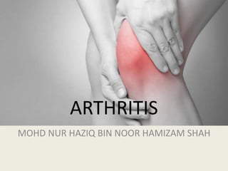
  • 1. ARTHRITIS MOHD NUR HAZIQ BIN NOOR HAMIZAM SHAH
  • 2. Arthritis is a disorder of the joint characterized by pain, swelling and limitation of movement. Arthralgia is pain of a joint, without any associated signs of inflammation. Essential Orthopaedics 4th edition
  • 3. ANATOMY OF A SYNOVIAL JOINT ARTICULARCARTILAGE: -‐ Covered by Hyaline cartilage. -‐ Covers weight-‐bearing surfaces of bones. -‐ Transmits load uniformly to bone. -‐ Decreasesfriction https://www.orthobullets.com/basic-science/9017/articular-cartilage?expandLeftMenu=true
  • 4.
  • 5. • ARTICULAR CARTILAGE: composedof -‐ 1. Extracellular matrix 2. Cells Extracellularmatrix - matrix of proteogylcans, collagen & glycoproteins - makes up the dry weight - mainly type II collagen & proteoglycan - provides compressive and tensile strength - atracts and retains water incartilage Water - about 65‐80%of the cartilage mass - Distribute nutrients to chondrocytes -providing lubrication Condrocytes • produce collagen, proteoglycans and enzyme • derive from chondroblasts that are trapped in lacunae and become chondrocytes. Sophia Fox, A. J., Bedi, A., & Rodeo, S. A. (2009). The basic science of articular cartilage: structure, composition, and function. Sports health, 1(6), 461-468.
  • 6. ARTICULAR CARTILAGE:nutrition-‐ -‐ Articular cartilage is avascular and aneural tissue. -‐ Nourished by diffusion from synovial fluid at surface -‐ Nourished by subchrondal bone at the base SYNOVIUM: -‐ Lining the cavity of the joint -‐ Highly vascularized structure -‐ Produces synovial fluid responsible for nutrition and lubrication -‐ Synovial fluid rich in hyaluronin and lubricin CAPSULE &LIGAMENTS: -‐ The tough fibrous material that covers the entire joint -‐ Stabilizers of joints -‐Prevent excessive movement in a joint -‐Have mechanoreceptors and free nerve endings that help with joint proprioception https://www.orthobullets.com/basic-science/9016/ligaments?expandLeftMenu=true
  • 7. OSTEOARTHRTIS • A chronic jointdisorder in which there is progressive softening and disintegration of articular cartilage accompanied by new growth of cartilage and bone at the joint margins and capsular fibrosis. Apley's System of Orthopaedics. and Fractures, Ninth Edition
  • 8. • Cardinal features are: 1. Progressive loss of articular cartilage thickness 2. Subarticular cyst formation and sclerosis 3. Remodelling of the bone ends and osteophytes formation 4. Synovial irritation 5. Capsular fibrosis OA is a dynamic phenomenon, which involves both destruction and repair Apley's System of Orthopaedics. and Fractures, Ninth Edition PATHOPHYSIOLOGY
  • 9. Softening of the articular cartilage (normally smooth and glistening surface become frayed and fibrillated) Articular cartilage worn away and expose the underlying bone Subarticular bone changes (attempts to remodel) High stress area – cyst appear and surrounding trabecular thickened or sclerotic, vascular congestion and increased intraosseous pressure rise Low stress area – cartilage proliferate and ossifies producing bony outgrowth (osteophytes) Shedding of the fibrilated articular cartilage as well as release of enzyme from damaged cells lead to synovitis Later stage, capsular fibrosis lead to joint stiffness Apley's System of Orthopaedics. and Fractures, Ninth Edition
  • 10. PREVALENCE & RISK FACTORS • Patients usually present after middle age, though cartilage changes start 10 - 20 years before. • Most affectedjoints: – Knee – Hip – Hand – Spine Apley's System of Orthopaedics. and Fractures, Ninth Edition
  • 11. RISK FACTORS • Non-modifiable • • Advancing age • • Female • • Genetic influence on hand and knee OA in women • Heberden’s nodes in hand • Modifiable • Previous knee injury/articular trauma • Occupation, repetitive knee bending • Muscle weakness • BMI >25kg/m2 CPG Management of Osteoarthritis ( Second Edition) 2013
  • 12. CLASSIFICATION PRIMARY No identifiable cause. A diagnosis by exclusion. SECONDARY Has an identifiable cause. -‐Dysplastic joint. -‐Trauma/ Infection/ Inflammatory arthritis. MONO/PAUCI-‐ARTICULAR-‐ Involves only1-‐2joints. -‐Large weight-‐bearingjoints. (hip, knees) -‐Has an underlying bone dysplasia. POLYARTICULAR-‐Multiplesmall jointsinvolved. eg: hands, MTP, knees, lumbar facet Apley's System of Orthopaedics. and Fractures, Ninth Edition
  • 13. SYMPTOMS • JOINT PAIN – Insidious onset – intermittent & relapsing – increased by joint use & impact – relieved by rest – night pain in severe OA • Stiffness – due to due to capsular fibrosis, muscle spasms – only last a few minutes; always < 30minutes – usually occurs after inactivity CPG Management of Osteoarthritis ( Second Edition) 2013
  • 14. • Swelling – with or without associated warmth & loss of function – Bony swelling: in hand OA, hypertrophic bone formation in the IP joint may result in reduced dexterity & difficulty in performing fine movements such as sewing – OA of the first CMC joint may result in writing difficulties • Gait disturbance – on weight-bearing joints (hip/knee) – can produce a prominent limp – impaired function of a weight-bearing joint will cause added stress on the contralateral weight-bearing joint e.g. a patient with impaired right knee function & pain will have difficulty with the left hip & vice versa CPG Management of Osteoarthritis ( Second Edition) 2013
  • 15. • Loss of muscle bulk – inactivity secondary to pain, may lead to significant weakness & loss of quadriceps muscle bulk • Limb deformity – knock knees’ (valgus) or ‘bowing’ (varus) • Clicking/ grinding sensation – clicking or grinding sensation with joint motion resulting in discomfort or pain • Instability – instability in the knee or hip may cause the patient to seek assistance in ambulation e.g. using a cane or crutch CPG Management of Osteoarthritis ( Second Edition) 2013
  • 16. SIGNS OF OA – Look • Alignment,symmetry • Muscle wasting • Swelling • Scar • Deformities (Heberden’s nodes, Bouchard’s nodes, genu varus,genu valgus) • Gait – Feel • Tenderness • Warmness • Jointeffusions • Crepitus – Move • Limited joint movement • Locking CPG Management of Osteoarthritis ( Second Edition) 2013
  • 17. • SWELLING- • Fluctuant (effusion)-‐ fluid shift/ patella tap/ fluid thrill. • Doughy (synovial hypertrophy). • Hard immobile (osteophytes). DEFORMITY-‐HAND-‐ Heberden’s nodes/ Bouchard’s nodes.
  • 18.
  • 19. DIAGNOTIC CRITERIA • The diagnostic criteria for classification of OA are based on the American College of Rheumatology (ACR) criteria
  • 20. CPG Management of Osteoarthritis ( Second Edition) 2013
  • 21. CPG Management of Osteoarthritis ( Second Edition) 2013
  • 22.
  • 23. KNEE OA • can also be diagnosed using evidence based recommendations by European League Against Rheumatism (EULAR) 2010
  • 24. CPG Management of Osteoarthritis ( Second Edition) 2013
  • 25. • In the presence of 3/10 at least 1 point from Domain II along with all entry criteria, the diagnosis of Knee OA can be established 2016 ACR REVISED CRITERIA FOR EARLY DIAGNOSIS OF KNEE OA http://aperito.org/uploads/pdf/ADTAOA-3-118.pdf
  • 26. INVESTIGATIONS • Diagnosis of OA is mainly clinical • Blood investigations - ESR/CRP normal • Synovial fluid analysis - normal & seldom required except to exclude septic, inflammatory & crystal arthropathy • Plain radiography is the investigation of choice – should be done in weight bearing position CPG Management of Osteoarthritis ( Second Edition) 2013
  • 27. PLAIN RADIOGRAPH • Classical features of OA – narrowed joint space – subchondral bone sclerosis – osteophytes – subchondral cysts CPG Management of Osteoarthritis ( Second Edition) 2013
  • 28.
  • 29.
  • 30.
  • 31.
  • 32.
  • 33.
  • 34.
  • 35. MANAGEMENT • NON-PHARMACOLOGICAL TREATMENT – Patient Education • nature of the disease • therapeutic options • importance of patient participation – Lifestyle Modification • weight loss programs • exercise – Physiotherapy – Occupational Therapy – Orthoses CPG Management of Osteoarthritis ( Second Edition) 2013
  • 36.
  • 37. Physiotherapy: – improve muscle strength, balance, coordination and joint mobility TENS: Trancutaneous Electrostimulation • patients with chronic moderate to severe pain who are not suitable for total knee arthroplasty. (ACR 2012) OCT: – To improve health, prevent disability & help individuals to achieve their optimum functional level & independence in performing ADL – Joint protection exercises, splinting, assistive devices (walking stick, tap turners)
  • 38. PHARMACOLOGICAL • Oral medication • Simple analgesics – paracetamol (acetaminophen) • Weak opioid analgesics – tramadol (combined with pcm or nsaids) • NSAIDs and COX-2 Inhibitors (diclofenac, celecoxib) • Nutraceutical - glucosamine, chondroitin, diacerein • Intraarticular injections • Steroids – for short term pain relief • Viscosupplementation – to replace hyaluronic acid in joints, controversial • Topical cream • NSAIDS and LMS cream CPG Management of Osteoarthritis ( Second Edition) 2013
  • 39.
  • 40. SURGICAL MANAGEMENT • Considered if symptoms are significant: – Pain – Limitation of ADL – Psychosocial health (psychological well being) – Economic impact – Recent deterioration
  • 41. OPERATIVE • Arthroscopic surgery – indicated in patients with OA associated with mechanical symptoms such as locking, catching or giving way of the joint caused by presence of loose bodies or flaps of meniscus or cartilage • High Tibial Osteotomy • Total Joint Replacement • Partial Joint Replacement • Arthrodesis
  • 42. - Osteotomised at its upper end and repositioned - To realign the mechanical axis of the limb away from the diseased area - Allows the joint to glide freely and distribute weight evenly In isolated medial compartment knee OA or varus malalingment • <50 years old • Good knee ROM (>120⁰) • Realign to correct mechanical axis • Load distributed more on normal knee compartment High Tibial Osteotomy
  • 43. ARTHROPLASTY -‐ Replacing the joint with implant -‐ Results in dramatic reduction of pain and -‐ Causes significant improvement in ADL -‐ Active infection is absolute contraindication INDICATIONS FOR JOINT REPLACEMENT • Pain • Severe disability • Deformity • Limited function (ADL) • Fail conservative treatment CPG Management of Osteoarthritis ( Second Edition) 2013
  • 44. ARTHRODESIS • Surgical fusion of the involvedjoint in optimal position • Provides pain relief &stability despite stiff joint Indications: • Young active labourer with unilateral painful OA • Severe OA with combined ligamentous injury • Uncontrolled septic arthritis & complete joint destruction • Failed and/or infected arthroplasty Advantages: • Stable pain free knee allowing long-term ambulation Disadvantages: • Stiff joint • Abnormal gait after surgery • Late back pain • Conversion to TKR or THR give unpredictable result CPG Management of Osteoarthritis ( Second Edition) 2013
  • 46. RHEUMATOID ARTHRITIS • Rheumatoid arthritis (RA) is a chronic inflammatory disease characterized by joint swelling, joint tenderness, and destruction of synovial joints, leading to severe disability and premature mortality Aletaha, D., Neogi, T., Silman, A. J., Funovits, J., Felson, D. T., Bingham, C. O., ... & Combe, B. (2010). 2010 rheumatoid arthritis classification criteria: an American College of Rheumatology/European League Against Rheumatism collaborative initiative. Arthritis & Rheumatology, 62(9), 2569-2581.
  • 47. RISK FACTORS 1. Gender Women are affected 3 times more often than men 2. Genetic factors Human Leucocyte Antigen (HLA)- DR4 and HLA-DR1 confer susceptibility to RA and are associated with development of more severe erosive disease 3. Agents such as mycoplasma, clostridium and EB virus
  • 48. Exposure of genetically predisposed individual to infectious agent Formation of immune complexes with IgM antibodies in the serum Immune complexes deposited in synovial membrane and initiate chronic granulamatous inflamation Synovium is edematous and filled with fibrin exudates and cellular infiltrates Synovium becomes hypertrophied and has effusion (reversible) Synovium surrounds articular cartilage to form pannus which cause reduction in joint space, early cartilage erosions and soft tissue destruction (controllable but irreversible) Pannus ultimately destroys articular cartilage, burrows into the subchondral bone to erode it (irreversible) Fibrous ankylosis and joint deformity Essential Orthopaedics 4th edition
  • 49.
  • 50. CLINICAL FEATURES Symptoms • insidious onset • morning stiffness >1 hour • polyarthropathy • usually affects hands and feet – (DIP joint of hand is usually spared) • may also affect other joints https://www.orthobullets.com/basic-science/9085/rheumatoid-arthritis
  • 51. ARTICULAR SIGNS • Red, warm, tender and swollen small joints of the hands and feet, excluding DIP, first MTP, and first CMC joint. • Symmetrical • Polyarthritic. Atlas of rheumatoid arthritis 2015
  • 53. Forefoot -Hyperpronation of rheumatoid foot -Metatarsalgia -Claw toes -Hallux valgus Midfoot -Talonavicular arthritis -Midfoot hyperpronation Hindfoot -Calcaneovalgus Atlas of rheumatoid arthritis 2015
  • 54. EXTRA-ARTICULAR MANIFESTATION • Systemic symptoms • fever • fatigue • malaise • weight loss • Musculoskeletal • osteoporotic # • tenosynovitis and bursitis • rupture of tendons & ligaments Face and neck -Mouth dryness -Atlanto axial subluxation Eyes -Conjuctival pallor -Schirmer’s filter paper test -Episcleritis -Scleritis -Cataracts -Keratoconjuctivitis sicca Chest -Pericardial rub +- effusion -Myocarditis -Pleural rub +- effusion -Fine crepitus -Vasculitis Abdomen -Splenomegaly Legs -Mononeuritis multiplex Atlas of rheumatoid arthritis 2015
  • 55.
  • 56. DIAGNOSTIC CRITERIA JOINT DISTRIBUTION (0-5) 1 large joint 0 2-10 large joints 1 1-3 small joints (large joints not counted) 2 4-10 small joints (large joints not counted) 3 >10 joints (at least one small joint) 5 SEROLOGY (0-3) Negative RF AND negative ACPA 0 Low positive RF OR low positive ACPA 2 High positive RF OR high positive ACPA 3 SYMPTOM DURATION (0-1) <6 weeks 0 ≥6 weeks 1 ACUTE PHASE REACTANTS (0-1) Normal CRP AND normal ESR 0 Abnormal CRP OR abnormal ESR 1 2010 ACR/ EULAR CLASSIFICATION CRITERIA ≥6 = Definite RA Aletaha, D., Neogi, T., Silman, A. J., Funovits, J., Felson, D. T., Bingham, C. O., ... & Combe, B. (2010). 2010 rheumatoid arthritis classification criteria: an American College of Rheumatology/European League Against Rheumatism collaborative initiative. Arthritis & Rheumatology, 62(9), 2569-2581.
  • 57. Definition of “JOINT INVOLVEMENT” - Any swollen or tender joint (excluding DIP of hand and feet, 1st MTP, 1st CMC) - Additional evidence from MRI / US may be used for confirmation of the clinical findings Definition of “SMALL JOINT” MCP, PIP, MTP 2-5, thumb IP, wrist NOT: DIP, 1st CMC, 1st MTP Definition of “LARGE JOINT” Shoulder, elbow, hip, knee, ankles Definition of “>10 JOINTS” - At least one small joint - Additional joints include: temporomandibular, sternoclavicular, acromioclavicular, and others (reasonably expected in RA) Definition of “SEROLOGY” Negative: ≤ULN (for the respective lab) Low positive: >ULN but ≤3xULN High positive: >3xULN Definition of “SYMPTOM DURATION” Refers to the patient’s self-report on the maximum duration of signs and symptoms of any joint that is clinically involved at the time of assessment.
  • 60. Useful for diagnosis • normochromic normocytic anaemia • elevated ESR • elevated CRP • anti-CCP (most sensitive & specific) • positive RF titer • elevated in 75-80% of patients with RA • joint fluid testing • decreased complement • may have elevated RF levels Useful for differential diagnosis • serum uric acid • antinuclear antibodies Useful to detect complications • chest xray Investigations for follow up and treatment • ESR • FBC • RP • LFT • Chest xray
  • 61. Categories of Synovial Fluid Based upon Clinical and Laboratory Findings CPG Management of Osteoarthritis ( Second Edition) 2013 There is no single diagostic test for RA. Investigationsare used largely to support clinical diagnosis and negative results does not exclude the diagnosis of RA.
  • 62. MANAGEMENT • Principle of treatment • Induction of remission and it’s maintenance: disease activity is brought under control by drugs • Preservation of joint functions and prevention of deformities during the activity of the disease and thereafter, by physiotherapy and splinting • Repair of joint damage which already exists, if it will relieve pain or facilitate functions
  • 63. TREATMENT MODALITIES FOR RA • NSAIDS • Steroids • DMARDs • Biological therapies • Surgery • Physiotherapy • Occupational therapy • Rehabilitation
  • 64. • Treatment with DMARD should be initiated as soon as a diagnosis of RA is made, with the aim of reaching a target of remission or low disease activity • EULAR recommends rheumatologists administer methotrexate (MTX) or combination therapy of MTX with other conventional synthetic DMARDs. • Low-dose glucocorticoids should also be considered in combination with DMARDs for up to six months, but should be tapered as soon as clinically feasible EULAR 2013 Rheumatoid Arthritis Management Recommendations, EULAR Data on File, 2013.
  • 65.
  • 66.
  • 67. SURGERY Preventive surgery: – To prevent damage to the joint and nearby tendons by the inflamed, hypertrophied synovium. It consists of synovectomy of the wrist, knee and MP joints Palliative surgery : – Done in situations where condition of the patient doesn’t permit corrective surgery, but some relief can be provided by bone block operations or tendon lengthening Reconstructive surgery : – This has revolutionised the rehabilitation of patients with deformed and painful joints. It includes tendon transfers and total joint replacement.
  • 68.
  • 69.
  • 70. SEPTIC ARTHRITIS Septic arthritis refers to infection in a joint; it is usually caused by bacteria but can be caused by fungi or mycobacteria. https://www.uptodate.com/contents/septic-arthritis-in-adults
  • 71. RISK FACTORS Adults: • Age > 80 years • Medical conditions • diabetes • rheumatoid arthritis • cirrhosis • HIV • History of crystal arthropathy • Endocarditis or recent bacteremia • IV drug user • Recent joint surgery Neonates: • Prematurity • Cesarean section • Patients treated in the NICU • Invasive procedures leading to transient bacteremia https://www.orthobullets.com/trauma/1058/septic-arthritis--adult?expandLeftMenu=true
  • 73. PATHOPHYSIOLOGY Routes of inoculation Direct inoculation from trauma or surgery Hematogenous seeding (bacteremia) Extension from adjacent bone (osteomyelitis) https://www.orthobullets.com/trauma/1058/septic-arthritis--adult?expandLeftMenu=true
  • 74. Apley's System of Orthopaedics. and Fractures, Ninth Edition a) In the early stage, there is an acute synovitis with a purulent joint effusion b) Soon the articular cartilage is attacked by bacterial and cellular enzyme. c) If infection is not arrested , the cartilage may be completely destroyed d) Healing then leads to ankylosis
  • 75. If left untreated, it will spread to the underlying bone and out of joint to form abscess and sinus. Healing with: 1.Complete resolution 2.Partial loss of articular cartilage and fibrosis of joint 3.Loss of articular cartilage and bony ankylosis 4.Bony destruction and permanent deformity
  • 76.
  • 77. PHYSICAL EXAMINATIONS • i)Temp. and vital signs • Fever • ii)Look • Localized swelling and erythema • Affected joint lies in a position of ease Joint Position of ease Shoulder Adduction, internal rotation Elbow Flexion, mid pronation Wrist Flexion Hip Flexion, abduction, ext rotation (FABER) Knee Flexion Ankle Plantar-flexion iii) Feel Effusion Warm to touch Tenderness iv) Move Severe pain with passive motion Unwillingness to move joint (pseudoparalysis)
  • 78. INVESTIGATION 1. WBC raised (>10k with left shift) 2. ESR raised (>30) • ESR is often elevated but may be normal early in process • rises within 2 days of infection and can rise 3-5 days after initiation of appropriate antibiotics, and returns to normal 3-4 weeks 3. CRP raised (>5) • best way to judge efficacy of treatment, as CRP rises within few hours of infection, and may normalize within 1 week of treatment 4. Joint aspiration (gold standard) – should be analyzed for • cell count with differential • gram stain • culture • glucose level • crystal analysis
  • 79. More than 50k is considered diagnostic
  • 80. 5. USG (most reliable) -widening of space between capsule and bone >2mm -able to confirm effusion in large joint such as hip -can be used in guiding aspiration 6. X-ray (AP & lateral of the joint, or frog leg lateral pelvic xray in paeds) -normal in early stages -widening of radiographic joint space or effusion -subluxation or dislocation -bone lesions (assc. osteomyelitis) 7. MRI -detects effusion and also adjacent bone involvement -helpful in determining operative treatment
  • 81. MANAGEMENT 1. URGENT surgical I&D 2. IV Antibiotics • Initiate empiric therapy prior to C&S based on age and risk factors • Transition to organism specific antibiotics after cultures obtained • Monitor efficacy of treatment using WBC, ESR and CRP • Paeds: Penicillinase-resistant penicillins+ 3rd generation cephalopsporins • Adults: Cloxacillin, or Flucloxacillin and Furisidic acid 3. Analgesia 4. Splintage: joint rested

Editor's Notes

  1. Articular cartilage is one of five forms of cartilage hyaline or articular cartilage fibroelastic cartilage (meniscus) fibrocartilage (at tendon and ligament insertion into bone) elastic cartilage (trachea) physeal cartilage (growth plate)
  2. exclusion criteria moderate to significant knee synovitis hot/ red knee hx / physical examination findings compatible with the internal derangement of knee
  3. Gull wing appearance Erosion with osteophytes
  4. The management of OA involves a multidisciplinary approach with the aim to relieve symptoms & improve joint function. It involves both non-pharmacological & pharmacological approaches.
  5. Tramadol is a synthetic opioid analgesic NSAIDs and COX-2 inhibitors reduce production of prostaglandin by inhibiting the enzyme cyclo-oxygenase. They vary in their selectivity for inhibiting different types of cyclo-oxygenase. They are a class of drugs that provide analgesic and anti-pyretic effects and in higher doses, antiinflammatory effects. COX-2 inhibitors selectively inhibit COX-2 and thus improve gastro-intestinal tolerance. Celecoxib 100 mg and 200 mg BID are as efficacious as diclofenac 50 mg BID and naproxen 500 mg BID in the treatment of hip, knee, or hand OA. GLUCOSAMINE-1 Amino sugar & prominent precursor in the biochemical synthesis of glycosylated proteins, lipids & glycosaminoglycans Glucosamine sulfate 1500 mg per day is more efficacious in pain reduction compared to placebo. Black C et al., 2009, level I; Towheed T et al., 2005, level I Pain relieving effect of glucosamine sulfate can be seen by three months after its initiation. Chondroitin sulfate is a sulfated GAG which is usually found attached to proteins as part of a proteoglycan Chondroitin sulfate 800 mg as a single dose is more efficacious than placebo in pain reduction, improving hand function & morning stiffness in patients with hand OA DIACEREIN-1 Purified anthraquinone derivative Inhibits production of interleukin (IL)-1beta which is a major proinflammatory cytokine Reduce articular cartilage destruction The commonly used topical treatment includes NSAIDs, capsaicin & methylsalicylate.
  6. HIP ARTHROPLASTY Hemiarthroplasty  Thompson  Austin Moore Bipolar hemiarthroplasty Total hip arthroplasty (THR) total hip arthroplasty (THA) indications end-stage, severe osteoarthritis arthritis preferred treatment for older patients (>50) and those with advanced structural changes total knee arthroplasty indications failed non-operative treatments
  7. Periarticular osteopenia, joint space narrowing and subchondral erosion
  8. citric citrullinated peptide
  9. NSAIDS reduce pain and swelling by inhibitting COX do not alter course of the disease chronic use should be minimized most common side effect related to GI tract Systemic Corticosteroids as bridge therapy rheumatic flares in pregnancy when other DMARDs cannot be used DMARDs drugs that actually alter the disease course should be used as soon as diagnosis is made appearance of benefit delayed for weeks to months Hydroxycloroquine ; 200mg BD x3/12, then OD. Methotrexate ; 7.5-25 mg once a week po, s/c, im Sulphasalazine ; 2 g OD Leflunomide ; 100mg OD x3/7, then 10-20mg OD
  10. If no remission after x3/12 after starting DMARDs, biological treatment should be added to the th/
  11. release of proteolytic enzymes (matrix metalloproteinases) from inflammatory and synovial cells, cartilage, and bacteria which may cause articular surface damage within 8 hours 
  12. acute inflammation is characterized by 5 cardinal signs: rubor (redness), calor (increased heat), tumor (swelling), dolor (pain), and functio laesa (loss of function) hip rests in a position of flexion, abduction, and external rotation (FABER)  hip capsular volume is maximized with flexion, abduction, and external rotation and is the position of comfort for hip septic arthritis
  13. treatment can be monitored by following serum WBC, ESR, and CRP levels during treatment