Downloaded 939 times


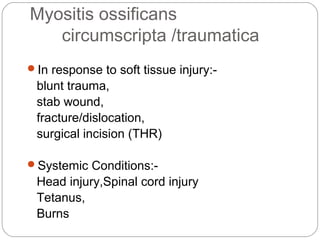


Myositis ossificans is the formation of bone within muscle tissue or within the soft tissues. It is usually caused by injury but can also occur spontaneously. There are two main types - myositis ossificans circumscripta which occurs as a localized mass in response to injury, and fibrodysplasia ossificans progressiva which is a rare inherited condition causing progressive heterotopic ossification throughout the body that is ultimately disabling. Diagnosis is based on clinical features and radiographic findings of ossification, and treatment focuses on prevention of further ossification through avoiding re-injury and surgical procedures when possible.


