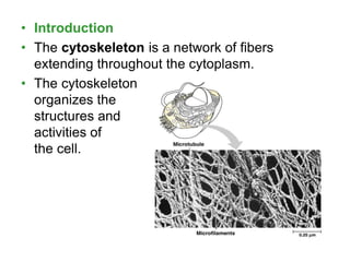
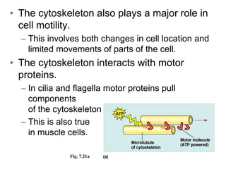
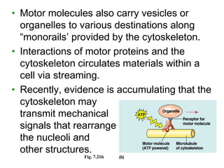
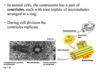
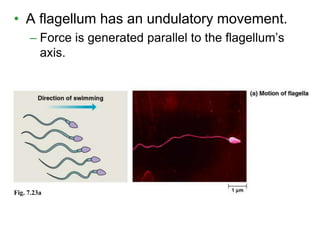
Cytoskeleton
Providing structural support to the cell,
the cytoskeleton also functions in cell
motility and regulation
Structural Support
• Mechanical support
– Maintains shape
• Fibers act like a geodesic dome to
stabilize and balance opposing forces
• Provides anchorage for organelles
• Dynamic
– Dismantles in one spot and reassembles in
another to change cell shape
• Introduction
• The cytoskeleton is a network of fibers
extending throughout the cytoplasm.
• The cytoskeleton
organizes the
structures and
activities of
the cell.
• The cytoskeleton also plays a major role in
cell motility.
– This involves both changes in cell location and
limited movements of parts of the cell.
• The cytoskeleton interacts with motor
proteins.
– In cilia and flagella motor proteins pull
components
of the cytoskeleton past each other.
– This is also true
in muscle cells.
Fig. 7.21a
• Motor molecules also carry vesicles or
organelles to various destinations along
“monorails’ provided by the cytoskeleton.
• Interactions of motor proteins and the
cytoskeleton circulates materials within a
cell via streaming.
• Recently, evidence is accumulating that the
cytoskeleton may
transmit mechanical
signals that rearrange
the nucleoli and
other structures.
Fig. 7.21b
• There are three main types of fibers in the
cytoskeleton: microtubules,
microfilaments, and intermediate
filaments.
• Microtubules, the thickest fibers, are hollow rods
about 25 microns in diameter.
– Microtubule fibers are constructed of the
globular protein, tubulin, and they grow or
shrink as more tubulin molecules are added or
removed.
• They move chromosomes during cell division.
• Another function is
as tracks that guide
motor proteins
carrying organelles
to their destination.
Fig. 7.21b
• In many cells, microtubules grow out from a
centrosome near the nucleus.
– These microtubules resist compression to the
cell.
Fig. 7.22
• In animal cells, the centrosome has a pair of
centrioles, each with nine triplets of microtubules
arranged in a ring.
• During cell division the
centrioles replicate.
• Microtubules are the central structural
supports in cilia and flagella.
– Both can move unicellular and small
multicellular organisms by propelling water
past the organism.
– If these structures are anchored in a large
structure, they move fluid over a surface.
• For example, cilia sweep mucus carrying trapped
debris from the lungs.
Fig. 7.2
• Cilia usually occur in large numbers on the
cell surface.
– They are about 0.25 microns in diameter and
2-20 microns long.
• There are usually just one or a few flagella
per cell.
– Flagella are the same width as cilia, but 10-200
microns long.
• A flagellum has an undulatory movement.
– Force is generated parallel to the flagellum’s
axis.
Fig. 7.23a
Fig. 7.23b
• Cilia move more like oars with alternating
power and recovery strokes.
– They generate force perpendicular to the cilia’s
axis.
• In spite of their differences, both cilia and
flagella have the same ultrastructure.
– Both have a core of microtubules sheathed by
the plasma membrane.
– Nine doublets of microtubules arranged around
a pair at the center, the “9 + 2” pattern.
– Flexible “wheels” of proteins connect outer
doublets to each other and to the core.
– The outer doublets are also connected by
motor proteins.
– The cilium or flagellum is anchored in the cell
by a basal body, whose structure is identical
to a centriole.
Fig. 7.24
• The bending of cilia and flagella is driven by
the arms of a motor protein, dynein.
– Addition to dynein of a phosphate group from
ATP and its removal causes conformation
changes in the protein.
– Dynein arms alternately
grab, move, and release
the outer microtubules.
– Protein cross-links limit
sliding and the force is
expressed as bending.
Fig. 7.25
• Microfilaments, the thinnest class of the
cytoskeletal fibers, are solid rods of the
globular protein actin.
– An actin microfilament consists of a twisted
double chain of actin subunits.
• Microfilaments are designed to resist
tension.
• With other proteins, they form a three-
dimensional network just inside the plasma
membrane.
Fig. 7.26 The shape of the
microvilli in this intestinal cell
are supported by microfilaments,
anchored to a network of
intermediate filaments.
• In muscle cells, thousands of actin filaments are
arranged parallel to one another.
• Thicker filaments, composed of a motor protein,
myosin, interdigitate with the thinner actin fibers.
– Myosin molecules walk along the actin filament,
pulling stacks of actin fibers together and
shortening
the cell.
Fig. 7.21a
• In other cells, these actin-myosin aggregates are less
organized but still cause localized contraction.
– A contracting belt of microfilaments divides the
cytoplasm of animals cells during cell division.
– Localized contraction also drives amoeboid movement.
• Pseudopodia, cellular extensions, extend and
contract through the reversible assembly and
contraction of actin subunits into microfilaments.
Fig. 7.21b
• In plant cells (and others), actin-myosin
interactions and sol-gel transformations
drive cytoplasmic streaming.
– This creates a circular flow of cytoplasm in the
cell.
– This speeds the distribution of materials within
the cell.
Fig. 7.21c
• Intermediate filaments,
intermediate in size at 8 -
12 nanometers, are
specialized for bearing
tension.
– Intermediate filaments
are built from a diverse
class of subunits from a
family of proteins called
keratins.
• Intermediate filaments are
more permanent fixtures of
the cytoskeleton than are
the other two classes.
• They reinforce cell shape
and fix organelle location. Fig. 7.26

cytoskeleton -Eukaryotic cell 123456.ppt

  • 1.
    Cytoskeleton Providing structural supportto the cell, the cytoskeleton also functions in cell motility and regulation
  • 2.
    Structural Support • Mechanicalsupport – Maintains shape • Fibers act like a geodesic dome to stabilize and balance opposing forces • Provides anchorage for organelles • Dynamic – Dismantles in one spot and reassembles in another to change cell shape
  • 3.
    • Introduction • Thecytoskeleton is a network of fibers extending throughout the cytoplasm. • The cytoskeleton organizes the structures and activities of the cell.
  • 4.
    • The cytoskeletonalso plays a major role in cell motility. – This involves both changes in cell location and limited movements of parts of the cell. • The cytoskeleton interacts with motor proteins. – In cilia and flagella motor proteins pull components of the cytoskeleton past each other. – This is also true in muscle cells. Fig. 7.21a
  • 5.
    • Motor moleculesalso carry vesicles or organelles to various destinations along “monorails’ provided by the cytoskeleton. • Interactions of motor proteins and the cytoskeleton circulates materials within a cell via streaming. • Recently, evidence is accumulating that the cytoskeleton may transmit mechanical signals that rearrange the nucleoli and other structures. Fig. 7.21b
  • 6.
    • There arethree main types of fibers in the cytoskeleton: microtubules, microfilaments, and intermediate filaments.
  • 8.
    • Microtubules, thethickest fibers, are hollow rods about 25 microns in diameter. – Microtubule fibers are constructed of the globular protein, tubulin, and they grow or shrink as more tubulin molecules are added or removed. • They move chromosomes during cell division. • Another function is as tracks that guide motor proteins carrying organelles to their destination. Fig. 7.21b
  • 9.
    • In manycells, microtubules grow out from a centrosome near the nucleus. – These microtubules resist compression to the cell.
  • 10.
    Fig. 7.22 • Inanimal cells, the centrosome has a pair of centrioles, each with nine triplets of microtubules arranged in a ring. • During cell division the centrioles replicate.
  • 11.
    • Microtubules arethe central structural supports in cilia and flagella. – Both can move unicellular and small multicellular organisms by propelling water past the organism. – If these structures are anchored in a large structure, they move fluid over a surface. • For example, cilia sweep mucus carrying trapped debris from the lungs. Fig. 7.2
  • 12.
    • Cilia usuallyoccur in large numbers on the cell surface. – They are about 0.25 microns in diameter and 2-20 microns long. • There are usually just one or a few flagella per cell. – Flagella are the same width as cilia, but 10-200 microns long.
  • 13.
    • A flagellumhas an undulatory movement. – Force is generated parallel to the flagellum’s axis. Fig. 7.23a
  • 14.
    Fig. 7.23b • Ciliamove more like oars with alternating power and recovery strokes. – They generate force perpendicular to the cilia’s axis.
  • 15.
    • In spiteof their differences, both cilia and flagella have the same ultrastructure. – Both have a core of microtubules sheathed by the plasma membrane. – Nine doublets of microtubules arranged around a pair at the center, the “9 + 2” pattern. – Flexible “wheels” of proteins connect outer doublets to each other and to the core. – The outer doublets are also connected by motor proteins. – The cilium or flagellum is anchored in the cell by a basal body, whose structure is identical to a centriole.
  • 16.
  • 17.
    • The bendingof cilia and flagella is driven by the arms of a motor protein, dynein. – Addition to dynein of a phosphate group from ATP and its removal causes conformation changes in the protein. – Dynein arms alternately grab, move, and release the outer microtubules. – Protein cross-links limit sliding and the force is expressed as bending. Fig. 7.25
  • 18.
    • Microfilaments, thethinnest class of the cytoskeletal fibers, are solid rods of the globular protein actin. – An actin microfilament consists of a twisted double chain of actin subunits. • Microfilaments are designed to resist tension. • With other proteins, they form a three- dimensional network just inside the plasma membrane.
  • 19.
    Fig. 7.26 Theshape of the microvilli in this intestinal cell are supported by microfilaments, anchored to a network of intermediate filaments.
  • 20.
    • In musclecells, thousands of actin filaments are arranged parallel to one another. • Thicker filaments, composed of a motor protein, myosin, interdigitate with the thinner actin fibers. – Myosin molecules walk along the actin filament, pulling stacks of actin fibers together and shortening the cell. Fig. 7.21a
  • 21.
    • In othercells, these actin-myosin aggregates are less organized but still cause localized contraction. – A contracting belt of microfilaments divides the cytoplasm of animals cells during cell division. – Localized contraction also drives amoeboid movement. • Pseudopodia, cellular extensions, extend and contract through the reversible assembly and contraction of actin subunits into microfilaments. Fig. 7.21b
  • 22.
    • In plantcells (and others), actin-myosin interactions and sol-gel transformations drive cytoplasmic streaming. – This creates a circular flow of cytoplasm in the cell. – This speeds the distribution of materials within the cell. Fig. 7.21c
  • 23.
    • Intermediate filaments, intermediatein size at 8 - 12 nanometers, are specialized for bearing tension. – Intermediate filaments are built from a diverse class of subunits from a family of proteins called keratins. • Intermediate filaments are more permanent fixtures of the cytoskeleton than are the other two classes. • They reinforce cell shape and fix organelle location. Fig. 7.26