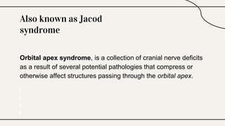
Orbital Apex
Syndrome
By Arish Bharathi R, CRRI
KMC, Chennai
Also known as Jacod
syndrome
Orbital apex syndrome, is a collection of cranial nerve deficits
as a result of several potential pathologies that compress or
otherwise affect structures passing through the orbital apex.
Anatomy
– The orbital apex is anatomically the posterior
part of the orbit positioned at the craniofacial
junction located where the four orbital walls
converge.
– The orbital apex incorporates the
optic canal and the superior orbital fissure.
– The optic canal transmits the optic nerve CN II (surrounded by meninges) and
the ophthalmic artery to the cranial fossa.
– The superior orbital fissure is anatomically lateral to the optic canal which transmits,
Lacrimal nerve (CN V1)
Frontal nerve (CN V1)
Trochlear nerve (CN IV)
Superior branch of the ophthalmic vein
Recurrent meningeal artery
Nasociliary nerve (CN V1)
Abducens nerve (CN VI)
Superior and inferior branches of CN III.
Inferior branch of the ophthalmic vein.
Common tendinous ring of Zinn
Pathophysiology
Involves damage to
● Optic nerve (II)
● Oculomotor nerve (III)
● Trochlear nerve (IV)
● Abducens nerve (VI)
● Ophthalmic branch of the trigeminal nerve (V1)
Clinical Presentation
● CN III, IV, and VI: ophthalmoplegia (diplopia), proptosis,
and ptosis
● Ophthalmic (V1) division of the trigeminal nerve: ipsilateral
hypoaesthesia of the forehead, upper eyelid, and cornea
● Optic nerve (II): visual deficit that can lead to
blindness
Etiology
● Trauma: usually fractures involving the orbital apex
● Infection: Invasive fungal sinusitis, orbital cellulitis, subperiosteal
abscess of the orbit
● Inflammatory: Tolosa-Hunt syndrome, sarcoidosis
● Vascular lesions: ophthalmic artery aneurysm, carotid-cavernous
fistula
● Neoplastic: extension of intracranial tumours or intraorbital tumours,
lymphoma, and metastasis
● Others: sino-nasal mucocele, foreign body, iatrogenic, intra-orbital
dermoid, intra-orbital epidermoid
Differential diagnosis
– Superior Orbital Fissure Syndrome:
Clinical presentation same as OAS
but the optic nerve is spared.
– Cavernous Sinus Syndrome:
Involves CN III, IV, VI, V1, V2 &
Oculosympathetic fibers; with optic
nerve spared.
Management
● Management of OAS is aimed at the underlying cause.
● Primary management - includes observation, surgical biopsy and
corticosteroids (if cause can not be determined.)
● If visual loss and ophthalmoplegia progresses repeat neuroimaging
and a biopsy.
● Neurosurgical, neurophysician, internal medicine, otolaryngologic and
radiological consultation must be required.
● Following traumatic and iatrogenic operative injury, corticosteroids
and surgical intervention will be considered.
Thanks

Orbital Apex Syndrome.pptx

  • 1.
    Orbital Apex Syndrome By ArishBharathi R, CRRI KMC, Chennai
  • 2.
    Also known asJacod syndrome Orbital apex syndrome, is a collection of cranial nerve deficits as a result of several potential pathologies that compress or otherwise affect structures passing through the orbital apex.
  • 3.
    Anatomy – The orbitalapex is anatomically the posterior part of the orbit positioned at the craniofacial junction located where the four orbital walls converge. – The orbital apex incorporates the optic canal and the superior orbital fissure.
  • 4.
    – The opticcanal transmits the optic nerve CN II (surrounded by meninges) and the ophthalmic artery to the cranial fossa. – The superior orbital fissure is anatomically lateral to the optic canal which transmits, Lacrimal nerve (CN V1) Frontal nerve (CN V1) Trochlear nerve (CN IV) Superior branch of the ophthalmic vein Recurrent meningeal artery Nasociliary nerve (CN V1) Abducens nerve (CN VI) Superior and inferior branches of CN III. Inferior branch of the ophthalmic vein. Common tendinous ring of Zinn
  • 5.
    Pathophysiology Involves damage to ●Optic nerve (II) ● Oculomotor nerve (III) ● Trochlear nerve (IV) ● Abducens nerve (VI) ● Ophthalmic branch of the trigeminal nerve (V1)
  • 6.
    Clinical Presentation ● CNIII, IV, and VI: ophthalmoplegia (diplopia), proptosis, and ptosis ● Ophthalmic (V1) division of the trigeminal nerve: ipsilateral hypoaesthesia of the forehead, upper eyelid, and cornea ● Optic nerve (II): visual deficit that can lead to blindness
  • 7.
    Etiology ● Trauma: usuallyfractures involving the orbital apex ● Infection: Invasive fungal sinusitis, orbital cellulitis, subperiosteal abscess of the orbit ● Inflammatory: Tolosa-Hunt syndrome, sarcoidosis ● Vascular lesions: ophthalmic artery aneurysm, carotid-cavernous fistula ● Neoplastic: extension of intracranial tumours or intraorbital tumours, lymphoma, and metastasis ● Others: sino-nasal mucocele, foreign body, iatrogenic, intra-orbital dermoid, intra-orbital epidermoid
  • 8.
    Differential diagnosis – SuperiorOrbital Fissure Syndrome: Clinical presentation same as OAS but the optic nerve is spared. – Cavernous Sinus Syndrome: Involves CN III, IV, VI, V1, V2 & Oculosympathetic fibers; with optic nerve spared.
  • 9.
    Management ● Management ofOAS is aimed at the underlying cause. ● Primary management - includes observation, surgical biopsy and corticosteroids (if cause can not be determined.) ● If visual loss and ophthalmoplegia progresses repeat neuroimaging and a biopsy. ● Neurosurgical, neurophysician, internal medicine, otolaryngologic and radiological consultation must be required. ● Following traumatic and iatrogenic operative injury, corticosteroids and surgical intervention will be considered.
  • 10.