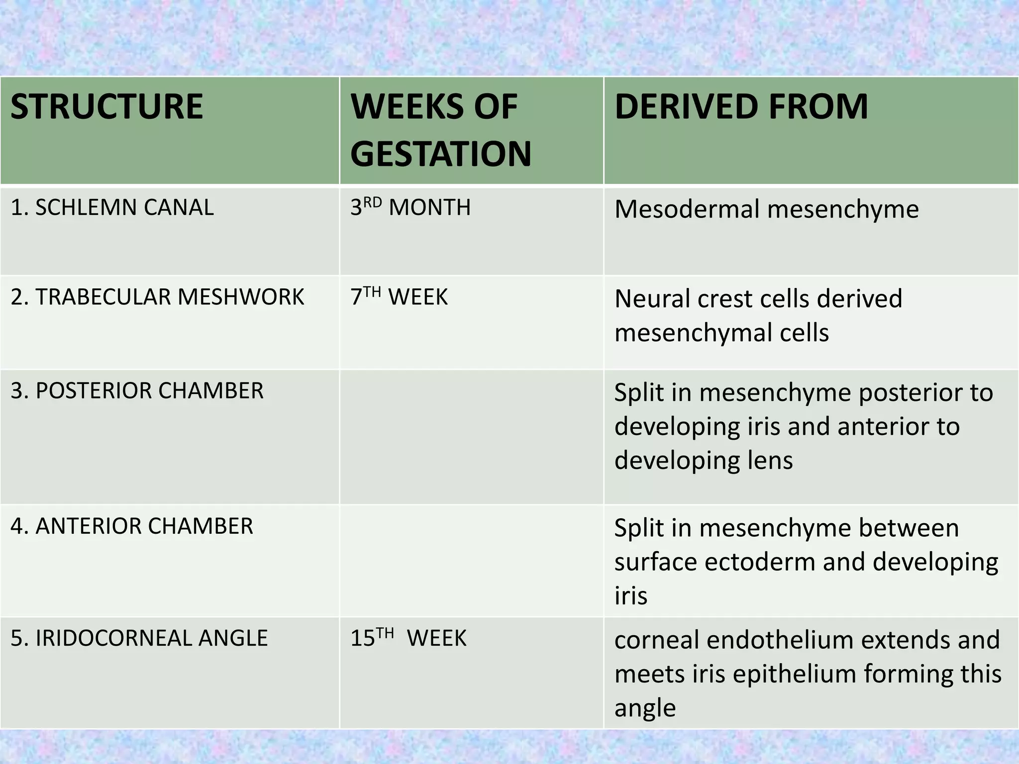
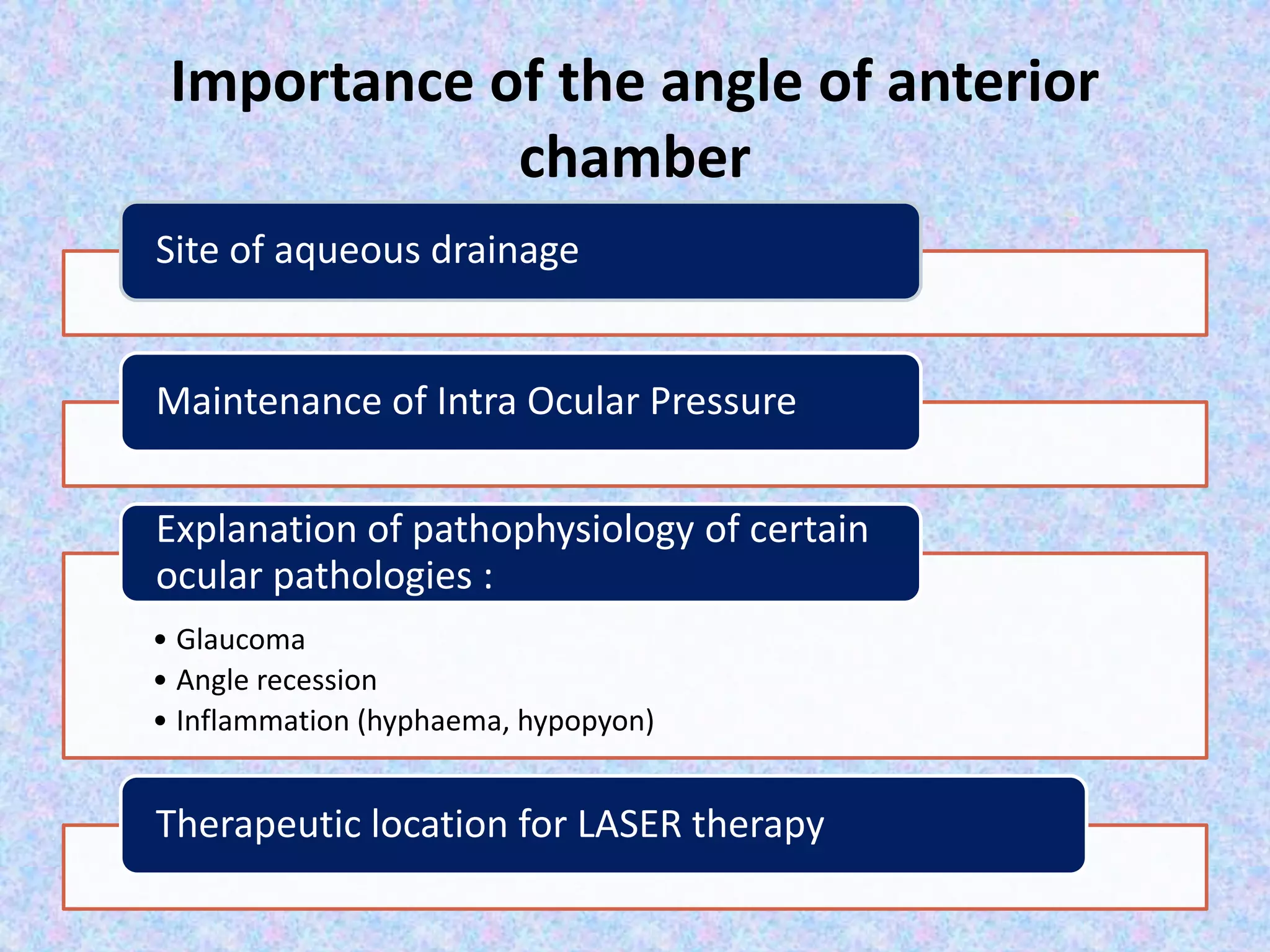
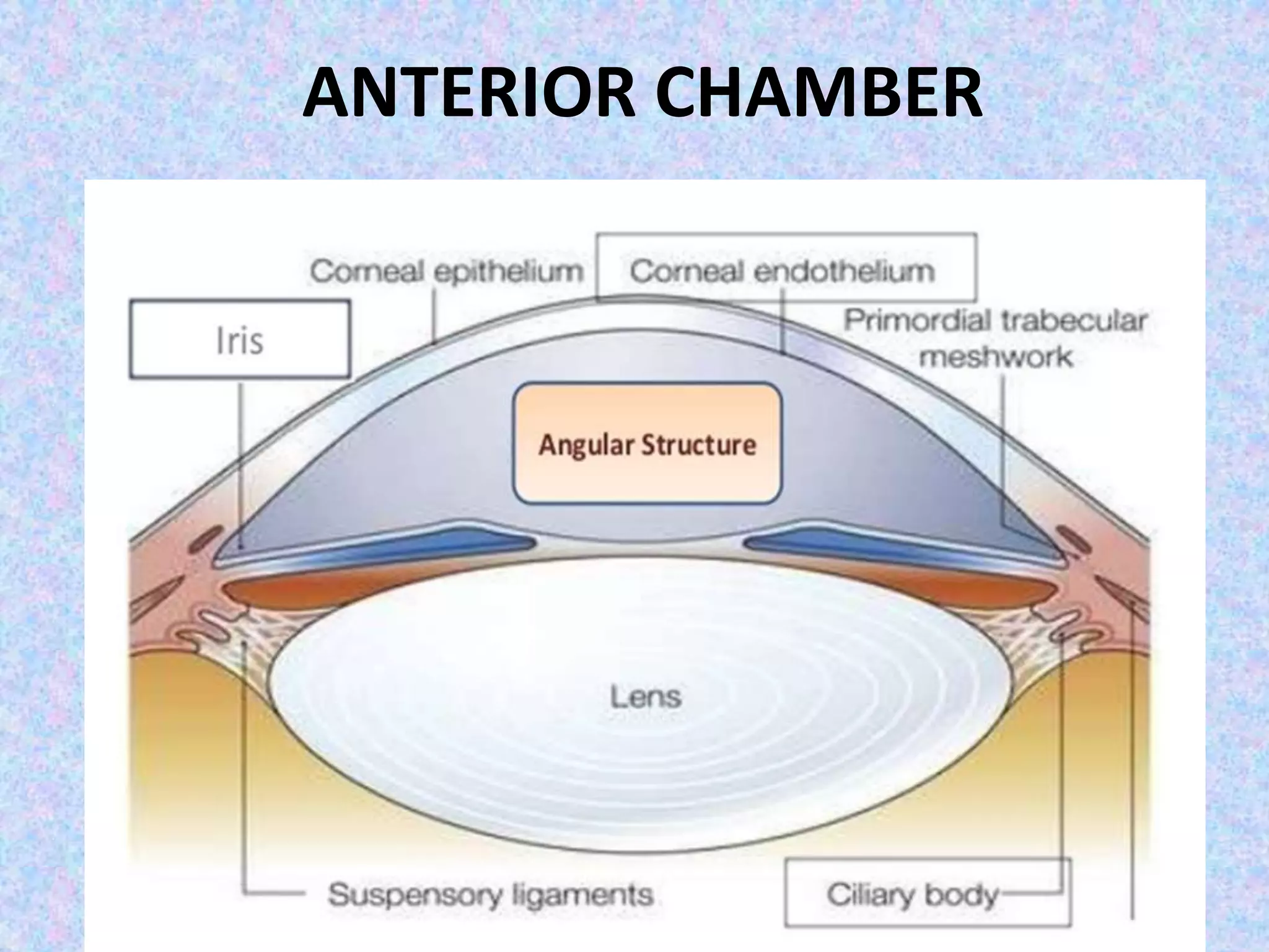
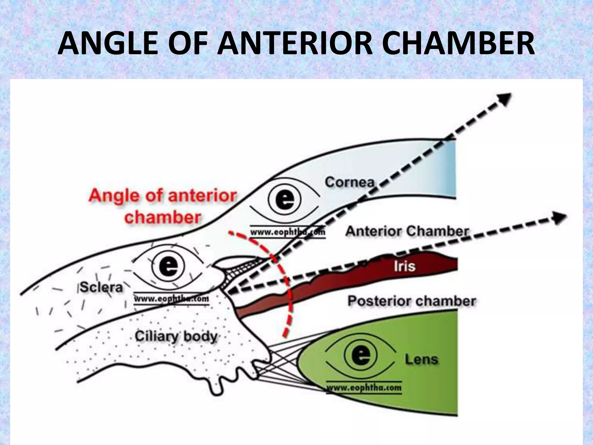
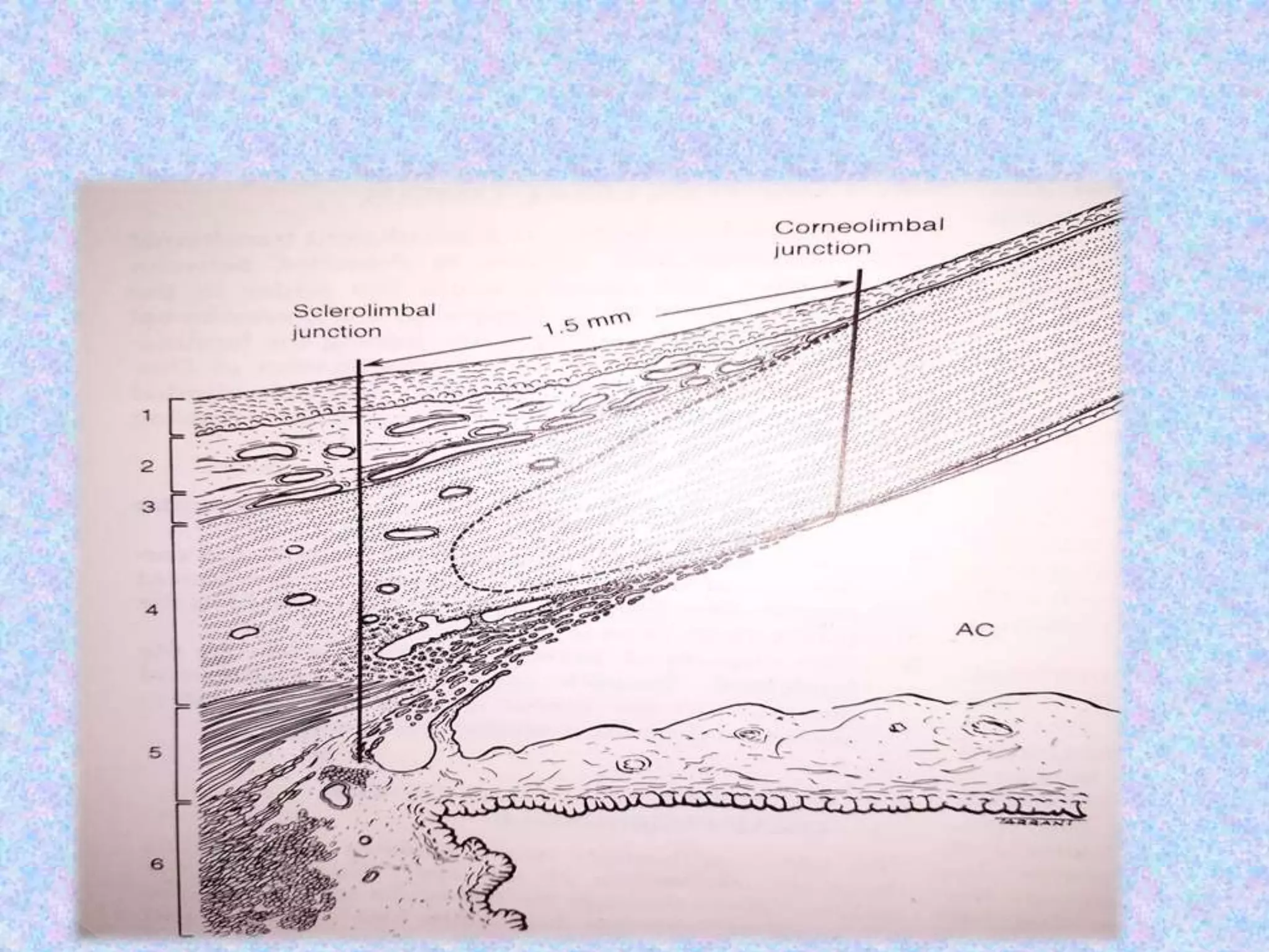
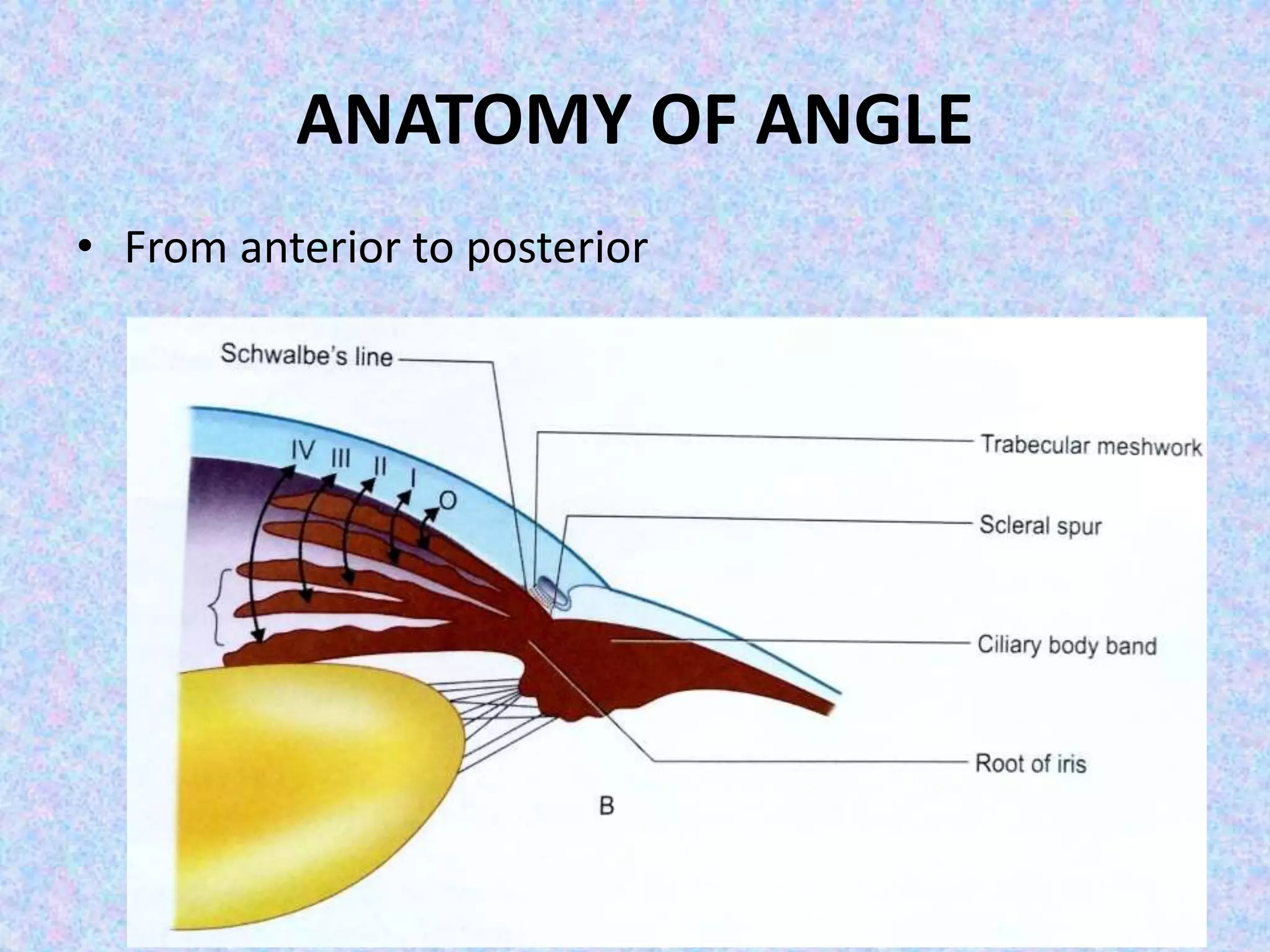
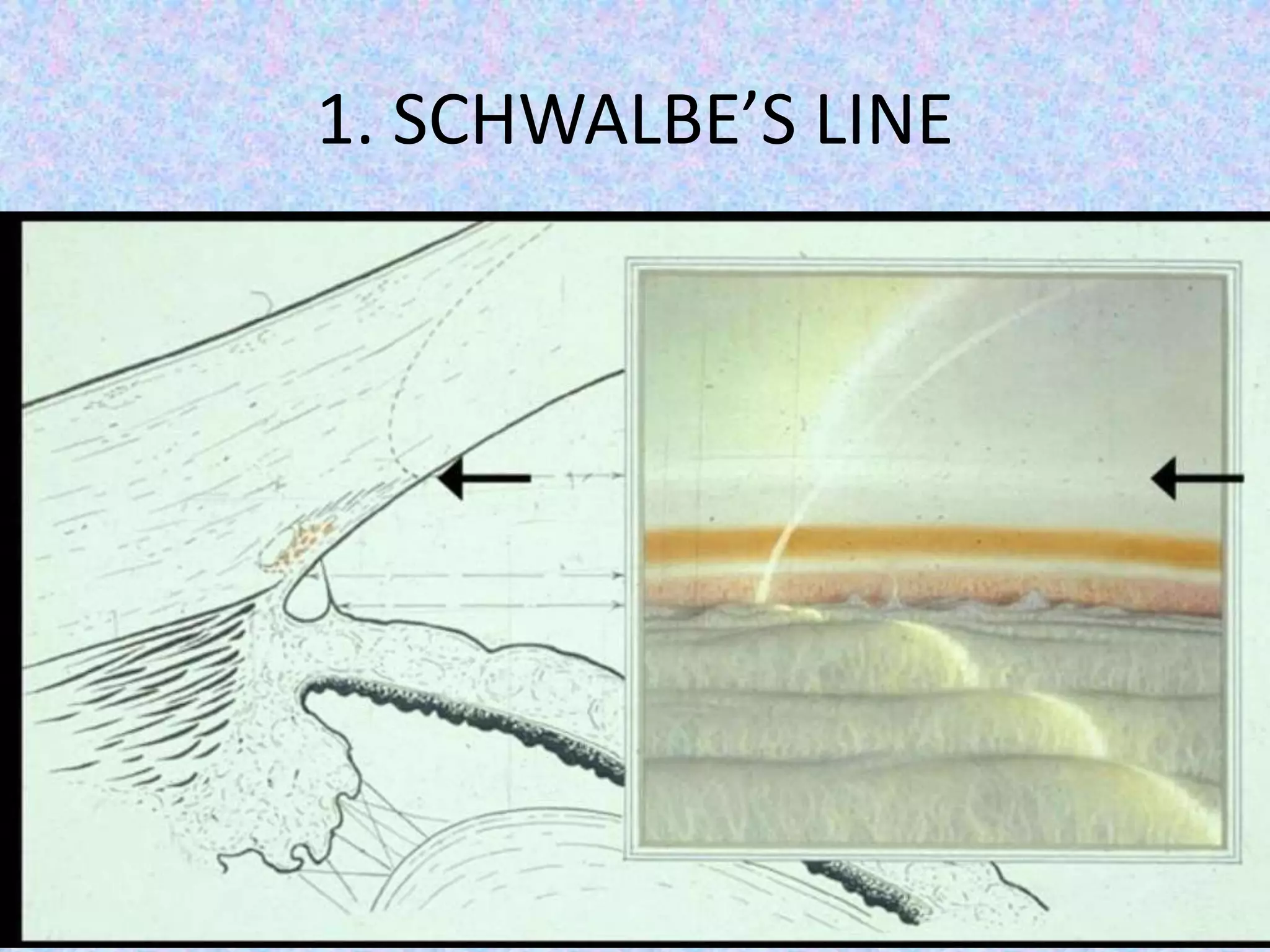
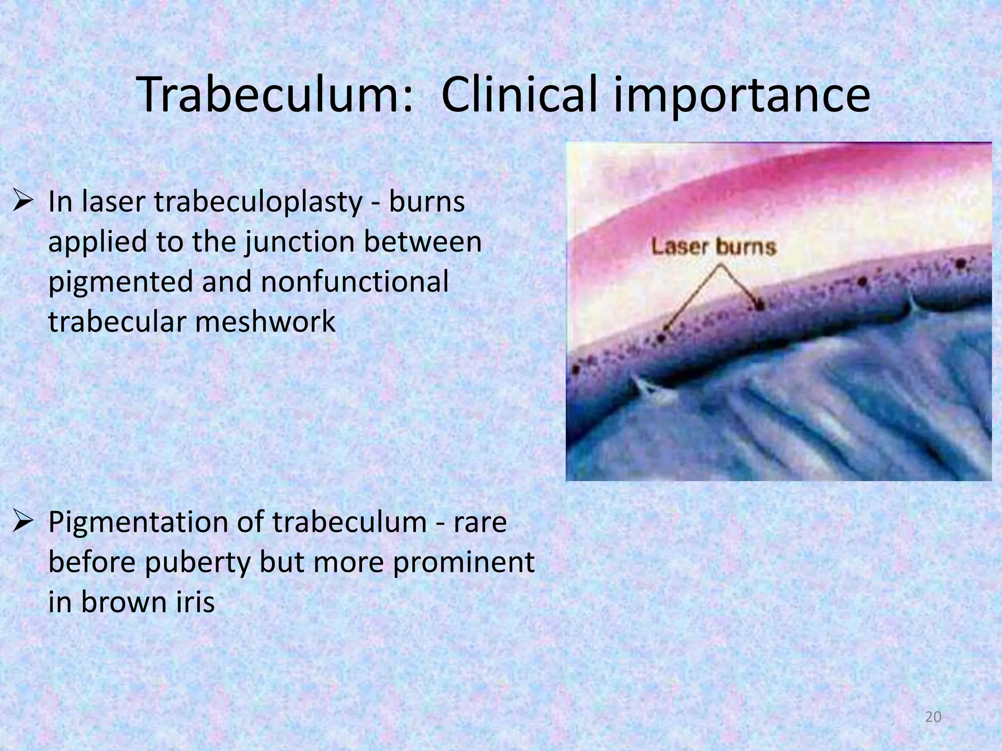
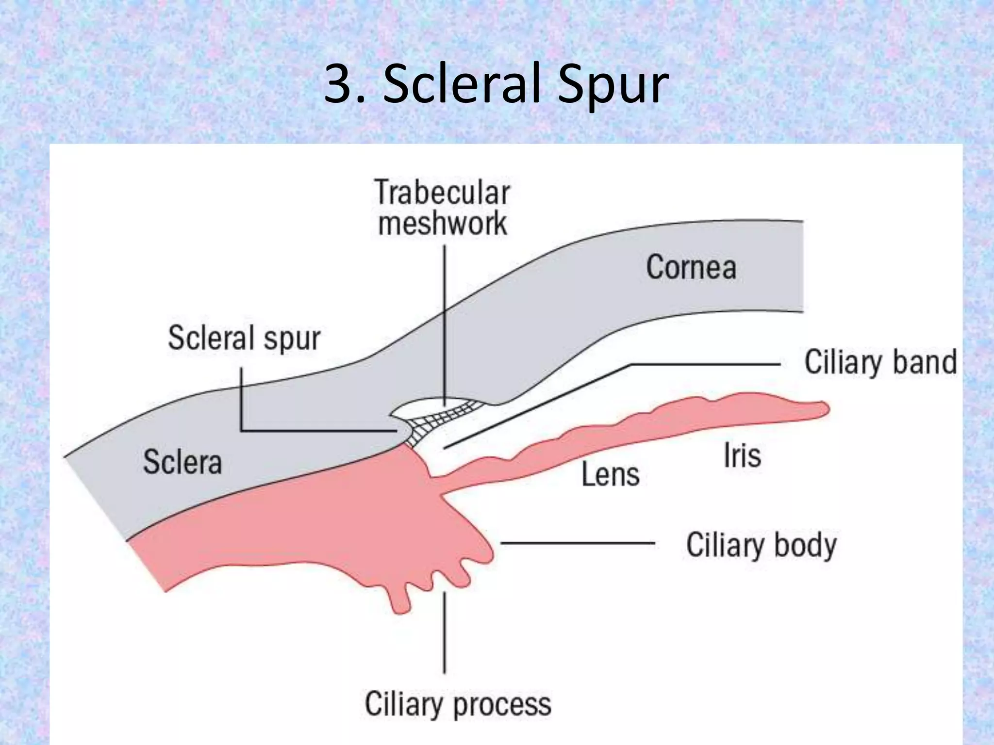
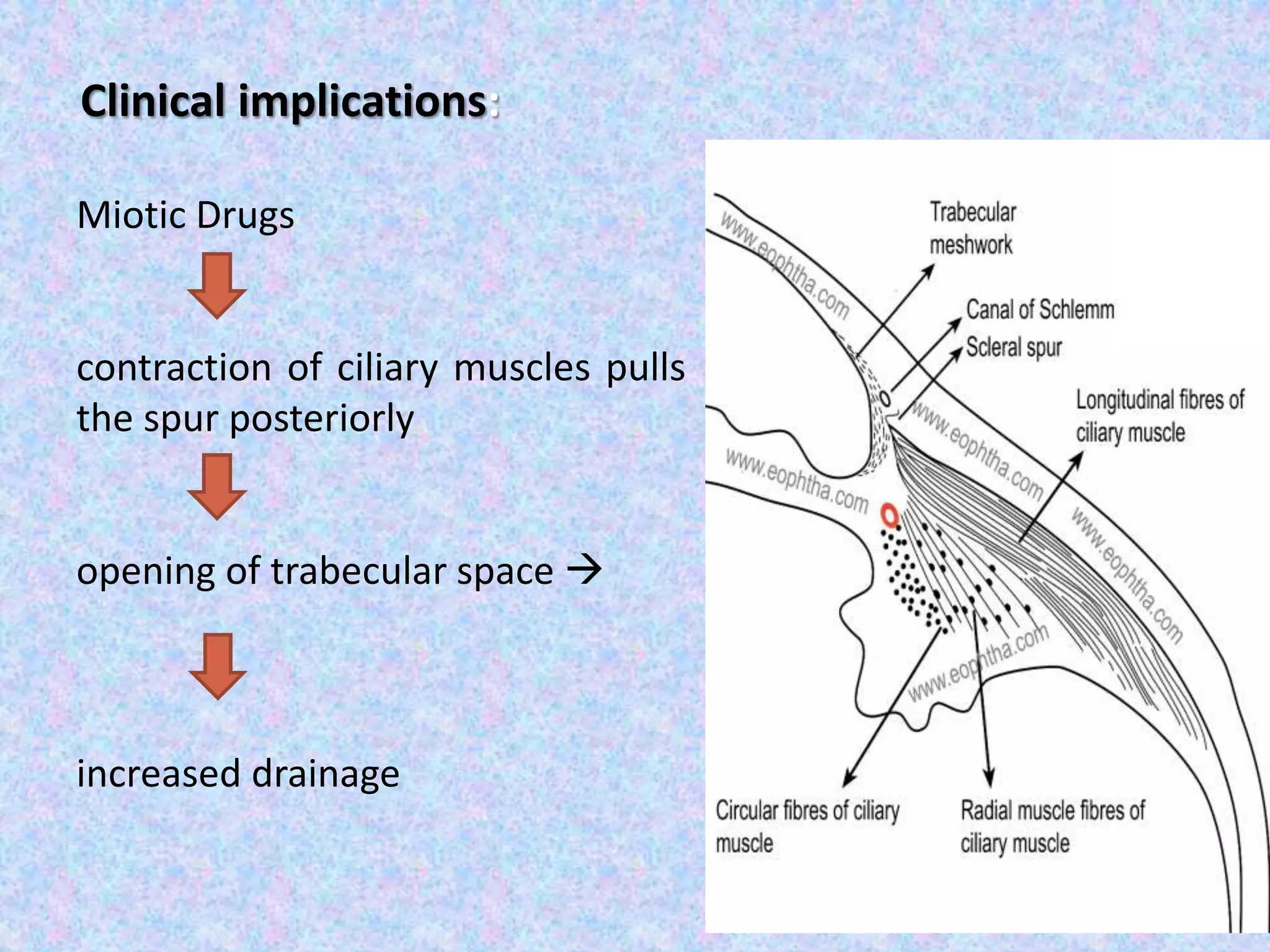
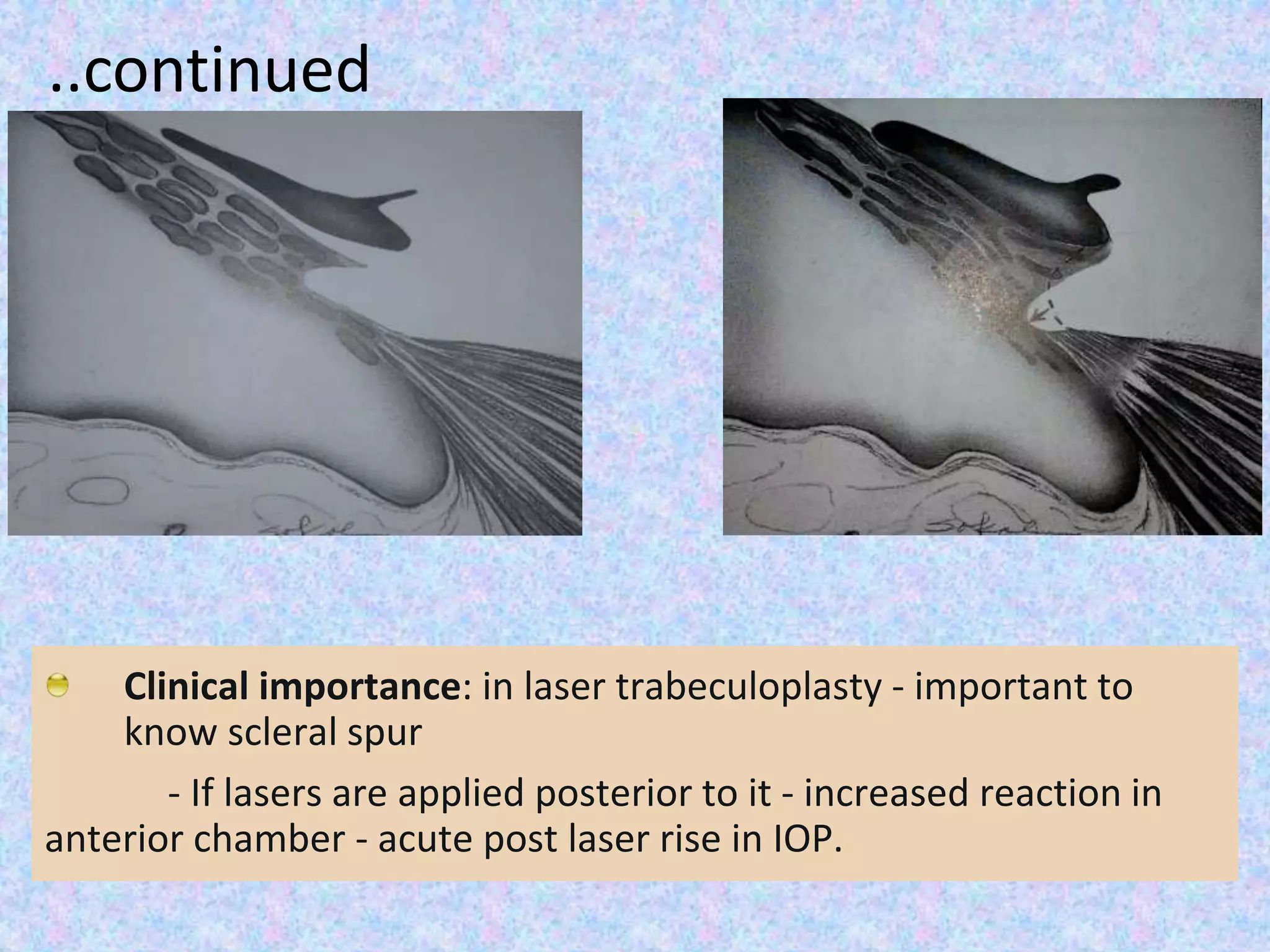
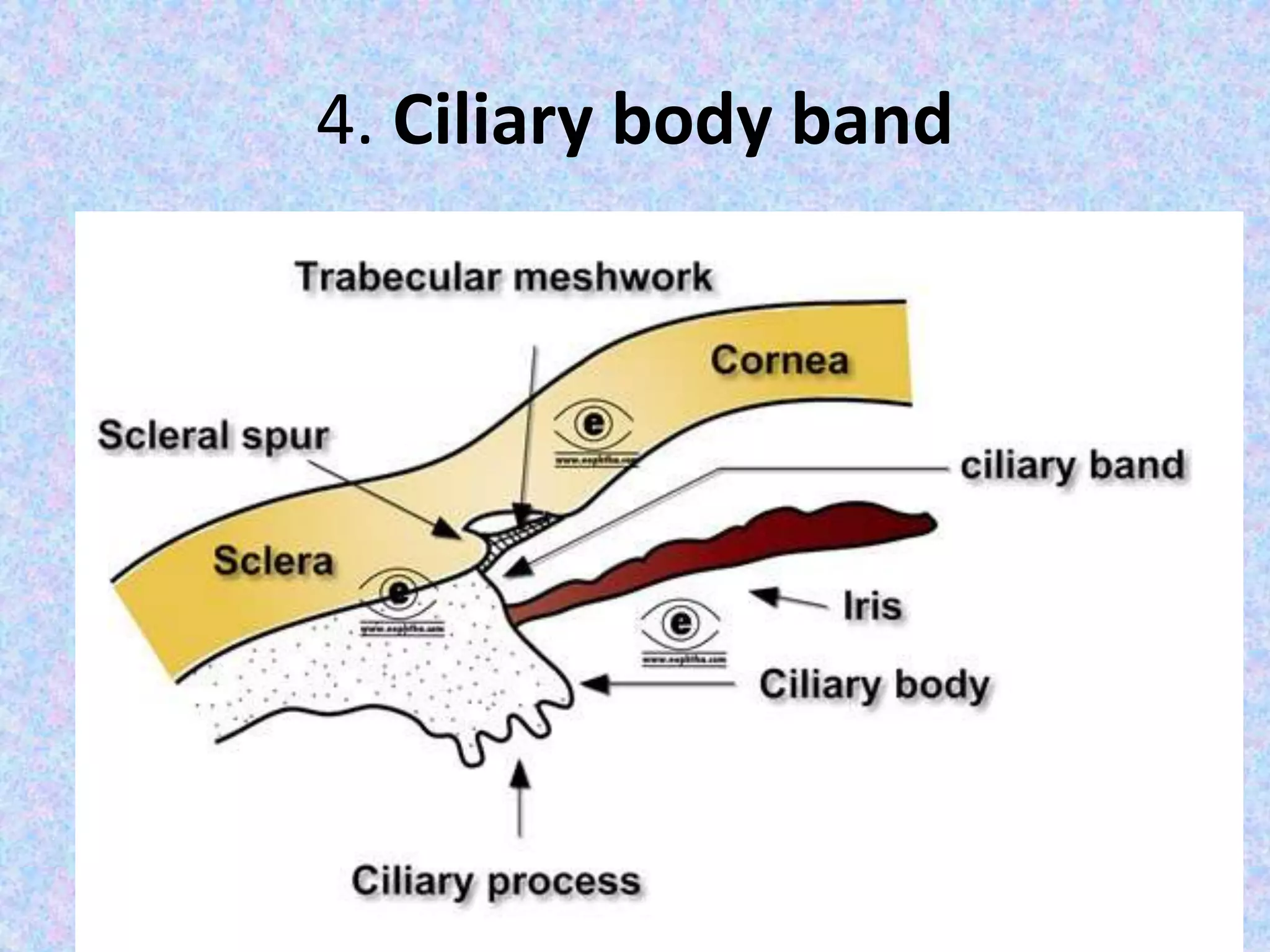
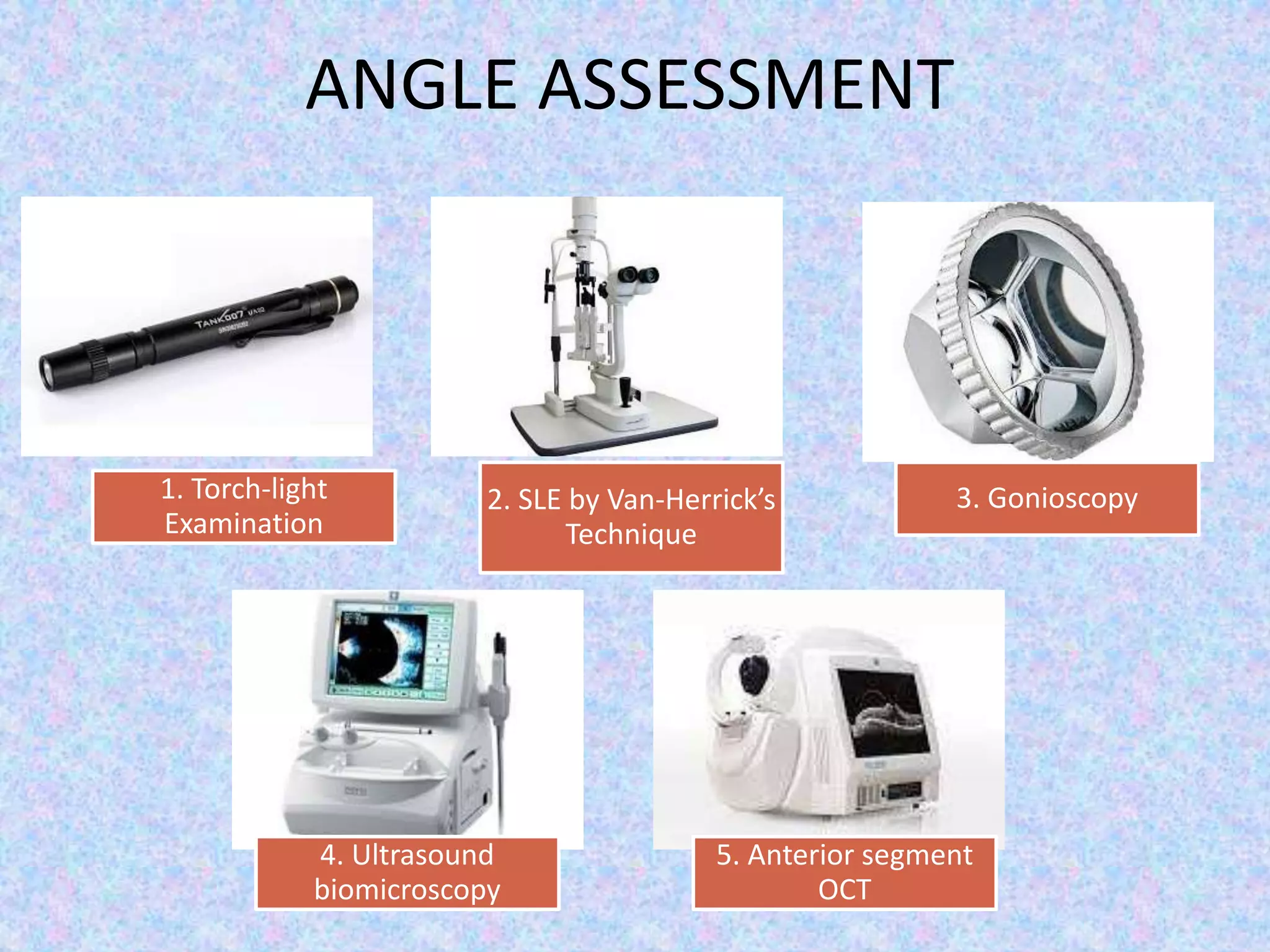
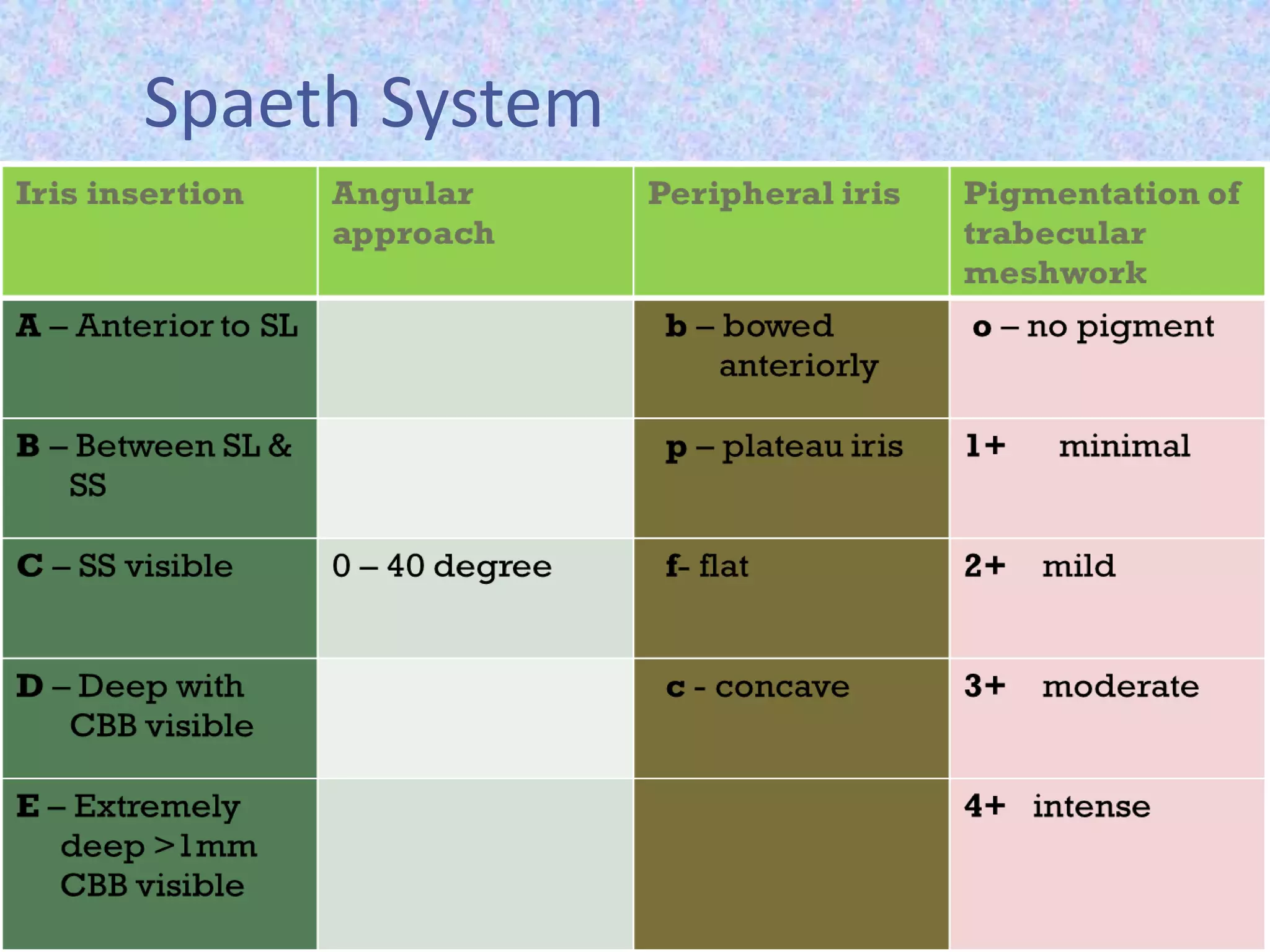
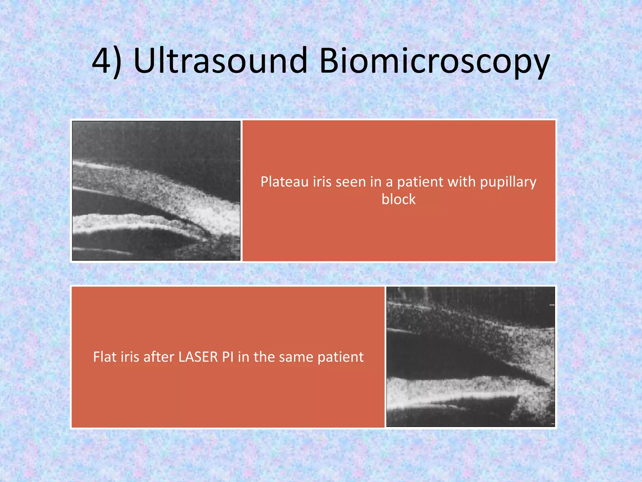
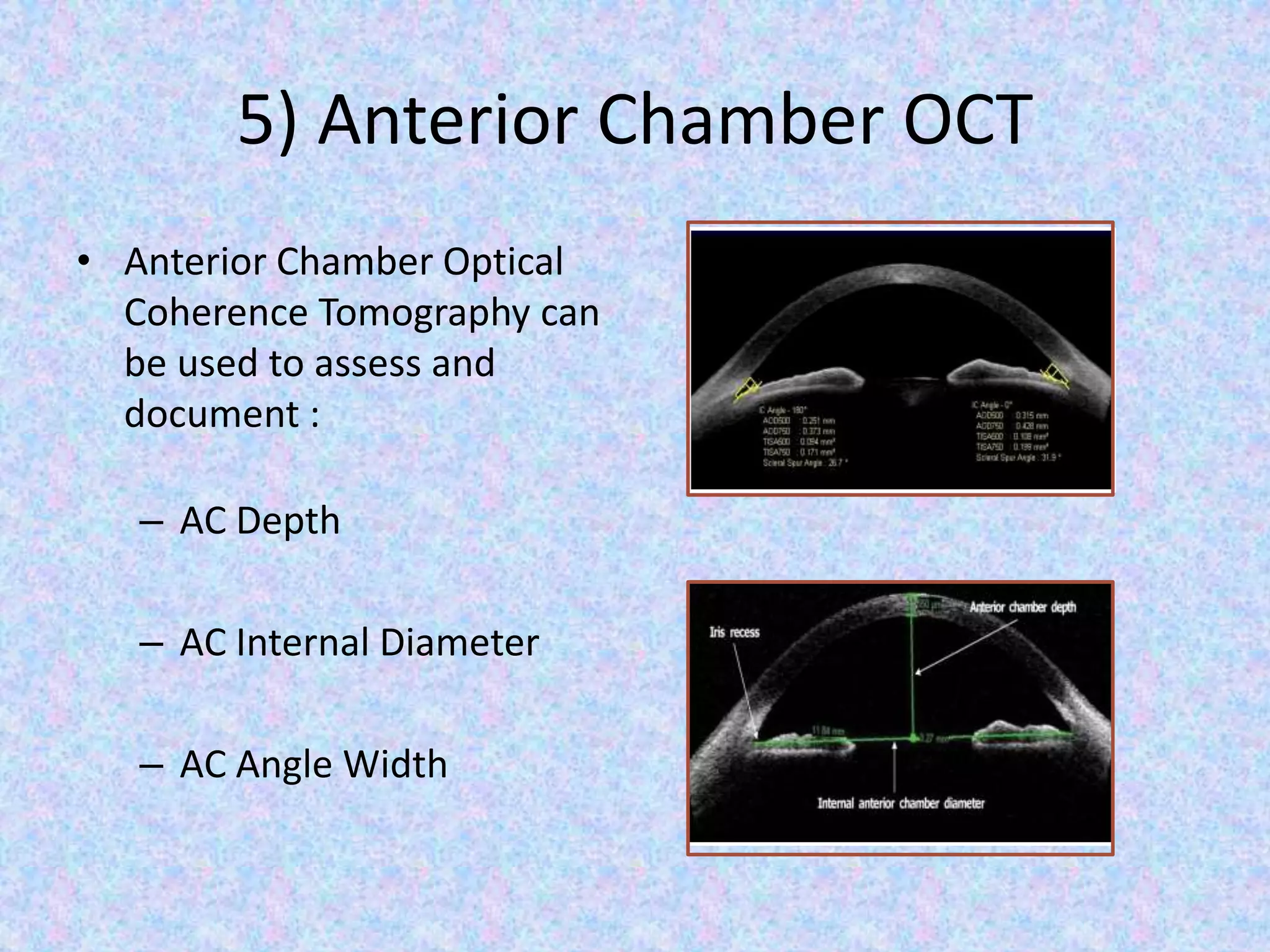
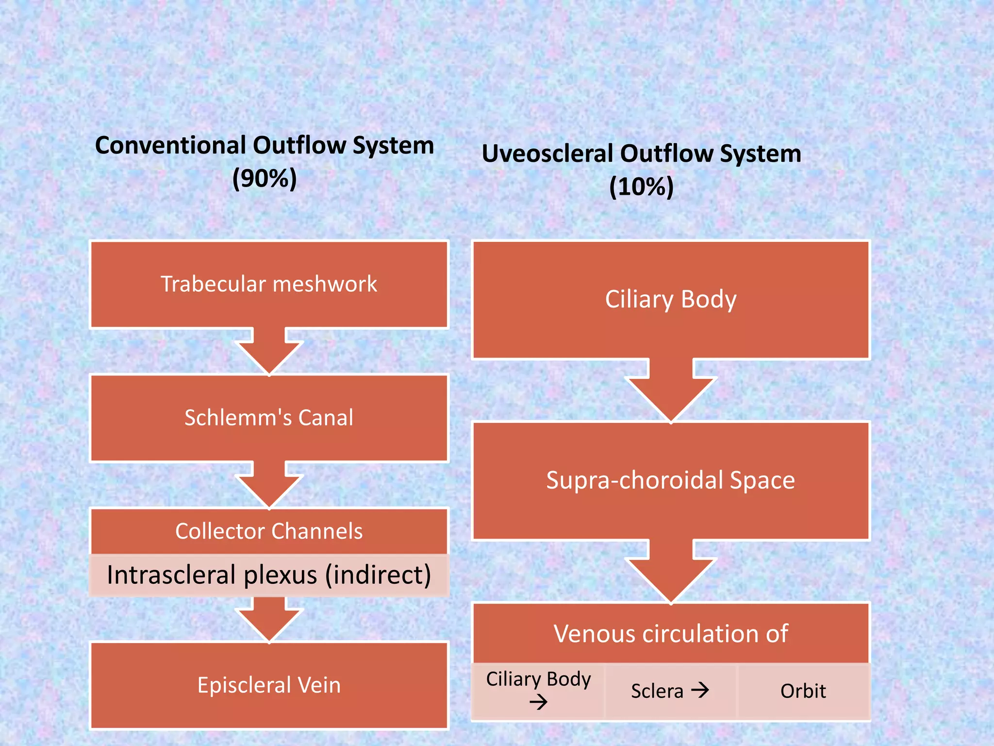
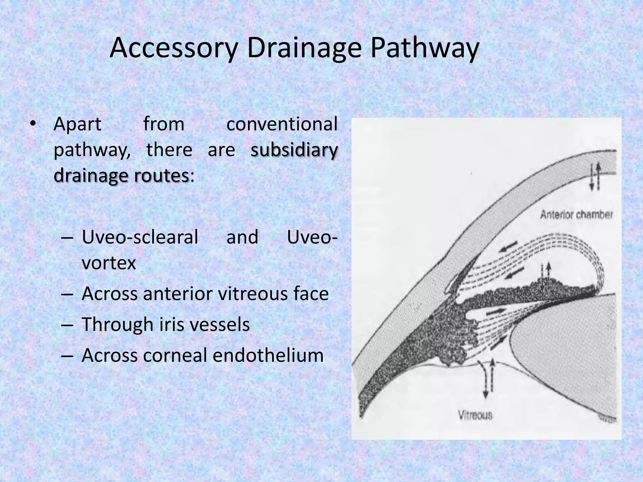
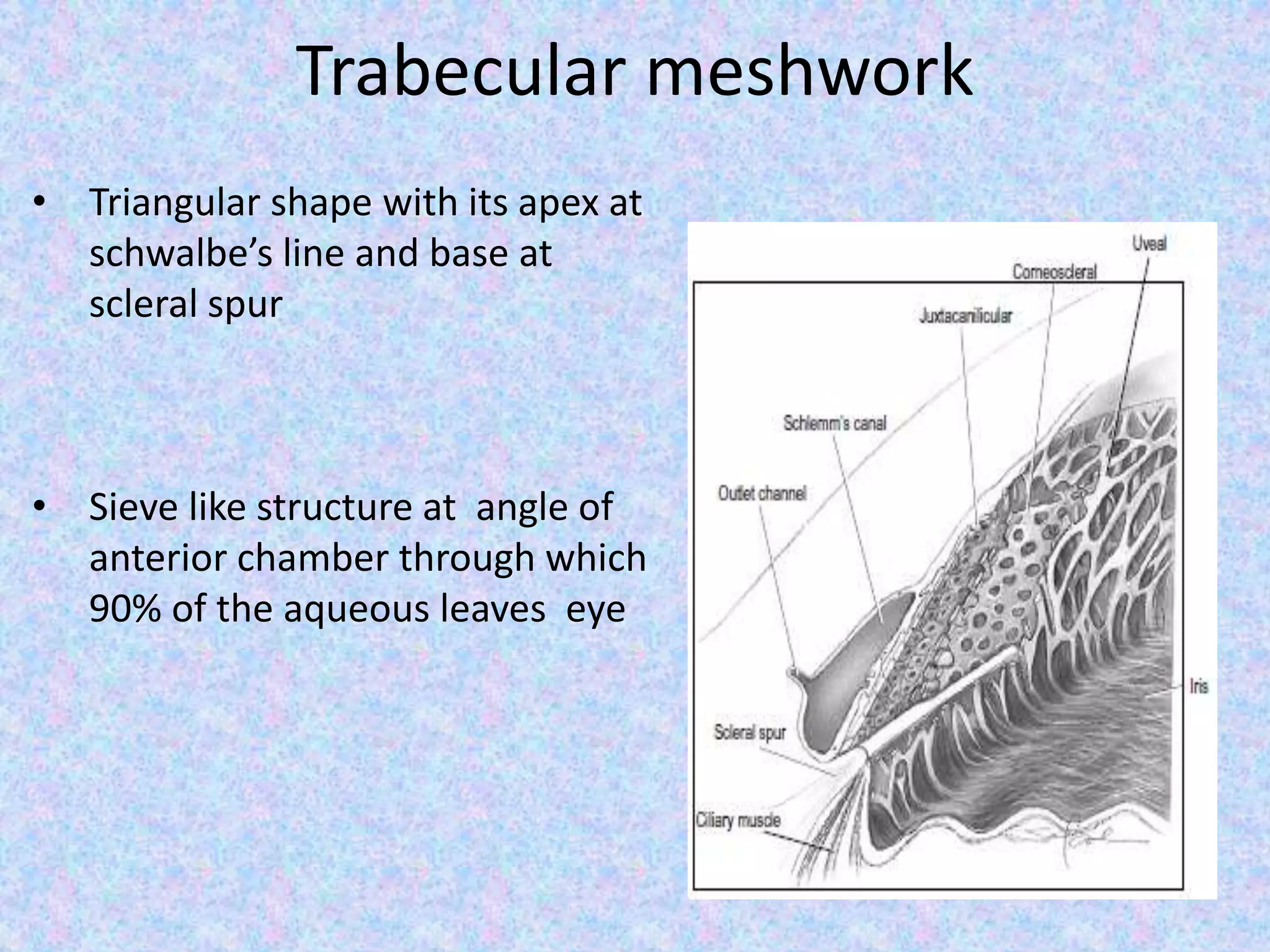
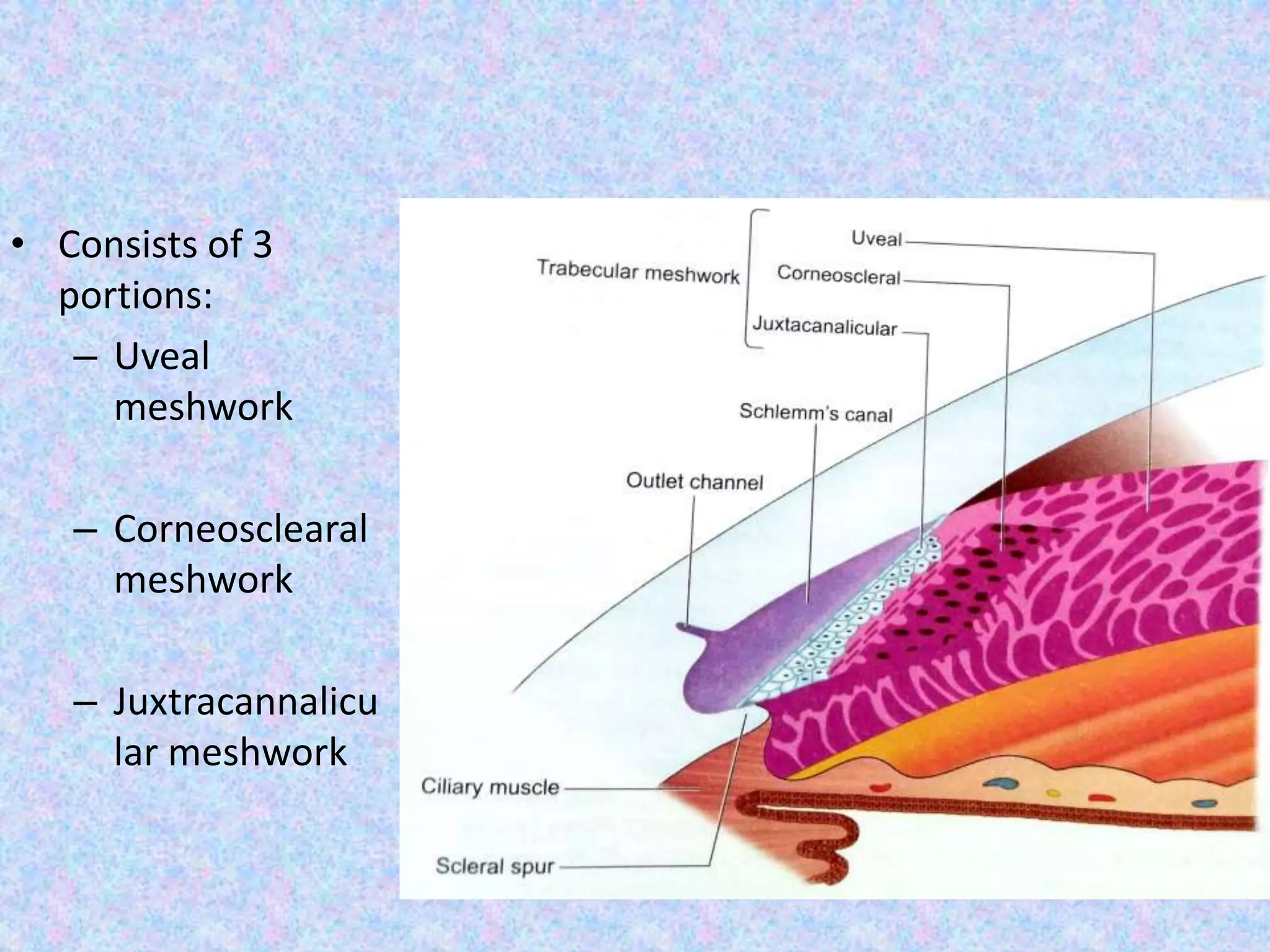
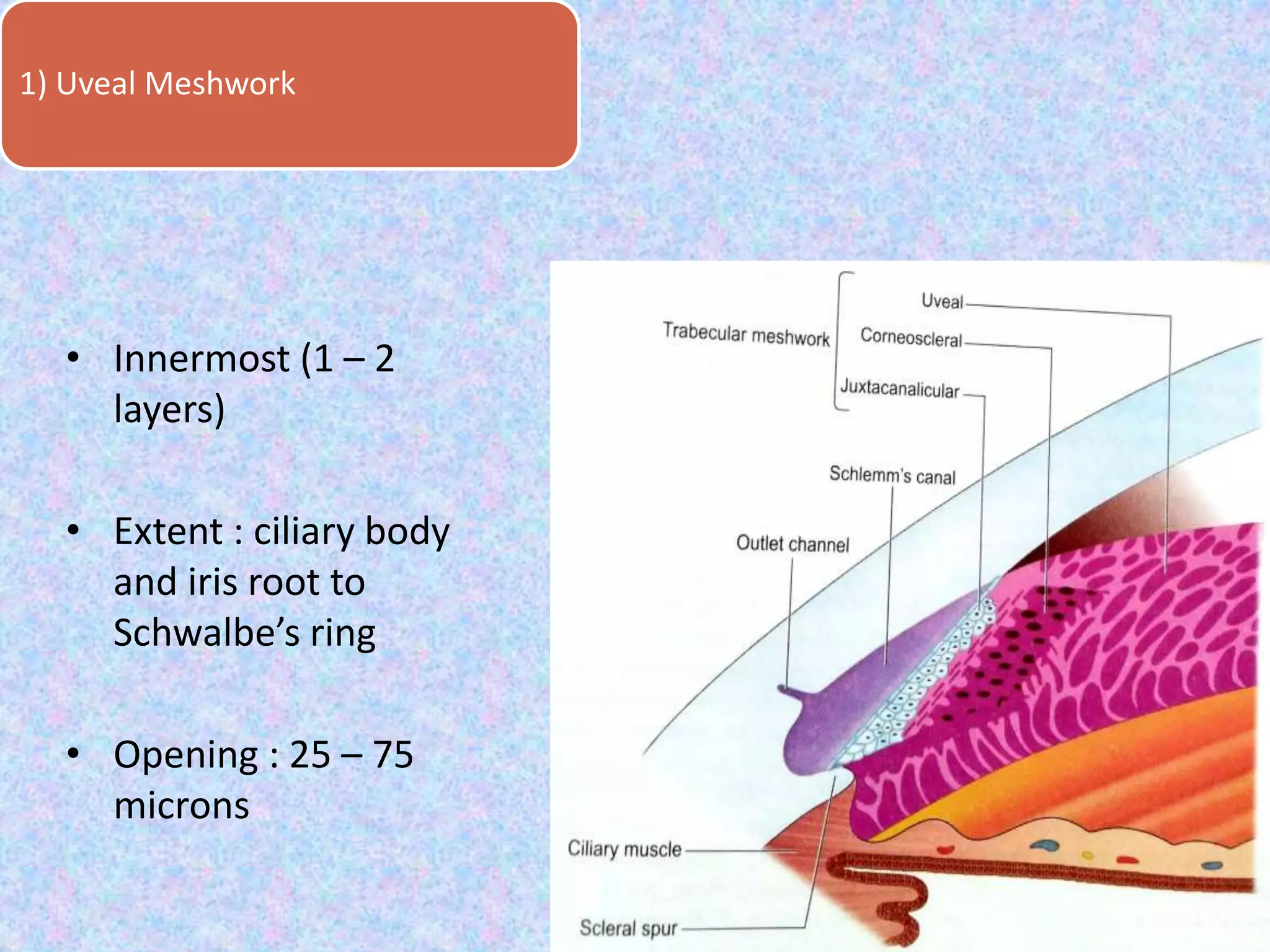
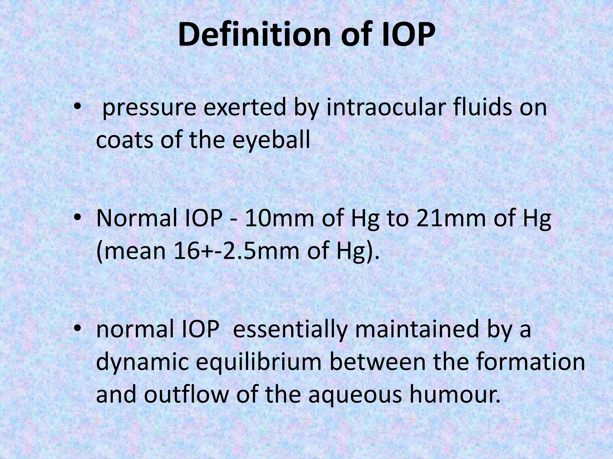
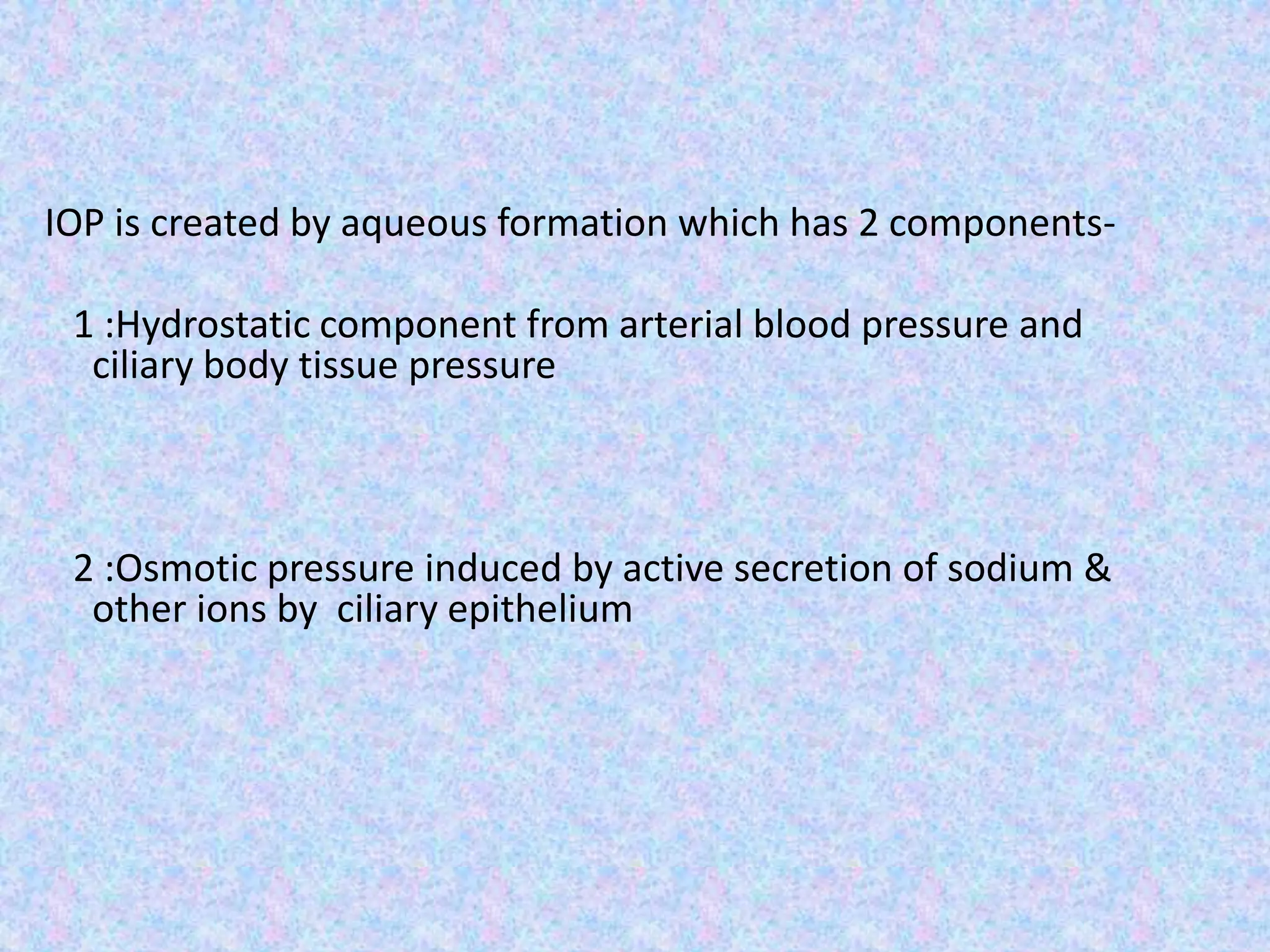
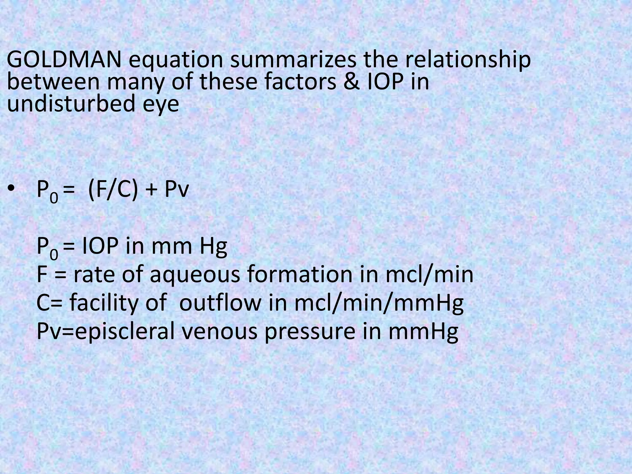
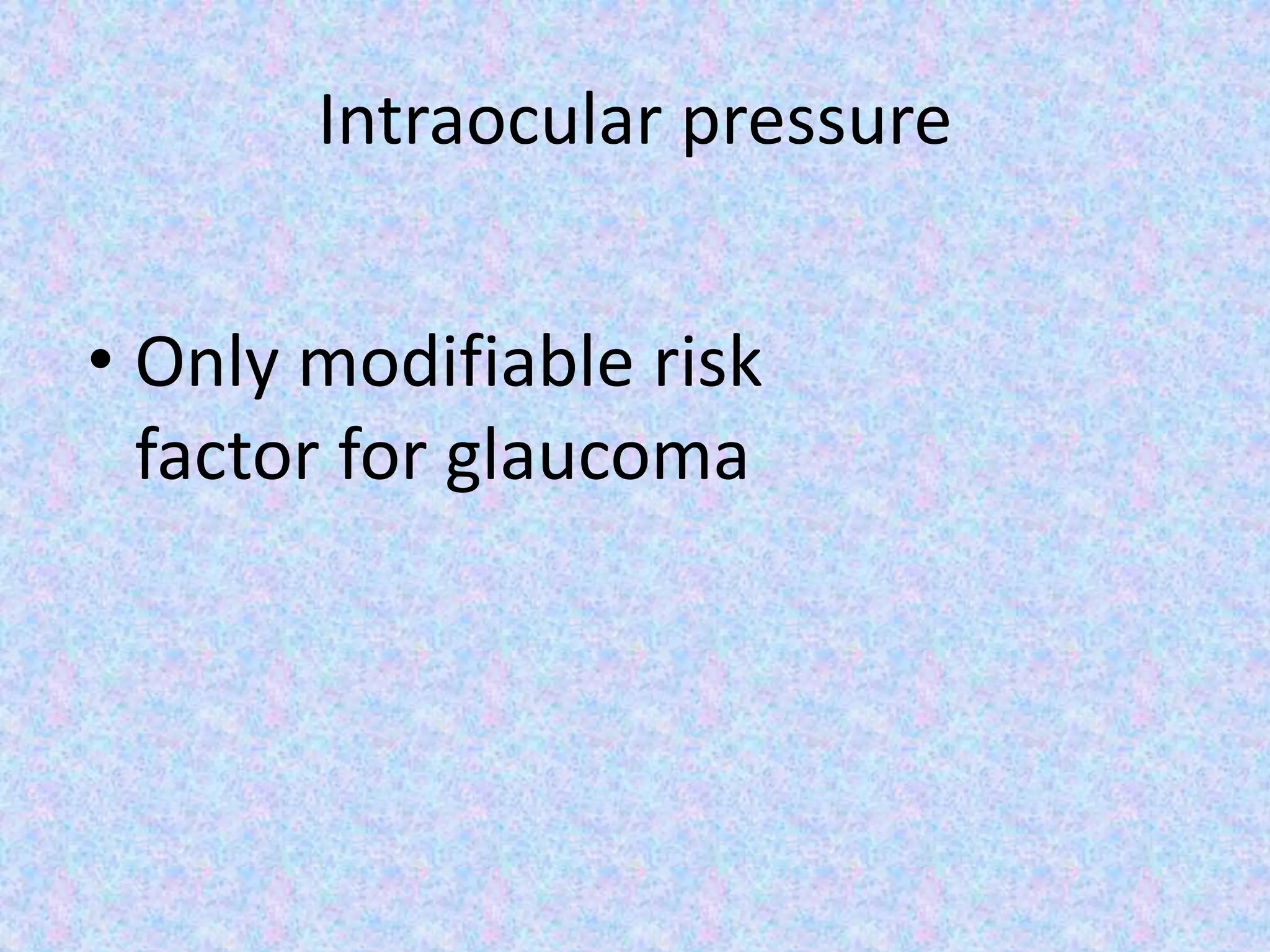
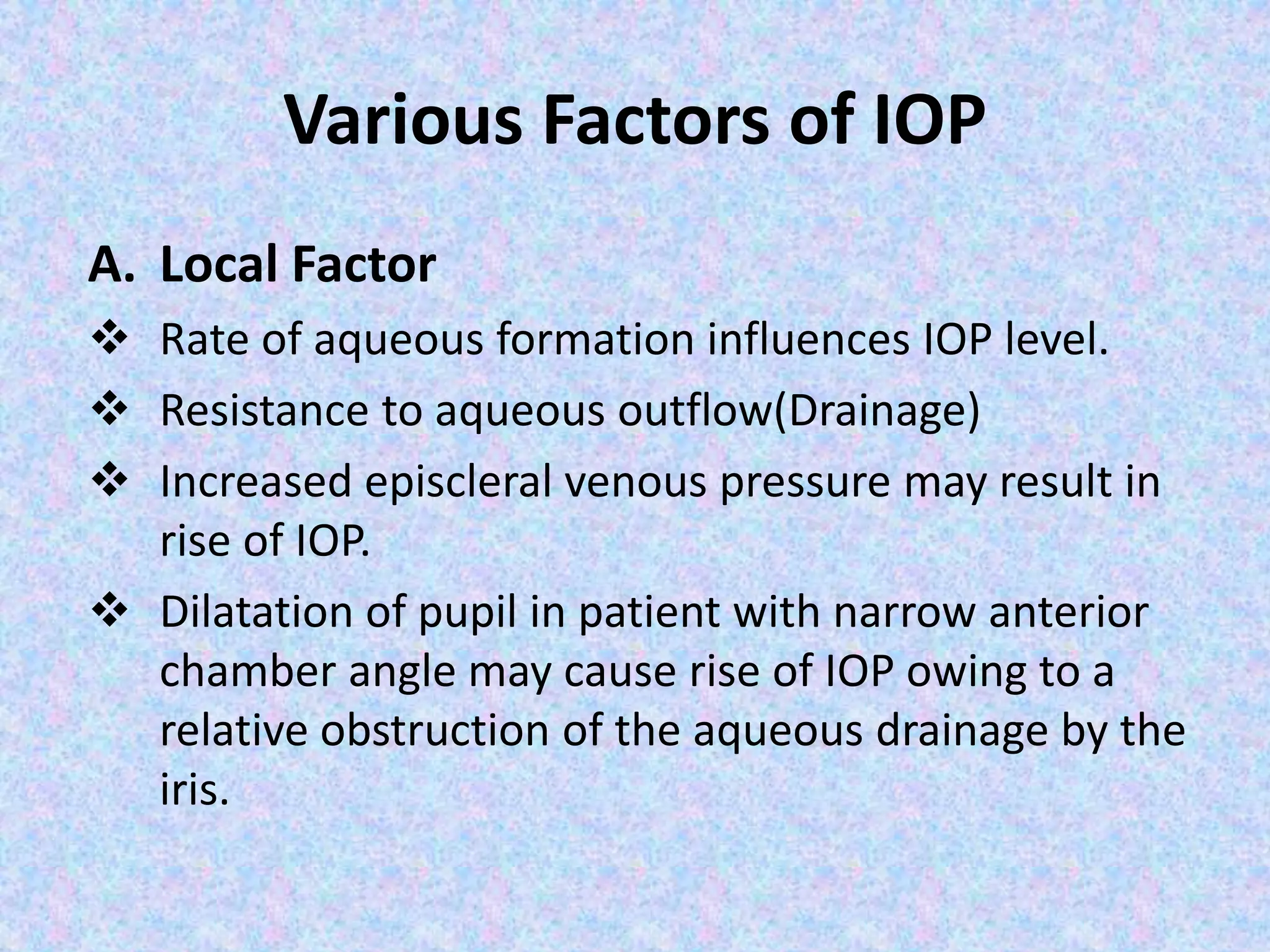
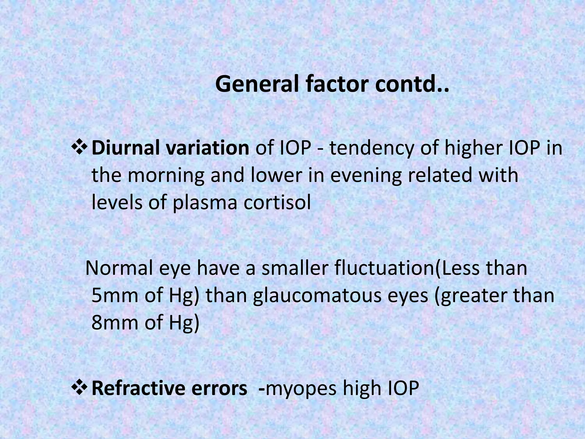
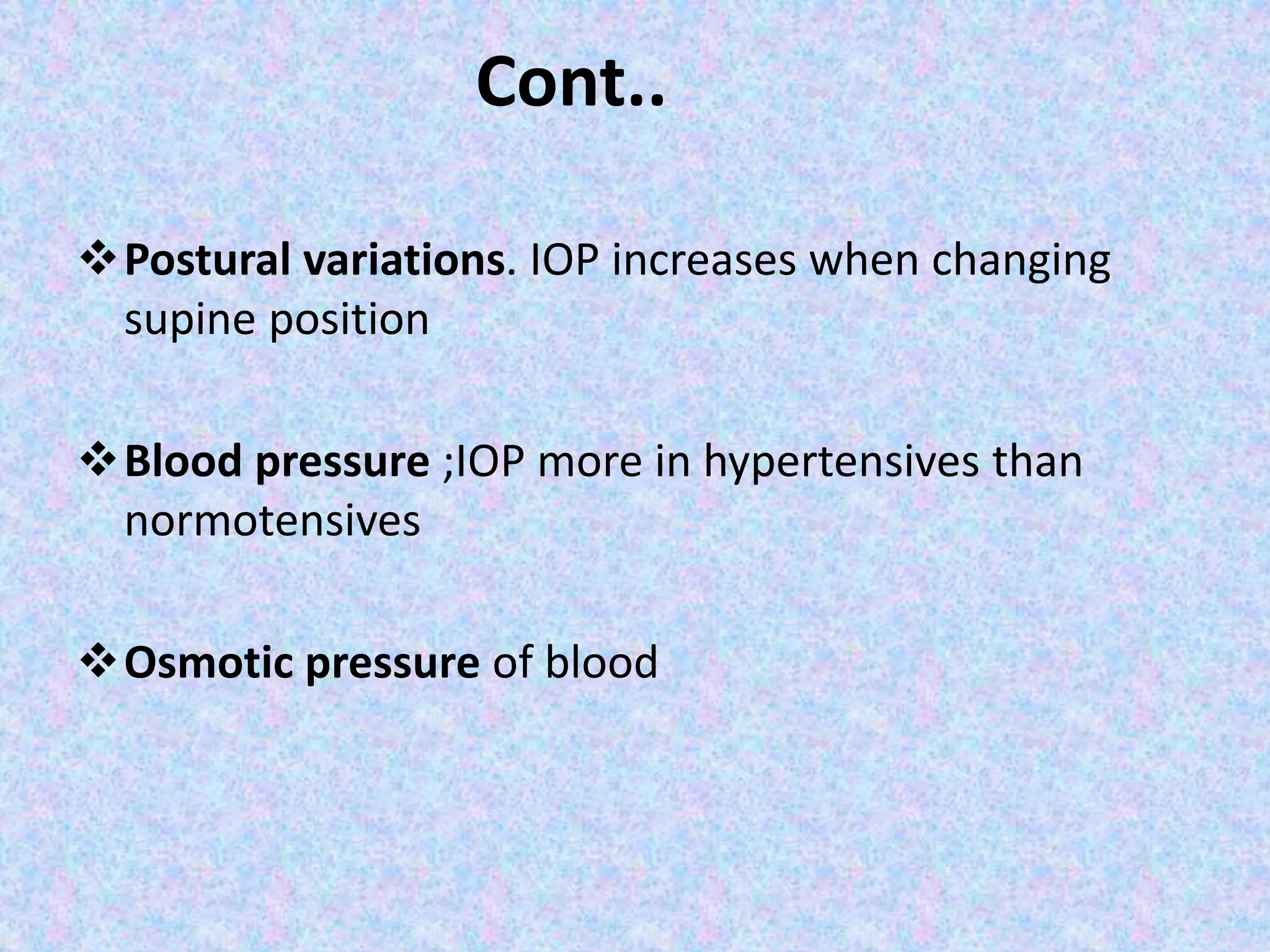
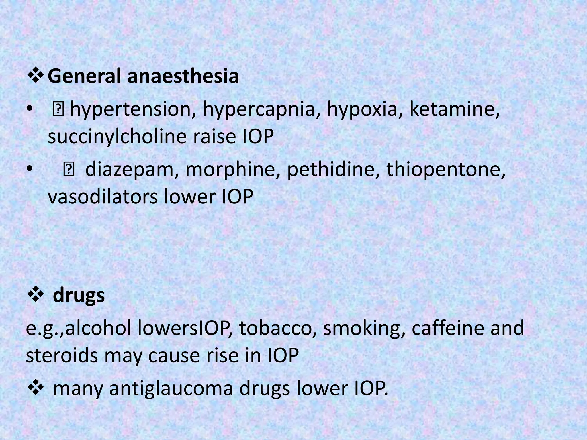
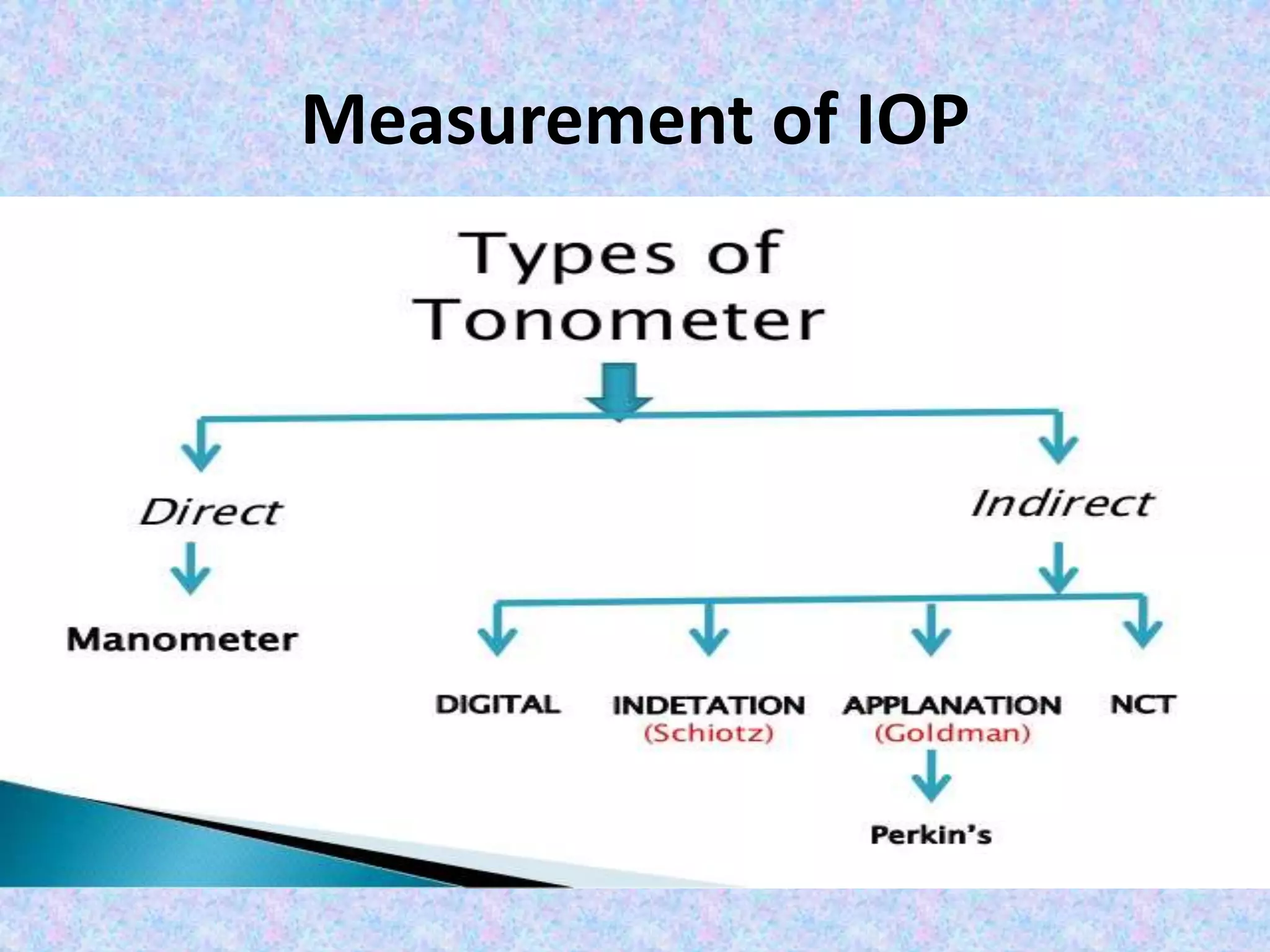
4. STRUCTURE WEEKS OF
GESTATION
DERIVED FROM
1. SCHLEMN CANAL 3RD MONTH Mesodermal mesenchyme
2. TRABECULAR MESHWORK 7TH WEEK Neural crest cells derived
mesenchymal cells
3. POSTERIOR CHAMBER Split in mesenchyme posterior to
developing iris and anterior to
developing lens
4. ANTERIOR CHAMBER Split in mesenchyme between
surface ectoderm and developing
iris
5. IRIDOCORNEAL ANGLE 15TH WEEK corneal endothelium extends and
meets iris epithelium forming this
angle
5. Importance of the angle of anterior
chamber
Site of aqueous drainage
Maintenance of Intra Ocular Pressure
⢠Glaucoma
⢠Angle recession
⢠Inflammation (hyphaema, hypopyon)
Explanation of pathophysiology of certain
ocular pathologies :
Therapeutic location for LASER therapy
8. Anatomy of Anterior chamber
Anterior boundary:
Posterior surface of cornea and peripherally by
trabecular meshwork
Posterior boundary:
In pupillary area ď anterior surface of lens
In peripheral area ď anterior surface of iris
Volume - 0.25ml
9. âŚ.continued
⢠chamber depth decreases by 0.01mm/year of life
⢠Shallower in hypermetropes than myopes
⢠chamber depth deepens by 0.06mm for each
diopter of myopia.
⢠decreases during accomodation
11. Anatomy of posterior chamber
⢠Volume: 0.06ml
Anterior boundary:
posterior surface of the iris and
part of ciliary body
Posterior Boundary:
lens and its zonules
Laterally
ciliary body
16. 16
1. Schwalbe line:
⢠Most anterior structure
⢠Signifies termination of descemetâs membrane
⢠Lie in plane of posterior corneal surface
â˘Transition between corneal endothelium and trabecular cells
⢠Appears as fine scalloped line (ring) in gonioscopy
17. Schwalbe line: Clinical importance
⢠Mid limbal incision during
cataract surgery
corresponds to schwalbe
line.
⢠In 15-20 % of normal
individuals - appears as
hypertrophied glistering
ridge - Posterior
Embryotoxon
17
19. Trabeculum
ď Anterior to scleral spur
ď Extends from schwalbe line to Scleral spur.
ď 600 micron width.
ď Anterior non functional part adjacent to schwalbe line-whitish.
ď Posterior functional pigmented part âgrayish blue translucent
appearance.
19
20. Trabeculum: Clinical importance
ď In laser trabeculoplasty - burns
applied to the junction between
pigmented and nonfunctional
trabecular meshwork
ď Pigmentation of trabeculum - rare
before puberty but more prominent
in brown iris
20
24. ..continued
Contain mechanoreceptors nerve ending to
measure stress at scleral spur caused by
change in IOP and ciliary muscle contraction.
Clinical importance: in laser trabeculoplasty - important to
know scleral spur
- If lasers are applied posterior to it - increased reaction in
anterior chamber - acute post laser rise in IOP.
26. Ciliary Body Band
⢠Posterior-most landmark
⢠Formed by anterior part of ciliary body between scleral spur
and root of iris.
⢠Width depends on the level of iris insertion
â Narrow in hyperopics
â Wide in myopia & aphakia, and in angle recession
and cyclodialysis
30. 2.Van-Herrickâs Grading using Slit-
Lamp
⢠Optical section
⢠60° between observation and illumination
⢠Full slit length
⢠Magnification approximately x 15
⢠Low to medium illumination
31. 2) Van-Herrickâs Grading using Slit-
Lamp
Grade III
(Mild
narrow)
PACD = Âź
- ½ CT
Grade II
(Moderat
e narrow)
PACD = Âź
CT
Grade I
(Extremel
y Narrow)
PACD < Âź
CT
Grade 0
(Closed
angle)
PACD = Nil
32. 3) Gonioscopic Assessment
⢠Oldest system based on visible posterior angle structure
⢠Higher the grade, narrower the angle
Scheie Classification (1957)
⢠Widely used based on angulation between posterior cornea and
iris root
⢠Higher the grade, wider the angle
Shafferâs System (1960)
⢠Newest system
⢠Complex 3D evaluation
Spaeth System
33.
34. Scheie Classification(1957)
Classification Structure visible
Wide open All structures visible
Grade I Iris root visible
Grade II Ciliary body obscured
GradeIII Posterior trabeculum obscured
Grade IV Only Schwalbe´s line visible
36. Grade Angle width Configuration Chances of
closure
Structure visible
IV 35-45 degree Wide open Nil Schwalbes line to
ciliary body
III 20-35 degree Open angle Nil Schwalbes to scleral
spur
II 20 Degree Mod narrow Possible Schwalbes to
trabecular meshwork
I 10 degree Very narrow High Schwalbes only
O 0 degree Closed Closed none
Shaffer system of grading
40. 5) Anterior Chamber OCT
⢠Anterior Chamber Optical
Coherence Tomography can
be used to assess and
document :
â AC Depth
â AC Internal Diameter
â AC Angle Width
42. Conventional Outflow System
(90%)
Episcleral Vein
Collector Channels
Intrascleral plexus (indirect)
Schlemm's Canal
Trabecular meshwork
Uveoscleral Outflow System
(10%)
Venous circulation of
Ciliary Body
ď
Sclera ď Orbit
Supra-choroidal Space
Ciliary Body
43. Accessory Drainage Pathway
⢠Apart from conventional
pathway, there are subsidiary
drainage routes:
â Uveo-sclearal and Uveo-
vortex
â Across anterior vitreous face
â Through iris vessels
â Across corneal endothelium
45. Trabecular meshwork
⢠Triangular shape with its apex at
schwalbeâs line and base at
scleral spur
⢠Sieve like structure at angle of
anterior chamber through which
90% of the aqueous leaves eye
46. ⢠Consists of 3
portions:
â Uveal
meshwork
â Corneosclearal
meshwork
â Juxtracannalicu
lar meshwork
47. 1) Uveal Meshwork
⢠Innermost (1 â 2
layers)
⢠Extent : ciliary body
and iris root to
Schwalbeâs ring
⢠Opening : 25 â 75
microns
48. ⢠Larger middle portion (8 â 15 layers)
⢠Extent : scleral spur to Schwalbeâs ring
⢠Openings : elliptical measuring
5 â 50 microns with openings getting smaller
as they approach
51. Trabecular cells
⢠Elongated cells
⢠Length â 120 um ; thickness â 4 to 8 um
⢠Long cytoplasmic processes
⢠Apposed cells â macula adherentes and gap
junctions
⢠Function:
-synthetic activites (collagen and GAG)
-phagocytic activity
52. ⢠Cortical zone:
-surrounds trabecular cells
-attached to trabecular cells by hemidesmosomes
-contains collagen
⢠Core:
-formed by type I, II, IV collagen,fibronectin,
elastic tissue and glycosaminoglycan
-elastic tissue imparts a recoil to the elements of
the meshwork
53. Schlemm's Canal (Sinus Venosus Sclerae)
Endothelial lined oval channel
present circumferentially in
scleral sulcus.
⢠Inner wall ď lined by
endothelial cells which are
irregular, spindle shaped and
containing giant vacuoles.
⢠Outer wall ď lined by
endothelium which are
smooth and flat. Contain
numerous openings for
collector channels.
54. Collector channels
⢠Also called intrascleral aqueous vessels
⢠25-35 in number
⢠leave the schlemâs canal at oblique angles to terminate ultimately
into episcleral veins
Lined by endothelium similar to outer wall of schlemâs canal
Direct system Indirect system
Terminate direct into Terminate into episcleral vein
the episcleral vein via 3 interconnecting venous
plexuses ( deep & mid -
intrascleral & episcleral) 54
56. Episcleral Veins
⢠They drain into :
â˘
Anterior ciliary vein
Superior ophthalmic vein
Cavernous sinus
57. Resistance to aqueous outflow
⢠Juxtacanalicular system
⢠Endothelial layer of Schlemm´s
⢠extracellular matrix and cellular elements like GAG
⢠GAG trap larger volume of water reducing functional
diameter of flow channel
58. Ageing eye and Open angle glaucoma
Ageing eye provide increased resistance to aqueous outflow due
to:
- Thickening and fusion of trabecular sheets
- Loss of endothelial cells
- Accumulation and alteration of extracellular matrix
- Decrease in number of gaint vacuoles
All the above changes when exaggerated leads to Open angle
glaucoma.
59. Definition of IOP
⢠pressure exerted by intraocular fluids on
coats of the eyeball
⢠Normal IOP - 10mm of Hg to 21mm of Hg
(mean 16+-2.5mm of Hg).
⢠normal IOP essentially maintained by a
dynamic equilibrium between the formation
and outflow of the aqueous humour.
60. IOP is created by aqueous formation which has 2 components-
1 :Hydrostatic component from arterial blood pressure and
ciliary body tissue pressure
2 :Osmotic pressure induced by active secretion of sodium &
other ions by ciliary epithelium
61. GOLDMAN equation summarizes the relationship
between many of these factors & IOP in
undisturbed eye
⢠P0 = (F/C) + Pv
P0 = IOP in mm Hg
F = rate of aqueous formation in mcl/min
C= facility of outflow in mcl/min/mmHg
Pv=episcleral venous pressure in mmHg
63. Intraocular pressure
⢠Glaucoma screening based solely on IOP> 21
mm of Hg misses half of people with glaucoma
and optic nerve damage
⢠Normal IOP defined as that pressure which
does not lead to glaucomatous damage of
optic nerve head
64. Various Factors of IOP
A. Local Factor
ďś Rate of aqueous formation influences IOP level.
ďś Resistance to aqueous outflow(Drainage)
ďś Increased episcleral venous pressure may result in
rise of IOP.
ďś Dilatation of pupil in patient with narrow anterior
chamber angle may cause rise of IOP owing to a
relative obstruction of the aqueous drainage by the
iris.
65. Sex
⢠no major effect on IOP in 20-40 year age group
⢠in older age rise in mean IOP with increasing age in women
Age
⢠pediatric cohort showed trend of increasing IOP with age and
approached adult levels by age of 12 years
⢠with aging reduced facility of aqueous outflow and
uveoscleral outflow
⢠episcleral venous pressure does not change significantly with
advancing age
B. General Factor
66. Genetics
- twin studies, IOP highly correlated between monozygotic
than dizygotic twins
- several loci on chromosomes linked to IOP but no âIOP
genesâ reported
Environment
- reduced gravity causes sudden and marked increase in
IOP
- exposure to cold air reduces IOP
67. General factor contd..
ďśDiurnal variation of IOP - tendency of higher IOP in
the morning and lower in evening related with
levels of plasma cortisol
Normal eye have a smaller fluctuation(Less than
5mm of Hg) than glaucomatous eyes (greater than
8mm of Hg)
ďśRefractive errors -myopes high IOP
68. Cont..
ďśPostural variations. IOP increases when changing
supine position
ďśBlood pressure ;IOP more in hypertensives than
normotensives
ďśOsmotic pressure of blood
69. ďśGeneral anaesthesia
⢠hypertension, hypercapnia, hypoxia, ketamine,
succinylcholine raise IOP
⢠diazepam, morphine, pethidine, thiopentone,
vasodilators lower IOP
ďś drugs
e.g.,alcohol lowersIOP, tobacco, smoking, caffeine and
steroids may cause rise in IOP
ďś many antiglaucoma drugs lower IOP.
73. References
⢠Wolffâs Anatomy
⢠BCSC section 10 (2016-2017), Glaucoma -AAO
series
⢠Becker and Shafferâs Diagnosis and Therapy of
Glaucoma-7th Edition
⢠Clinical Ophthalmology- Jack J Kanski-8th
Edition
⢠Khuranaâs Anatomy and Physiology Of Eye- 3RD
Edition
Editor's Notes
Three waves of tissue come forward betwn surface ectoderm n developing lens from undifferentiated mesenchymal mass of neural crest origin and contribute to formation of structures of anterior segments of eyeball
Developmental anomaly like aniridia, Rieger anomalyâŚTo note extent of neovascularisation
Measures 2.5 to 3.0 mm deep in the center in normal adult⌠Communicates with the posterior chamber through the pupil
Chamber becomes shallower with accommodation due to..Increased lens curvature.. Forward translocation of the lens
An angle recess formed in between posterior surface of cornea and anterior surface of irisâŚ. bounded by:(anterior to posterior)1. cornea, 2.sclera, 3.ciliary body 4.the iris.
This angle can be reached from outside to inside as 1. conjunctival epithelium 2.conjunctival stroma 3.tenonâs capsule n episcleral 4.lcorneoscleral stroma-midlimbus 5.trabecular meshwork and schlemn canal-deep limbus
Contains collagen fibers with elastic fibers.. - insertion of trabecular meshwork into corneal stroma.
Fibres of ciliary muscle are inserted in SS which contract to pull the scleral spur posteriorly rotating the trabeculum inward , thus opening of the SC lumen and enlargement of intertrabecular spaces.
Projection of torch lightâŚFrom temporal side Parallel to the iris surface.. Observation of light Illumination pattern on iris Presence / absence of shadow.. If 2/3rd or more of the nasal iris is in shadow, the chamber is shallow
Narrowest and brightest beam of light is directed towards the temporal limbus at 60 degree.Periphral anterior chamber (PAC) depth is compared to corneal thickness (CT)
A-ant to sl,b-betn sl n ss ,c- ss visible,d-deep with cbb visible, d-extremely deep >1mm cbb visible. Peripheral iris: f-flat,b-bowed anteriorly,p-plateau iris, f-flat c-concave
Internal Anteriorly âSchwalbeâs line ..Posteriorly â Scleral spur..Accomodates canal of Schlemm ..trabecular meshwork(corneoscleral portion)
Large middle portion..8-15 layers thick,,Extent :anteriorly :merge with inner corneal lamellae..posteriorly : scleral spur..contains flattened perforated sheets..Intertrabecular space: 5-20 um
Cells attach to one another by macula occludentes, desmosomes, gap junctions..Intercellular space : 10 um ..Function: outflow resistance..phagocytic..consists of a layer of connective tissue lined on either side by endothelium
Arise from outer wall schlemmâs canal..25-35 in number..Valveless..Aqueous veins..episcleral veins.Deep intrascleral plexus..intrascleral plexus..episcleral veins