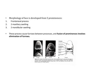
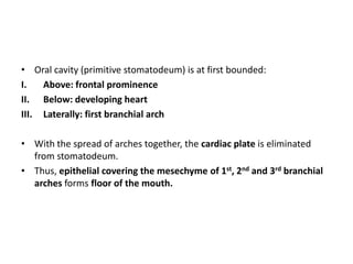
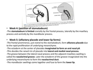
Central face begins to develop by 4th week, when olfactory placodes appear on both sides of the frontonasal process.
Gradually both placodes develop to form the median and lateral nasal process.
Upper lip is formed by 6th week by fusion of two median nasal processes in midline and the maxilllary process of the 1st branchial arch.
PRE-NATAL GROWTH AND DEVELOPMENT OF PALATEFormation of primary and secondary palate
Elevation of palatal shelves
Fusion of palatal shelves
Amelogenesis is the formation of enamel. During amelogenesis, the ameloblast (enamel-forming cells) undergo various stages i.e the life cycle of ameloblast.
For more content check out my blog: www.rkharitha.wordpress.com "a little about everything dental"
This Slide, gives a Brief introduction to the Anatomy of the tooth specifically the outer shell, the enamel, including the structures, development and abnormalities.
Created by Dr. Mohsen S. Mohamed
For Ozident.com
Tongue is a muscular structure that has the organs of taste reception. The organs for sense of taste are the taste buds. Tongue is located inside the mouth and is an important muscle. It does not have any bones. It is reddish-pink in color. The main function of tongue is taste, help in chewing food, in swallowing food and speech.
The Indian Dental Academy is the Leader in continuing dental education , training dentists in all aspects of dentistry and offering a wide range of dental certified courses in different formats.
Indian dental academy provides dental crown & Bridge,rotary endodontics,fixed orthodontics,
Dental implants courses.for details pls visit www.indiandentalacademy.com ,or call
00919248678078
Face develops in humans between 4th – 10th week of intrauterine life.
prenatal growth of the maxilla
DEVELOPMENT OF UPPER LIP
Development of lower lip
Development of nose
hare lip
OBLIQUE FACIAL CLEFT
macrostomia
lateral facial cleft
microstomia
PRENATAL GROWTH OF MANDIBLE
Occurs between the 4th and 7th week of intrauterine life.
4th week of intrauterine life
Formation of the head fold
Following which the developing brain and the pericardium form 2 prominent bulges on the ventral aspect of the embryo.
The 2 bulges are separated from each other by a shallow depression called stomatoedum (corresponding to the primitive mouth).
Floor of the stomatodeum is formed by the Buccopharyngeal membrane, which separates the stomatodeum from the foregut.Soon, mesoderm covering the developing forebrain proliferates, and forms a downward projection that overlaps the upper part of the stomatodeum – this downward projection is called frontonasal process.
Development of Face /certified fixed orthodontic courses by Indian dental ac...Indian dental academy
The Indian Dental Academy is the Leader in continuing dental education , training dentists in all aspects of dentistry and offering a wide range of dental certified courses in different formats.
Welcome to TechSoup New Member Orientation and Q&A (May 2024).pdfTechSoup
In this webinar you will learn how your organization can access TechSoup's wide variety of product discount and donation programs. From hardware to software, we'll give you a tour of the tools available to help your nonprofit with productivity, collaboration, financial management, donor tracking, security, and more.
Embracing GenAI - A Strategic ImperativePeter Windle
Artificial Intelligence (AI) technologies such as Generative AI, Image Generators and Large Language Models have had a dramatic impact on teaching, learning and assessment over the past 18 months. The most immediate threat AI posed was to Academic Integrity with Higher Education Institutes (HEIs) focusing their efforts on combating the use of GenAI in assessment. Guidelines were developed for staff and students, policies put in place too. Innovative educators have forged paths in the use of Generative AI for teaching, learning and assessments leading to pockets of transformation springing up across HEIs, often with little or no top-down guidance, support or direction.
This Gasta posits a strategic approach to integrating AI into HEIs to prepare staff, students and the curriculum for an evolving world and workplace. We will highlight the advantages of working with these technologies beyond the realm of teaching, learning and assessment by considering prompt engineering skills, industry impact, curriculum changes, and the need for staff upskilling. In contrast, not engaging strategically with Generative AI poses risks, including falling behind peers, missed opportunities and failing to ensure our graduates remain employable. The rapid evolution of AI technologies necessitates a proactive and strategic approach if we are to remain relevant.
Biological screening of herbal drugs: Introduction and Need for
Phyto-Pharmacological Screening, New Strategies for evaluating
Natural Products, In vitro evaluation techniques for Antioxidants, Antimicrobial and Anticancer drugs. In vivo evaluation techniques
for Anti-inflammatory, Antiulcer, Anticancer, Wound healing, Antidiabetic, Hepatoprotective, Cardio protective, Diuretics and
Antifertility, Toxicity studies as per OECD guidelines
Honest Reviews of Tim Han LMA Course Program.pptxtimhan337
Personal development courses are widely available today, with each one promising life-changing outcomes. Tim Han’s Life Mastery Achievers (LMA) Course has drawn a lot of interest. In addition to offering my frank assessment of Success Insider’s LMA Course, this piece examines the course’s effects via a variety of Tim Han LMA course reviews and Success Insider comments.
Acetabularia Information For Class 9 .docxvaibhavrinwa19
Acetabularia acetabulum is a single-celled green alga that in its vegetative state is morphologically differentiated into a basal rhizoid and an axially elongated stalk, which bears whorls of branching hairs. The single diploid nucleus resides in the rhizoid.
How to Make a Field invisible in Odoo 17Celine George
It is possible to hide or invisible some fields in odoo. Commonly using “invisible” attribute in the field definition to invisible the fields. This slide will show how to make a field invisible in odoo 17.
Palestine last event orientationfvgnh .pptxRaedMohamed3
An EFL lesson about the current events in Palestine. It is intended to be for intermediate students who wish to increase their listening skills through a short lesson in power point.
Operation “Blue Star” is the only event in the history of Independent India where the state went into war with its own people. Even after about 40 years it is not clear if it was culmination of states anger over people of the region, a political game of power or start of dictatorial chapter in the democratic setup.
The people of Punjab felt alienated from main stream due to denial of their just demands during a long democratic struggle since independence. As it happen all over the word, it led to militant struggle with great loss of lives of military, police and civilian personnel. Killing of Indira Gandhi and massacre of innocent Sikhs in Delhi and other India cities was also associated with this movement.
2. •
1.
2.
3.
•
Morphology of face is developed from 5 prominences:
Frontonasal process
2 maxillary swelling
2 mandibular swelling
These process cause furrows between processes, and fusion of prominences involves
elimination of furrows.
3. • Oral cavity (primitive stomatodeum) is at first bounded:
I.
Above: frontal prominence
II. Below: developing heart
III. Laterally: first branchial arch
• With the spread of arches together, the cardiac plate is eliminated
from stomatodeum.
• Thus, epithelial covering the mesechyme of 1st, 2nd and 3rd branchial
arches forms floor of the mouth.
4. • Week 4: (position of stomatodeum)
-The stomatodeum is limited cranially by the frontal process, laterally by the maxillary
process and ventrally by the mandibular process.
• Week 5: (olfactory placode and lower lip forms)
-The frontal prominence, just rostral to the stomatodeum, form olfactory placode due
to the rapid proliferation of underlying mesenchyme.
-The ectoderm at the center of placodes invaginated to form an oral nasal pit
-This devides the raised rim of placode into lateral and medial nasal process.
-The groove between the lateral nasal process and the adjacent maxillary swelling is
called the nasolarimal groove. Later the ectoderm of this groove invaginated into the
underlying mesenchyme to form the nasolacrimal duct.
-The mandibular swellings come together and fuse to form the lower lip.
5. • Week 6: (septum and philtrum formation)
-At the beginning of week 6, the lateral part of the face expands and broadens. This causes the
eyes and maxillary process which where initially located at the side of the face to come to the
front of the face.
-The medial nasal process migrates towards each other and fuse to form the septum of the
nose.
-The inferior tips of the medial nasal process expand laterally and inferiorly and fuse to form the
intermaxillary process. The tip of the maxillary swelling grows to meet the intermaxillary process
and fuse. This gives rise to the philtrum.
6. -By the time of face development, the epithelium at the inferior border of maxillary process and
superior border of mandibular process proliferate and thickens.
-When fuse they form a single primary epithelium band, joining the upper and lower jaw.
8. Nasal passage:
At the end of 6th week:
-The nasal pits deepens and fuse to form a single, enlarged,
ectodermal nasal sac lying super posterior to the
intermaxillary process.
At the end of 6th week to the beginning of the 7th
week:
-The floor and the posterior wall of the nasal sac proliferate to
form the nasal fin separating the nasal sac from oral cavity.
-Vacuoles develop in the nasal fin and fuse with nasal sac, the sac
enlarges and thinning the fin to a thin membrane called the
oronasal membrane.
-oral nasal membrane separate the sac from oral cavity.
During the 7th week:
-Oronasal membrane ruptures to form an opening called primitive
choana.
-The floor of the nasal cavity (primary palate) is formed by the
Posterior extension of the Intermaxillary process.
9. Secondary palate formation (week78):
-Formation involves the fusion of:
i. Right and left maxillary processes
(palatine shelf)
ii. Median nasal process
-The median nasal process grows
downward and forward to form the nasal
septum.
-The palatine shelves, at first grows
downward to the elevated the tongue.
-As a result of the enlargement of the
mandible, the tongue drops to the floor of the
stomatodeum.
-When the tongue is moved from the growing
lateral palatine processes, the processes are
straightened to a horizontal position and
grows medially towards and fuse with
each other with primary palate to form the
secondary palate
10. -The nasal septum fuse with the upper surface of the primary and secondary palates along
the midline.
-Now nasal cavity opens into the pharynx behind the secondary palate through an opening
called the definitive choana
12. Tongue development:
• Pharyngeal arch: 1, 3, and 4
• Occipital Somite Mesoderm
Begins late of 4th week
1st arch:
-Forms a median swelling called tuberculum impar.
-A pair of lateral swellings, the distal tongue buds. The lateral swellings quickly enlarge
and merge with each other and also with median swelling and form the anterior
two-third of the tongue.
2nd arch:
-Develop a midline swelling called
copula.
5th and 6th week:
a midline swelling of the 3rd and 4th arches called
hypopharyngeal eminence overgrows the copula
which disappears. Thus, forms the posterior onethird of the tongue.
(Hypo pharyngeal eminence expand mainly by the
growth of 3rd arch endoderm.)
A depression called foramen cecum is visible where the
median and terminal sulcus intersect.
13. 4th arch:
-Marks the development of epiglottis.
-Tongue separates from the floor of the mouth by a downward growth of ectoderm
around its periphery, subsequently degenerates to form lingual sulcus and gives the
tongue mobility.
14. Muscles of the tongue:
-Different origin.
-Arise from the mesoderm of the occipital somites, which have migrated forward
into the tongue area.
-With their nerve- 12th cranial nerve Hypoglossal nerve (motor).
Nerve supply (sensory) correspond to their origin:
i. 1st arch nerve 5th cranial (trigeminal) nerve supply anterior two-third.
ii. 3rd arch nerve 9th cranial nerve (glossopharyngeal) supply the posterior
one third.
16. Formation of mandible (week 6-10):
-Cartilage of 1st arch (meckel’s cartilage) is associated to this formation but makes no contribution
to it.
6th week: (meckel’s cartilage)
-Cartilage extend as a hyaline cartilagenous rod, surronded by fibrocellular capsule.
-The 2 cartilage (from left and right) do not meet at midline but are separated by a thin band
of mesenchyme.
-Mandibular nerve comes along 2/3
of the way along the length of cartilage.
At this point, it divides into lingual branch
(medial) and inferior alveolar branch
(lateral) which run along medial and
lateral aspect of the cartilage.
-Inferior alveolar further divides into
incisor and mental branches anteriorly.
-In the angle between incisor and mental
branch, condensation of mesenchyme
occurs.
17. 7th week:
•
First bone
-Intramembranous ossification begins in this condensation forming first bone.
-From this center, the bone spreads anteriorly to the midline and posteriorly to the point
where mandibular nerve divides.
-As it spreads anteriorly to midline, the bone forms a trough (drain) that consist of lateral and
medial plates that unite beneath the incisor nerve.
-It then comes into close approximation with similar trough formed in the adjoining
mandibular process.
-Trough is converted to a canal as bone form over the nerve , joining lateral and medial plate.
•
Backward extension of ossification
-Similar as forward but backward, it forms a
trough at the lateral aspect of meckel’s
cartilage.
-It is then converted to a canal that contains
inferior alveolar nerve.
•
Ramus of mandible
-Developed by a rapid spread of ossification
posteriorly into mesenchyme of 1st branchial
arch, turning away from the meckel’s cartilage.
-This point of diverge is marked by the lingula.
18. Fate of meckel’s cartilage:
1. Posterior extremity: Incus and malleus of inner ear.
2. Posterior extremity Sphenomalleolar ligament.
3. From sphenoid to the division of the mandibular nerve, cartilage is lost totally.
But its fibrous cellular capsule persist as the Sphenomandibular ligament.
4. From lingula forward to the division of the alveolar nerve: degrade, space
replace by new bone.
Further growth of mandible:
Three secondary growth cartilages appeared (Compare to primary cartilage):
i. Cells are larger
ii. Less intercellular matrix formed.
1. Condylar cartilage: appears during 12th week. Converted quickly to bone by
endochondral ossification.
2. Coronoid cartilage: anterior border and top of the condylar cartilage.
Disappears long before birth.
3. Symphyseal cartilage: two end of Meckel’s cartilage. Obliterated within the 1st
year after birth.
19. • Conclusion:
-Membrane bone is developed in relation to the nerve of 1st branchial arch and
entirely independent of meckel’s cartilage.
-It consist of neural, alveolar, and muscular element.
--It’s growth is assisted by development of secondary cartilage.